Library › Breast carcinoma pathology: invasive carcinoma, special types, biomarkers and reporting
Breast carcinoma pathology: invasive carcinoma, special types, biomarkers and reporting
Last reviewed 2026-11-10 by a board-certified pathologist. 109 references.
Invasive Breast Carcinoma of No Special Type, Invasive Lobular Carcinoma and Nottingham Grading
Definition. Invasive breast carcinoma of no special type (IBC-NST) is the invasive carcinoma that lacks the defining features of any special histologic type. Invasive lobular carcinoma (ILC) is the invasive carcinoma composed of discohesive cells, most often as a result of loss of E-cadherin function (McCart Reed et al. 2021). The 6th edition of the WHO Classification of Breast Tumours (2026) keeps a morphology-based classification of invasive carcinoma and adds separate sections on molecular classification, prognosis and prediction (Quinn et al. 2026). Histologic grade is assigned with the Nottingham combined histologic grade, the Elston–Ellis modification of the Scarff–Bloom–Richardson system (CAP 2026; Elston and Ellis 1991).
Classification terms for invasive breast carcinoma in the WHO 6th edition
Subtype. A tumor that differs from the main tumor type in at least one clinical, histologic or genetic dimension, preferably with a different treatment or outcome (Quinn et al. 2026).
Pattern. A tumor that differs from the main type in morphology only, with the same biology, molecular features, treatment and outcome (Quinn et al. 2026).
Variant. The 6th edition reserves this term for molecular or genetic alterations and discourages its use for morphologic differences (Quinn et al. 2026). The current CAP case summary retains the option label "Invasive lobular carcinoma, variant pattern" (CAP 2026).
Situation
Current CAP protocol (2026), based on the WHO 6th edition
5th edition rule
Pure favorable special type (tubular, mucinous, cribriform)
At least 90% special-type histology; otherwise classified as mixed (CAP 2026)
More than 90% special-type morphology (Cserni 2020)
Special type present in 10–90% of the tumor
Tumors with less than 90% special-type histology are classified as mixed, with types and percentages (CAP 2026)
Mixed IBC-NST and special type, with the estimated proportion stated (Cserni 2020)
Special type present in less than 10%
Not addressed in CAP 2026; CAP uses "Other histologic type" only for carcinomas with some features of a specific type that are not definitive, or rare tumors not otherwise listed (CAP 2026)
Classified as IBC-NST, with the minor special type mentioned in the description (Cserni 2020)
Features indeterminate between IBC-NST and ILC after immunohistochemistry
IBC-NST with "lobular features" or "lobular growth pattern" (Quinn et al. 2026)
No specific term; the 5th-edition ILC chapter called tumors with both IBC-NST and ILC features ductulolobular carcinoma (Cserni 2020)
Rule before the 5th edition
Not applicable
A special type had to form at least 50% of the tumor to be called mixed (Cserni 2020)
Morphologic patterns of invasive breast carcinoma of no special type
The 5th edition moved several former types into IBC-NST as special morphologic patterns (Cserni 2020). The classification of large cell neuroendocrine carcinoma in the 5th and 6th editions is given in Special-Type Breast Carcinomas.
Pattern of IBC-NST
Defining features
Source
Medullary pattern
High grade with predominant stromal tumor-infiltrating lymphocytes; formerly medullary carcinoma
Cserni 2020; CAP 2026
Neuroendocrine differentiation
Moderate to strong, or diffuse and weak but unequivocal, neuroendocrine marker expression
Quinn et al. 2026
Osteoclast-like stromal giant cells
Osteoclast-like giant cells in the stroma; rare
CAP 2026
Pleomorphic, choriocarcinomatous and melanocytic patterns
Subsets of IBC-NST since the 3rd edition
Cserni 2020
Oncocytic, lipid-rich, glycogen-rich clear cell and sebaceous patterns
Former special types that the 5th edition made patterns of IBC-NST
Cserni 2020
Medullary pattern. The 4th edition grouped medullary carcinoma, atypical medullary carcinoma and IBC-NST with medullary features as carcinomas with medullary features. These tumors share an association with BRCA1 germline mutation and a frequent triple-negative phenotype (Cserni 2020). The 5th edition eliminated medullary carcinoma as a type (Cserni 2020).
Neuroendocrine differentiation. The neuroendocrine classification, including routine staining and marker thresholds, is given in Special-Type Breast Carcinomas.
Clinical and gross features of invasive breast carcinoma of no special type and invasive lobular carcinoma
Frequency of ILC. ILC is the most common special type of breast cancer and accounts for up to 15% of breast cancers (McCart Reed et al. 2021). It is the second most prevalent histologic type of invasive breast cancer (Ciriello et al. 2015).
Imaging of ILC. Classic ILC infiltrates as single cells with little stromal response. Tumor size often exceeds the imaging size, and mammography is falsely negative in 19–43% of cases (McCart Reed et al. 2021).
Margins in ILC. The subtle infiltration of ILC makes clear surgical margins difficult to obtain (McCart Reed et al. 2021).
Circumscribed carcinomas. Mucinous, encapsulated papillary, medullary-pattern and some grade 3 IBC-NST can be circumscribed and mimic benign lesions on imaging (Cserni 2020).
Metastatic pattern of ILC. ILC metastasizes to liver, lung and bone and also to gastrointestinal and gynecologic sites. Peritoneal metastases are more frequent in ILC than in ER-positive IBC-NST (McCart Reed et al. 2021).
Long-term outcome of ILC. ILC has a good short-term prognosis. Its long-term prognosis is often worse than that of IBC-NST (McCart Reed et al. 2021).
Microscopic features of invasive carcinoma of no special type
Definition by exclusion. IBC-NST comprises carcinomas that cannot be classified as any special type. The category is heterogeneous in gross and microscopic appearance (Cserni 2020).
Architecture. IBC-NST can consist of irregular neoplastic glands and trabeculae that infiltrate the breast parenchyma (Quinn et al. 2026).
Grading features. Tubule formation, nuclear pleomorphism and mitotic count are assessed in every IBC-NST for the Nottingham grade (CAP 2026).
Microscopic features of invasive lobular carcinoma
Classic ILC. Classic ILC shows single-cell infiltration and a targetoid growth pattern around ducts, with minimal stromal response (McCart Reed et al. 2021). It is typically histologic grade 2 (McCart Reed et al. 2021).
Precursors. Atypical lobular hyperplasia and lobular carcinoma in situ (LCIS), together termed lobular neoplasia, are non-obligate precursors of ILC (McCart Reed et al. 2021).
Discohesion. Loss of cellular adhesion is frequently due to biallelic inactivation of CDH1, the gene that encodes E-cadherin (McCart Reed et al. 2021).
Mixed tumors. Carcinomas with both IBC-NST and ILC features form about 5% of breast carcinomas (Cserni 2020; McCart Reed et al. 2021).
ILC feature in the WHO 6th edition
Description
Source
Architectural patterns
Solid, alveolar and trabecular; patterns can overlap
Quinn et al. 2026
Trabecular pattern
Resembles IBC-NST and is probably under-recognized
Quinn et al. 2026
Solid papillary ILC
A form of the solid pattern with solid papillary architecture, a pseudocapsule and peripheral satellite foci of ILC
Quinn et al. 2026
Cytologic features
Signet ring cell, apocrine, histiocytoid and pleomorphic
Quinn et al. 2026
ILC with extracellular mucin (ILCEM)
Emerging ILC subtype with more aggressive behavior and less favorable prognosis than classic ILC or mucinous carcinoma
Quinn et al. 2026
Tubulolobular carcinoma
E-cadherin-negative tumor with lobular and tubular elements; regarded as ILC with a very good prognosis
Quinn et al. 2026
Mixed ILC and tubular carcinoma
E-cadherin-negative ILC elements with E-cadherin-positive tubular elements
Quinn et al. 2026
ILC with extracellular mucin, previous rule. The 5th edition described extracellular mucin in lobular carcinomas only in its introduction and mucinous carcinoma chapter. It did not list the finding under ILC (Cserni 2020).
Proposed "ILC with tubular elements". The 6th edition did not endorse this proposed term because evidence was insufficient (Quinn et al. 2026). In the 5th edition, tubulolobular carcinoma remained a pattern of ILC (Cserni 2020).
Pleomorphic ILC. Pleomorphic ILC shows the characteristic discohesion of ILC with high-grade pleomorphic nuclei and pink, foamy cytoplasm of apocrine type (McCart Reed et al. 2021). ERBB2 and ERBB3 mutations are more frequent in pleomorphic ILC than in classic ILC (McCart Reed et al. 2021).
Variant LCIS adjacent to ILC. Pleomorphic LCIS has nuclei more than 4 times the size of a lymphocyte or equivalent to high-grade DCIS nuclei. Florid LCIS expands the acini with little or no intervening stroma, or an involved space spans about 40–50 cells (CAP 2026).
E-cadherin and catenin immunohistochemistry in invasive lobular carcinoma
Role in typing. Carcinomas may be classified on H&E appearance without immunohistochemistry, and ER and HER2 status can sometimes help inform histologic type (CAP 2026). The 5th-edition-era protocol stated that special stains such as E-cadherin are not required for determining histologic type (CAP 2023). E-cadherin loss has never been a defining criterion of ILC in the WHO classifications (Cserni 2020).
Aberrant E-cadherin. About 15% of ILC and lobular neoplasia show aberrant E-cadherin staining: cytoplasmic, weak or focal membranous, or rarely marked membranous. Aberrant staining does not exclude ILC when the morphology is typical (Cserni 2020).
p120 catenin and β-catenin. Some ILC retain membranous E-cadherin and lose membrane staining for p120 catenin and β-catenin (Quinn et al. 2026).
E-cadherin loss outside ILC. Non-lobular carcinomas can lack E-cadherin expression (Cserni 2020).
Staining result in a morphologically discohesive carcinoma
E-cadherin membranous, p120 and β-catenin membrane staining lost
Consistent with ILC
Quinn et al. 2026
E-cadherin and catenins intact and membranous
Supports IBC-NST
Cserni 2020
Features remain indeterminate
IBC-NST with lobular features
Quinn et al. 2026
Molecular findings in invasive lobular carcinoma and invasive carcinoma of no special type
Intrinsic subtypes. Gene expression profiling separates ER-positive luminal, normal breast-like, HER2-overexpressing and basal-like carcinomas. The HER2 and basal-like groups have poor outcomes (Nielsen et al. 2004).
ILC-enriched alterations. Besides E-cadherin loss, mutations in PTEN, TBX3 and FOXA1 are enriched in ILC (Ciriello et al. 2015).
GATA3 mutations.GATA3 mutation and high GATA3 expression characterize luminal A ductal carcinoma (Ciriello et al. 2015).
Mixed ductal-lobular carcinomas. These tumors are classified molecularly as ILC-like or ductal-like, without true hybrid features (Ciriello et al. 2015).
Early genetic events and CDH1 mutation. Gain of 1q, loss of 16q and mutations in PIK3CA, AKT1 or PTEN are early events in lobular neoplasia. CDH1 mutation frequency in ILC is reported as 43–80% (McCart Reed et al. 2021).
Hormone receptors in classic ILC. About 90% show strong ER positivity and 60–70% strong PR expression. Most are HER2 negative (McCart Reed et al. 2021).
Treatment. No major treatment differences currently separate IBC-NST from ILC. Treatment follows biomarkers, predictive assays, stage and grade (Quinn et al. 2026).
Recurrence score in ILC. In 49,819 ILC tested by the 21-gene assay, 63.9% had a low, 33.6% an intermediate and 2.5% a high recurrence score. Classic ILC had a lower mean score (16.3) than IBC-NST (18.4) and ILC variants (18.2), and fewer high scores (2.5%, 10.7% and 8.4%) (McCart Reed et al. 2021).
Nottingham histologic grading of invasive breast carcinoma
All invasive breast carcinomas are graded with the Nottingham combined histologic grade (CAP 2026). Each of three components is scored 1 to 3, and the scores are added (CAP 2026). Microinvasion alone is not graded (CAP 2026). For multifocal carcinomas with different features, tumor site, histologic type, histologic grade and tumor size can be reported for each distinct invasive carcinoma (CAP 2026). The 5th-edition-era protocol assigned the grade of the largest area of invasion and recorded smaller foci of a different grade under additional findings (CAP 2023).
Component
Score 1
Score 2
Score 3
Tubule formation (percentage of tumor area forming glandular or tubular structures)
More than 75%
10–75%
Less than 10%
Nuclear pleomorphism
Nuclei less than 1.5 times benign epithelial nuclei; minimal pleomorphism; even chromatin; nucleoli inconspicuous
Nuclei 1.5–2 times benign epithelial nuclei; mild to moderate pleomorphism; small visible nucleoli
Nuclei more than 2 times benign epithelial nuclei; vesicular chromatin; marked variation; often prominent nucleoli
Counting mitoses. Mitoses are counted in 10 consecutive high-power fields in the most mitotically active part of the tumor. Only clearly identifiable mitotic figures are counted. Hyperchromatic, karyorrhectic and apoptotic nuclei are excluded (CAP 2026).
Calibrating the field. The field size is determined for each microscope with a micrometer or digital measurement. Eyepiece magnification × objective magnification × field diameter gives a constant, from which the diameter at another objective is calculated. The field area is π × r² (CAP 2026).
Field-size table. The CAP table converts mitotic counts per 10 fields to scores for field diameters from 0.40 mm to 0.69 mm (CAP 2026). The score-2 bands for fields of 0.47–0.69 mm are listed below.
Field diameter (mm)
Area (mm²)
Mitoses per 10 fields for score 2
0.47
0.173
7–12
0.48
0.181
7–13
0.49
0.189
7–13
0.50
0.196
8–14
0.51
0.204
8–14
0.52
0.212
8–15
0.53
0.221
9–16
0.54
0.229
9–16
0.55
0.238
9–17
0.56
0.246
9–17
0.57
0.255
10–18
0.58
0.264
10–19
0.59
0.273
10–19
0.60
0.283
11–20
0.61
0.292
11–21
0.62
0.302
12–22
0.63
0.312
12–22
0.64
0.322
12–23
0.65
0.332
13–24
0.66
0.342
13–24
0.67
0.353
13–25
0.68
0.363
14–26
0.69
0.374
14–27
Source: CAP 2026, Table 1.
Prognostic value. In the Nottingham/Tenovus study, grade was assessed in 1,831 patients. Grade 1 tumors had significantly better survival than grade 2 and 3 tumors (P < 0.0001) (Elston and Ellis 1991).
Nottingham Prognostic Index. Histologic grade is combined with tumor size and lymph node stage in the Nottingham Prognostic Index (Elston and Ellis 1991).
Grade and tubular carcinoma. Tubular carcinomas score 1-1-1 or at most 1-2-1 (Cserni 2020). A tubule score of 1 requires more than 75% tubule formation (CAP 2026). Tubular carcinoma requires at least 90% tubular morphology (CAP 2026).
Haphazard nests without myoepithelium or basement membrane; endothelial markers outline the spaces
Quinn et al. 2026
D2-40 in lymphovascular emboli. D2-40 labels lymphatic endothelium and also myoepithelial cells, so it alone does not separate lymphovascular emboli from DCIS (Quinn et al. 2026).
DCIS without myoepithelial cells. Absence of both myoepithelial markers and basement membrane does not establish invasion when no recognizable invasive pattern is present (Quinn et al. 2026).
Reporting histologic type and grade in the CAP protocol
Histologic type options. The CAP case summary lists IBC-NST (ductal), IBC-NST with a specific morphologic pattern, ILC classic, ILC variant pattern (for example pleomorphic or histiocytoid) and mixed histologic types with types and percentages (CAP 2026). The option label "variant pattern" retains the older term. The WHO 6th edition reserves "variant" for molecular or genetic alterations and describes these forms as architectural patterns and cytologic features of ILC (Quinn et al. 2026; CAP 2026).
WHO edition. The current CAP resection protocol (version 4.11.0.0, June 2026) incorporates the WHO 6th edition and states that the latest WHO criteria classify histologic type (CAP 2026).
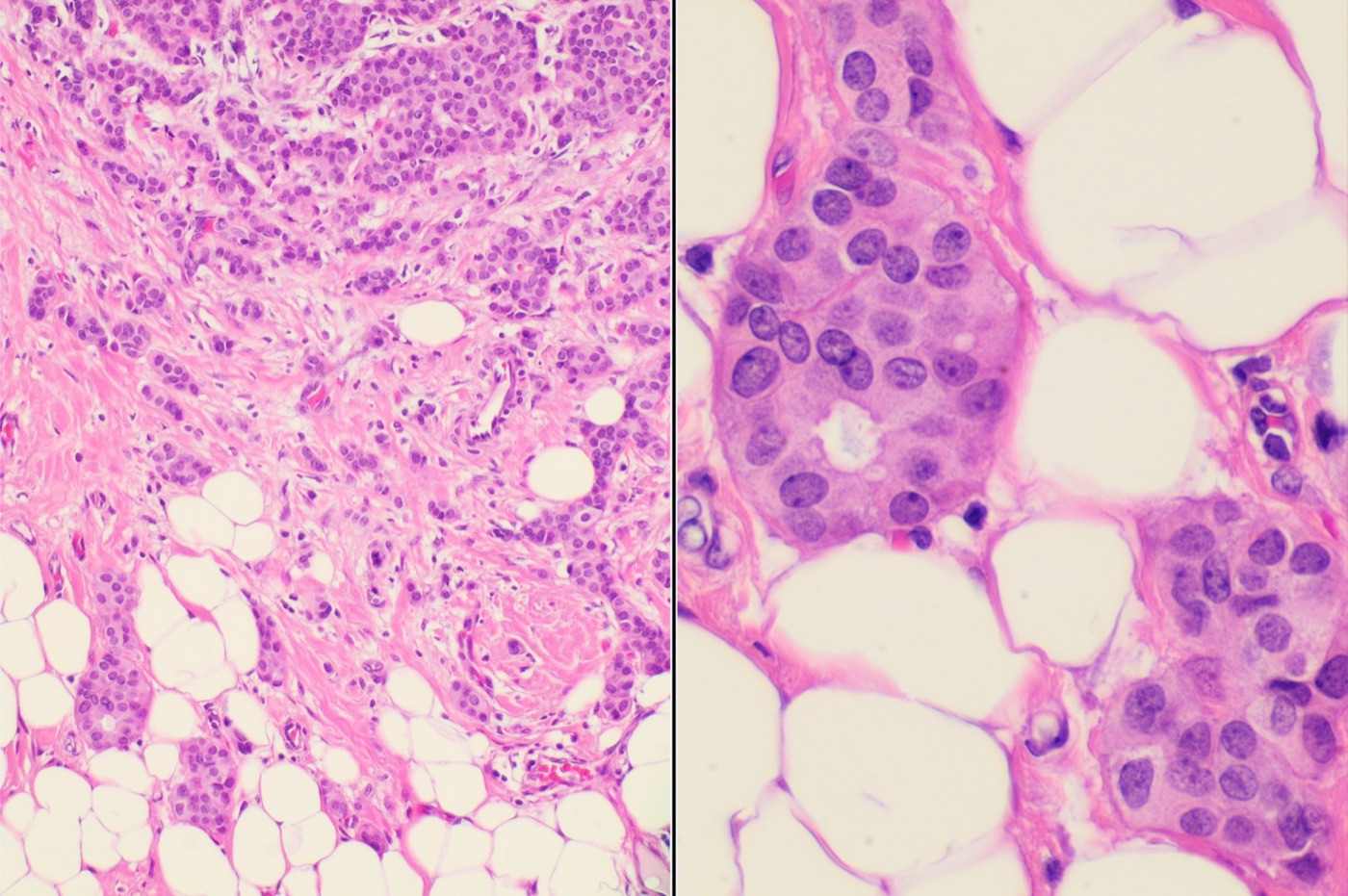
Invasive carcinoma of no special type. Left, nests, cords and trabeculae of tumor cells infiltrating desmoplastic stroma and fat, with no special-type pattern (low power). Right, tumor cells in fat with enlarged, vesicular nuclei, moderate pleomorphism and small nucleoli (high power). H&E, magnification not stated.Image: Mikael Häggström, M.D., Wikimedia Commons, CC0.
Special-Type Breast Carcinomas
Definition. Special-type breast carcinomas are invasive carcinomas with distinctive morphology, and often distinctive biology, that the WHO Classification of Breast Tumours recognizes as separate types (Quinn et al. 2026). Special types account for up to 25% of invasive breast cancers (Weigelt, Geyer and Reis-Filho 2010). Several show genotype–phenotype correlations: secretory carcinoma carries t(12;15) with ETV6::NTRK3, adenoid cystic carcinoma carries t(6;9) with MYB::NFIB, and lobular carcinoma shows CDH1 inactivation (Weigelt, Geyer and Reis-Filho 2010). The 6th edition (2026) renames acinic cell carcinoma, removes polymorphous adenocarcinoma, restricts mucinous carcinoma and revises the neuroendocrine and papillary categories (Quinn et al. 2026).
Very rare types in the CAP case summary. Invasive papillary carcinoma, neuroendocrine tumor, acinic cell-like carcinoma, mucoepidermoid carcinoma, mucinous cystadenocarcinoma and tall cell carcinoma with reversed polarity are reported under "other histologic type" with their WHO names (CAP 2026).
At least 90% tubular morphology; Nottingham scores 1-1-1 or 1-2-1
ER positive, HER2 negative
CAP 2026; Cserni 2020; CAP biomarker template 2025
Invasive cribriform carcinoma
At least 90% cribriform morphology
ER positive, HER2 negative
CAP 2026; Cserni 2020; CAP biomarker template 2025
Mucinous carcinoma
Mucin-secreting carcinoma with grade 1 or 2 morphology and a favorable biomarker profile
ER positive, HER2 negative
Quinn et al. 2026; CAP biomarker template 2025
Invasive micropapillary carcinoma
Cell clusters with inside-out polarity in clear stromal spaces
Mostly ER positive (71% in one series)
Cserni 2020; Nassar et al. 2001
Invasive apocrine carcinoma
Apocrine cytology and immunophenotype (5th edition definition)
ER negative
Cserni 2020
Metaplastic carcinoma
Squamous, spindle cell or heterologous mesenchymal differentiation
ER, PR and HER2 usually negative
Rakha et al. 2017; CAP 2026
Adenoid cystic carcinoma
Biphasic tumor with MYB::NFIB; indolent
ER negative or low, HER2 negative
Wetterskog et al. 2012; CAP biomarker template 2025
Secretory carcinoma
Microcystic, solid and tubular with secretory material; ETV6::NTRK3
ER, PR and HER2 negative
Tang et al. 2021
Tubular and invasive cribriform carcinoma
Tubular carcinoma. The more-than-90% purity rule for tubular carcinoma has applied in every WHO edition that lists the type (Cserni 2020). Highly atypical nuclei exclude the diagnosis (Cserni 2020).
Prognosis of tubular carcinoma. Tubular carcinomas have a better prognosis than grade 1 invasive breast carcinoma of no special type (IBC-NST). Adjuvant systemic and axillary treatment can be omitted in most (Cserni 2020).
Invasive cribriform carcinoma. The 3rd edition introduced the more-than-90% rule and allowed the diagnosis when more than 90% of the tumor showed mixed tubular and cribriform patterns with cribriform predominance. The 5th edition abandoned this allowance and requires more than 90% cribriform morphology (Cserni 2020).
Tubular versus cribriform predominance. The original description of invasive cribriform carcinoma called mixed tumors with tubular predominance tubular carcinoma. The Royal College of Pathologists retains this rule. The WHO editions never adopted it (Cserni 2020).
Mucinous carcinoma and other mucin-producing carcinomas
6th edition definition. Mucinous carcinoma is reserved for mucin-secreting carcinomas with grade 1 or 2 morphology and a favorable biomarker profile (Quinn et al. 2026).
Mucin-producing carcinomas excluded. Mucinous tumors with high nuclear grade, ER negativity or HER2 positivity are classified as IBC-NST with mucin production (Quinn et al. 2026). The 5th edition defined mucinous carcinoma by substantial extracellular mucin, without these biomarker exclusions (Cserni 2020; Quinn et al. 2026). The 5th-edition-era CAP protocol required at least 90% pure pattern for special types (CAP 2023).
Hypocellular and hypercellular forms. The 3rd to 5th editions recognized a hypocellular (Capella type A) and a cellular (type B) form. The distinction has no current clinical relevance (Cserni 2020). Hypercellular mucinous carcinoma can show neuroendocrine morphology and is a special type distinct from neuroendocrine neoplasms (Quinn et al. 2026).
Micropapillary morphology in mucinous carcinoma. Peripheral EMA and MUC1 staining is frequent in both classic mucinous carcinoma and tumors with prominent micropapillary morphology, so the staining pattern does not separate them. A prominent micropapillary pattern is noted in the report and is not designated as a subtype (Quinn et al. 2026). The 5th edition discussed mucinous carcinoma with micropapillary features under both the mucinous and micropapillary chapters (Cserni 2020).
Metastasis. The 6th edition emphasizes exclusion of a metastasis from another primary site before mucinous carcinoma is diagnosed (Quinn et al. 2026).
Morphology. Cohesive tumor cell clusters lie within prominent clear spaces that resemble dilated lymphovascular channels (Nassar et al. 2001).
Reversed polarity. The clusters show inside-out polarity, with a microvillous secretory surface facing the stroma and a gap between stroma and tumor cells. EMA (MUC1) highlights the reversed polarity (Cserni 2020).
Frequency. A micropapillary growth pattern was present in 6% of breast carcinomas in one institutional series. It was usually a focal component of IBC-NST (Nassar et al. 2001).
Nodal and skin involvement. Axillary metastases were present in 77% of cases and were usually multiple. Skin invasion occurred in 22% (Nassar et al. 2001).
Proportion and outcome. Outcome did not depend strongly on the amount of micropapillary component and did not differ from IBC-NST of similar nodal status (Nassar et al. 2001).
Purity rule. The CAP case summary lists invasive micropapillary carcinoma as pure or greater than 90% (CAP 2026).
Feature
Invasive micropapillary carcinoma
Lymphovascular invasion by IBC-NST
Source
Spaces around tumor clusters
Clear stromal spaces without endothelial lining
Spaces lined by endothelium
Nassar et al. 2001; Quinn et al. 2026
D2-40
No endothelial lining highlighted
Lymphatic endothelium highlighted
Quinn et al. 2026
EMA (MUC1)
Stroma-facing (inside-out) staining
Not used for the diagnosis; the diagnosis requires an endothelial lining
Cserni 2020; Quinn et al. 2026
Carcinoma with apocrine differentiation
5th edition definition. Apocrine cells have distinct cell borders, abundant granular eosinophilic or vacuolated cytoplasm, large round to oval nuclei and prominent nucleoli. The immunophenotype is GCDFP-15 positive, ER negative, PR negative and AR positive (Cserni 2020).
Earlier definition. The 3rd edition required apocrine cells in more than 90% of the tumor and described the profile as GCDFP-15 positive, bcl2 negative, usually ER and PR negative and often AR positive (Cserni 2020).
Luminal androgen receptor subtype. The luminal androgen receptor (LAR) subtype of triple-negative breast cancer is characterized by androgen receptor signaling (Lehmann et al. 2011).
Current CAP term. The CAP case summary lists the type as invasive apocrine carcinoma (CAP 2026). The 6th-edition summary does not discuss apocrine carcinoma (Quinn et al. 2026).
Metaplastic carcinoma
Subtypes. The WHO recognizes spindle cell carcinoma, carcinoma with heterologous differentiation or matrix production, squamous cell carcinoma and mixed forms. Low-grade adenosquamous carcinoma and fibromatosis-like metaplastic carcinoma are the two favorable types (CAP 2026).
Reporting. The specific subtype is reported because outcomes differ widely. Mixed metaplastic carcinomas are reported with types and percentages (CAP 2026).
Amount of metaplastic component. The 5th-edition chapter gave no percentage requirement and stated that a very small metaplastic component in IBC-NST is noted in the report (Cserni 2020).
Origin in adenomyoepithelioma. Malignant transformation of the myoepithelial component of an adenomyoepithelioma usually produces metaplastic spindle cell carcinoma. Squamous, low-grade adenosquamous and matrix-producing carcinomas also occur (Quinn et al. 2026).
Marker group
Frequency in metaplastic carcinoma
Source
Broad-spectrum cytokeratins (AE1/AE3, MNF116)
About 80%
Rakha et al. 2017
Basal cytokeratins (34βE12, CK5/6, CK14, CK17)
About 70%
Rakha et al. 2017
Luminal cytokeratins (CK8/18, CK7, CK19)
About 30–60%
Rakha et al. 2017
Myoepithelial markers, particularly p63
Frequently positive
Rakha et al. 2017
ER, PR and HER2
Usually all negative
Rakha et al. 2017
CD34
Consistently negative; often positive in phyllodes tumors
Rakha et al. 2017
Panel approach. No single marker is positive in all metaplastic carcinomas, so a panel of antibodies is used (Rakha et al. 2017).
Keratin-negative sarcomatoid tumors. In a series of 140 metaplastic carcinomas, 69% were negative for one or more cytokeratins and 6% lacked all cytokeratins tested (Rakha et al. 2021).
Management of keratin-negative sarcomatoid breast tumors. After extensive work-up, sarcomatoid breast neoplasms without carcinomatous morphology or keratin expression can be managed as carcinomas. Evidence that they are primary breast sarcomas is lacking (Rakha et al. 2021).
Triple-negative and basal-like breast carcinoma
Basal-like immunophenotype. Basal-like carcinomas defined by gene expression are typically ER and HER2 negative and positive for basal cytokeratins, HER1 (EGFR) and/or c-KIT. A four-antibody panel (ER, HER1, HER2 and CK5/6) identifies them with high specificity (Nielsen et al. 2004).
Prognosis. Basal cytokeratin expression was associated with low disease-specific survival. HER1 was expressed in 54% of basal cytokeratin-positive tumors and independently predicted poor survival (Nielsen et al. 2004).
Transcriptional subtypes. Gene expression analysis of 587 triple-negative breast cancers identified two basal-like (BL1, BL2), immunomodulatory, mesenchymal, mesenchymal stem-like and luminal androgen receptor subtypes (Lehmann et al. 2011).
Triple-negative special type
Behavior
Source
Adenoid cystic carcinoma
Indolent; lymph node metastases rare
Wetterskog et al. 2012; Neilson et al. 2025
Secretory carcinoma
Most are slow-growing with favorable outcomes
Mortensen et al. 2021
Low-grade adenosquamous and fibromatosis-like metaplastic carcinoma
Favorable metaplastic types
CAP 2026
Acinic cell-like carcinoma
Molecular profile similar to conventional triple-negative carcinoma
Quinn et al. 2026
Spindle cell, squamous and heterologous metaplastic carcinoma
Outcomes differ by subtype
CAP 2026
Clinical relevance of typing. Adenoid cystic carcinoma is genomically distinct from triple-negative basal-like IBC-NST, which supports histologic subtyping of triple-negative carcinomas (Wetterskog et al. 2012).
Adenoid cystic carcinoma of the breast
Morphology and immunohistochemistry. Cribriform, tubular and solid-basaloid patterns surround true glandular and pseudoglandular spaces. Luminal cells express CK7, CK8/18, EMA and CD117. The pseudoglandular spaces are stromal invaginations lined by myoepithelial–basal cells that express CK5, CK5/6, CK14, CK17, p63, actin, calponin and S-100 (Neilson et al. 2025).
Fusion. The t(6;9)(q22-23;p23-24) translocation produces MYB::NFIB in adenoid cystic carcinomas of the breast and head and neck (Persson et al. 2009). The fusion was detected in all but 1 of 13 breast adenoid cystic carcinomas (Wetterskog et al. 2012).
Genome. Breast adenoid cystic carcinomas have few copy number aberrations and lack the 1q gains and 16q losses of grade-matched IBC-NST (Wetterskog et al. 2012).
Hormone receptors. About 75% lack ER, PR and HER2 (Neilson et al. 2025).
Grade. Breast adenoid cystic carcinoma is graded with the Nottingham system and is usually grade 1 or 2 (Neilson et al. 2025).
Behavior. Axillary involvement occurs in less than 5% and metastatic disease at presentation in less than 2%. In a cohort of 933 patients, 5-year overall survival was 88% (Ghabach et al., as reported by Neilson et al. 2025).
Site comparison. Adenoid cystic carcinoma of the breast has a better prognosis than adenoid cystic carcinoma of the salivary glands (Cserni 2020).
Types. The 5th-edition CAP list included classic, solid-basaloid and high-grade-transformed adenoid cystic carcinoma (CAP 2023). The 2026 case summary lists classic adenoid cystic carcinoma (CAP 2026).
Secretory carcinoma of the breast
Frequency and age. Secretory carcinoma forms less than 0.15% of invasive breast carcinomas. It was first described in children and adolescents with an average age of 9 years and also occurs in adults of both sexes (Tang et al. 2021).
Morphology. Solid, microcystic and tubular structures contain abundant intracellular and extracellular secretory material that is positive with Alcian blue or PAS. Cells have low-grade round to oval nuclei, scant mitoses and abundant granular eosinophilic cytoplasm (Tang et al. 2021).
Immunohistochemistry. Tumor cells are S100 positive and negative for ER, PR and HER2 (Tang et al. 2021).
Fusion.ETV6::NTRK3 from t(12;15)(p13;q25) was found in 12 of 13 secretory carcinomas (92%) and not in other ductal carcinomas (Tognon et al. 2002).
Targeted therapy. Larotrectinib and entrectinib are approved for advanced solid tumors with NTRK fusions (Mortensen et al. 2021).
Behavior. Most secretory carcinomas are slow-growing with favorable outcomes (Mortensen et al. 2021; Tang et al. 2021). Early local recurrence and brain metastases have been reported (Mortensen et al. 2021; Tang et al. 2021).
Papillary carcinomas of the breast
Entity
WHO 6th edition rule
5th edition rule
Source
Encapsulated papillary carcinoma (EPC)
Staged as ductal carcinoma in situ (DCIS) despite frequent lack of peripheral myoepithelial cells; very rare high-grade or ER-negative tumors may still be classified as EPC, with a report comment
EPC with high nuclear grade, increased mitoses, triple-negative or HER2-positive phenotype was graded, staged and managed as invasive carcinoma
Quinn et al. 2026
Solid papillary carcinoma (SPC) in situ
Circumscribed nodules with smooth contours, classified as in situ even without myoepithelial cells
SPC in situ recognized
Quinn et al. 2026; Cserni 2020
SPC in situ with invasion
SPC in situ with conventional invasive carcinoma, usually hypercellular mucinous carcinoma or IBC-NST
Grouped under invasive SPC; the 6th edition separated the two categories
Quinn et al. 2026
Invasive SPC
Solid papillary nodules with fine vessels, no myoepithelial rim, ragged jigsaw contours in desmoplastic stroma
Also included SPC in situ with conventional invasive carcinoma
Quinn et al. 2026; Cserni 2020
Invasive papillary carcinoma
Not addressed in the 6th-edition summary; reported by its WHO name under "other histologic type" (CAP 2026)
More than 90% papillary invasive component with fibrovascular cores; extremely rare
Cserni 2020; CAP 2026
Papillae versus micropapillae. True papillae have fibrovascular cores. Micropapillae are epithelial outgrowths without fibrovascular cores (Cserni 2020).
Biopsy samples. Distinction of EPC from EPC-like invasive carcinoma is difficult on core biopsy when neoadjuvant therapy is considered. Thorough excision sampling and multidisciplinary review are used in difficult cases (Quinn et al. 2026).
Biomarker testing. ER testing is recommended for EPC and SPC in situ without invasion. HER2 testing is not recommended for these lesions (CAP biomarker template 2025).
Neuroendocrine morphology, negative or focal weak markers
IBC-NST
IBC-NST; neuroendocrine differentiation without distinct and uniform features remained a pattern of IBC-NST
Quinn et al. 2026; Cserni 2020
Moderate to strong, or diffuse weak, neuroendocrine marker expression
IBC-NST with neuroendocrine differentiation; includes former large cell neuroendocrine carcinoma
Neuroendocrine tumor (grade 1 or 2) or neuroendocrine carcinoma (small or large cell) when features were distinct and uniform
Quinn et al. 2026; Cserni 2020
Typical neuroendocrine tumor morphology with strong diffuse markers
Primary neuroendocrine tumor, exceptionally rare; metastasis excluded first
Neuroendocrine tumor
Quinn et al. 2026; Cserni 2020
Small cell morphology
Small cell neuroendocrine carcinoma as a cytomorphologic growth pattern after metastasis is excluded
Neuroendocrine carcinoma, small cell type
Quinn et al. 2026; Cserni 2020
Routine staining. Routine staining of IBC-NST for neuroendocrine markers is not recommended. Neuroendocrine differentiation has no conclusive clinical relevance (Quinn et al. 2026).
Distinct special types. Solid papillary carcinoma and hypercellular mucinous carcinoma show neuroendocrine features and remain distinct special types (Quinn et al. 2026).
Terminology. The term "carcinoid tumour" is not recommended (Quinn et al. 2026).
Metastasis. A breast tumor with neuroendocrine tumor morphology requires exclusion of metastasis from the gastrointestinal tract or lung (Quinn et al. 2026). Metastases to the breast are covered in Angiosarcoma, Lymphoma and Metastases in the Breast.
Previous rule. The 5th edition adopted the 2018 IARC–WHO unified neuroendocrine neoplasm terminology. The 6th edition found it difficult to apply to the breast (Quinn et al. 2026).
Rare, salivary gland-type and biphasic carcinomas of the breast
Acinic cell-like carcinoma. The 6th edition renamed acinic cell carcinoma because breast tumors lack the t(4;9)(q13;q31) rearrangement and NR4A3 overexpression of salivary acinic cell carcinoma. Their molecular profile resembles conventional triple-negative carcinoma and carcinomas arising in microglandular adenosis (Quinn et al. 2026). The 5th edition used the name acinic cell carcinoma (Quinn et al. 2026).
Polymorphous adenocarcinoma. The 6th edition removed this type because evidence is insufficient (Quinn et al. 2026). The 5th edition listed it (Quinn et al. 2026; CAP 2023).
Tall cell carcinoma with reversed polarity. This type entered the WHO classification in the 5th edition. It was first described as a breast tumor resembling the tall cell variant of papillary thyroid carcinoma (Cserni 2020). Solid, circumscribed nodules contain fibrovascular cores (Chiang et al. 2016).
IDH2 in tall cell carcinoma with reversed polarity. In a series reported as solid papillary carcinoma with reverse polarity, 10 of 13 tumors (77%) had IDH2 R172 hotspot mutations. Eight of these 10 had concurrent PIK3CA or PIK3R1 mutations (Chiang et al. 2016).
Biphasic carcinomas. Carcinomas arising from both epithelial and myoepithelial cells of an adenomyoepithelioma are reported by component. The term epithelial–myoepithelial carcinoma is discouraged for them (Quinn et al. 2026).
HRAS Q61R. Immunohistochemistry for the HRAS Q61R mutant protein is moderately sensitive and highly specific for adenomyoepithelioma (Quinn et al. 2026).
Differential diagnosis of special-type breast carcinomas
Entity
Main mimic
Distinguishing finding
Source
Invasive micropapillary carcinoma
Lymphovascular invasion
No endothelial lining; inside-out EMA
Nassar et al. 2001; Cserni 2020
Mucinous carcinoma
IBC-NST with mucin production
High grade, ER negativity or HER2 positivity excludes mucinous carcinoma
Quinn et al. 2026
Mucinous carcinoma
ILC with extracellular mucin
Discohesive E-cadherin-negative cells
Quinn et al. 2026
Metaplastic spindle cell carcinoma
Malignant phyllodes tumor
Keratin and p63 expression; CD34 negative
Rakha et al. 2017
Adenoid cystic carcinoma
Invasive cribriform carcinoma
Biphasic luminal and myoepithelial–basal cells; ER negative or low
Clinical and imaging exclusion of a gastrointestinal or lung primary
Quinn et al. 2026
special-type breast carcinoma lookup
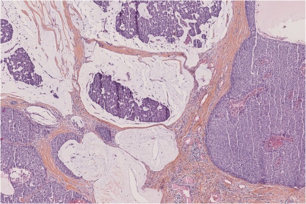
Mucinous carcinoma arising with solid papillary carcinoma: nests and clusters of tumor cells float in pools of pale extracellular mucin separated by thin fibrous septa (left and top), next to a solid papillary carcinoma nodule (right). H&E, low power (magnification not stated).Image: Quinn Cecily et al., PMC open access (Histopathology. 2026 Apr 21;89(2):199–218. doi: 10.1111/his.70149) Figure 5, CC BY.
Breast Cancer Biomarkers: ER, PR, HER2, Ki-67 and Multigene Assays
Definition. Breast cancer biomarkers are the tissue tests that predict response to endocrine, HER2-directed and other therapies and refine prognosis. Estrogen receptor (ER), progesterone receptor (PR) and HER2 testing is recommended on all primary invasive breast carcinomas and on recurrent or metastatic tumors (CAP biomarker template 2025). ER and PR testing follows the 2020 ASCO/CAP guideline update (Allison et al. 2020). HER2 testing follows the 2018 ASCO/CAP focused update, which the 2023 update affirmed (Wolff et al. 2018; Wolff et al. 2023). Ki-67 assessment follows the 2021 recommendations of the International Ki67 in Breast Cancer Working Group (IKWG) (Nielsen et al. 2021). The WHO 6th edition (2026) updates HER2 reporting categories after the DESTINY-Breast04 and DESTINY-Breast06 trials (Quinn et al. 2026).
Preanalytic requirements for breast biomarker testing
Specimen choice. Core needle biopsy samples are preferred at primary diagnosis. When several invasive foci are present, the largest is tested. Smaller carcinomas of different type or higher grade are also tested (CAP biomarker template 2025).
Cold ischemia time. One hour or less is preferable. Prolonged cold ischemia degrades antigen and causes false-negative ER and HER2 results (CAP biomarker template 2025).
Fixation. Fixation in buffered formalin for at least 6 hours is recommended. Prolonged fixation can also reduce immunoreactivity (CAP biomarker template 2025).
Fixation window (ASCO/CAP 2020). Samples for ER and PR testing are fixed in 10% neutral buffered formalin for 6 to 72 hours. Samples are sliced at 5-mm intervals and placed in a sufficient volume of formalin. The time from tissue acquisition to fixation is kept as short as possible (Allison et al. 2020).
Fixative pH. ER is degraded in acidic fixatives such as Bouin and B-5. Buffered formalin has a pH of 7.0–7.4 (CAP biomarker template 2025).
Decalcification. Decalcification can cause loss of immunoreactivity and degrades DNA for in situ hybridization (ISH). A standardized report comment identifies testing on decalcified tissue (CAP biomarker template 2025).
Recording. The time of tissue removal and the time of placement in fixative are communicated to the laboratory. Reporting these times in the pathology report is optional (CAP biomarker template 2025).
Preanalytic or analytic factor
Effect
Source
Cautery heat at the tumor edge
False-negative ER
CAP biomarker template 2025
Fixation shorter than 6 hours
Underfixation; false-negative results
CAP biomarker template 2025
Formalin fixation longer than 1 week
ISH failure
CAP biomarker template 2025
Dark hematoxylin counterstain
Obscures faint diaminobenzidine (DAB) staining
CAP biomarker template 2025
Edge artifact in core biopsies
False-positive HER2 IHC
CAP biomarker template 2025
Estrogen and progesterone receptor testing (ASCO/CAP 2020)
Method. ER status is determined by validated immunohistochemistry (IHC) on formalin-fixed, paraffin-embedded tissue. No other assay is recommended for predicting endocrine therapy benefit (Allison et al. 2020). Only nuclear staining counts (CAP biomarker template 2025).
Positive threshold. Cancers with 1–100% of tumor nuclei staining are ER positive (Allison et al. 2020).
ER Low Positive. Cancers with 1–10% staining are reported as ER Low Positive, with a recommended comment. Data on endocrine benefit in this group are limited (Allison et al. 2020). The category was introduced in the 2020 update (Allison et al. 2020).
Negative. Less than 1% or 0% nuclear staining is ER negative (Allison et al. 2020).
Controls. The status of controls is reported for cancers with 0–10% staining (Allison et al. 2020). A laboratory standard operating procedure confirms low or negative results (Allison et al. 2020).
PR. Similar principles apply to PR testing, which is used mainly for prognosis in ER-positive cancers (Allison et al. 2020).
Ductal carcinoma in situ (DCIS). ER testing of DCIS is recommended to estimate the benefit of endocrine therapy for risk reduction. PR testing of DCIS is optional (Allison et al. 2020).
ER result category
Criteria
Report content
Source
Positive
More than 10% of tumor nuclei stain
Percentage (or range) and average intensity
CAP biomarker template 2025
Low Positive
1–10% of tumor nuclei stain
Percentage, intensity, standardized comment and control status
Allison et al. 2020; CAP biomarker template 2025
Negative
Less than 1%
Control status
Allison et al. 2020; CAP biomarker template 2025
Cannot be determined
Internal controls also negative, or other reason stated
Repeat on another block or specimen
CAP biomarker template 2025
Low Positive biology. Cancers with 1–10% ER expression are heterogeneous and often have gene expression profiles closer to ER-negative cancers, with high grade and better response to neoadjuvant chemotherapy (CAP biomarker template 2025).
Frequency of Low Positive results. The category applies only to invasive carcinoma and represents a small minority of cases (less than 5–10%). It is not required for PR or DCIS (CAP biomarker template 2025).
Internal controls. Benign breast epithelium is the internal positive control (CAP biomarker template 2025).
No internal control. When a negative or Low Positive cancer has no internal control, the pathologist weighs histologic type and grade, cold ischemia and fixation times and external controls. A second review may be obtained (CAP biomarker template 2025).
Intensity and scores. Intensity is scored as weak (1+), moderate (2+) or strong (3+). Allred and H-scores are optional. Only the percentage determines the result category (CAP biomarker template 2025).
ER-negative, PR-positive cancers. A PR-positive result raises the possibility of a false-negative ER result. Confirmed ER-negative, PR-positive cancers are a rare and controversial phenotype (CAP biomarker template 2025).
Frequency of ER positivity. About 75–80% of invasive breast cancers are ER positive, including almost all grade 1 and most grade 2 cancers (CAP biomarker template 2025).
Breast biomarker results discordant with histologic type and grade
Histology
Expected result
Unusual or possibly discordant result
Source
Low-grade invasive carcinoma of no special type or invasive lobular carcinoma
Uniform ER staining; HER2 negative
Low or negative ER; HER2 positive
CAP biomarker template 2025
Pure mucinous, tubular or cribriform carcinoma
Uniform ER staining; HER2 negative
Low or negative ER; HER2 positive
CAP biomarker template 2025
Low-grade DCIS, encapsulated papillary carcinoma, solid papillary carcinoma in situ
Uniform ER staining; HER2 negative
Low or negative ER; HER2 positive
CAP biomarker template 2025
Adenoid cystic and other salivary gland-like carcinomas
ER negative or low; HER2 negative
High percentage of ER staining; HER2 positive
CAP biomarker template 2025
Response to a discordant result. The histologic type, grade and testing factors are rechecked, and repeat testing or second review is considered. A valid but unusual result is reported with a comment (CAP biomarker template 2025).
HER2 immunohistochemistry (ASCO/CAP 2018 and 2023)
HER2 (ERBB2) is amplified and overexpressed in 10–20% of breast cancers, depending on the population tested (CAP biomarker template 2025). HER2 status is both prognostic and predictive (CAP biomarker template 2025).
IHC score
Criteria
Result
Source
0 (absent membrane staining)
No staining
Negative
CAP biomarker template 2025
0 (0+, with membrane staining)
Incomplete, faint or barely perceptible membrane staining in 10% or fewer tumor cells
Negative
CAP biomarker template 2025
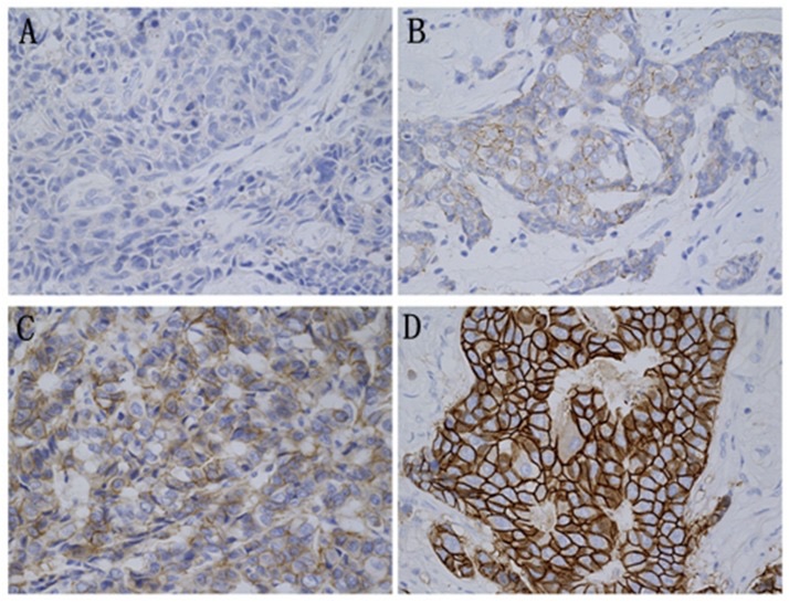
1+
Incomplete, faint or barely perceptible membrane staining in more than 10% of tumor cells
Negative
CAP biomarker template 2025
2+
Weak to moderate complete membrane staining in more than 10% of tumor cells
Equivocal
Wolff et al. 2018; CAP biomarker template 2025
3+
Complete, intense circumferential membrane staining in more than 10% of tumor cells, seen at low power in a homogeneous contiguous population
Positive
CAP biomarker template 2025
Less common 2+ patterns. Moderate to intense incomplete basolateral staining is scored 2+. This pattern occurs in some HER2-amplified micropapillary carcinomas (CAP biomarker template 2025).
Equivocal results. A 2+ result is followed by reflex ISH on the same specimen or by a new test on another specimen (CAP biomarker template 2025).
Clustered heterogeneity. A discrete 3+ population forming more than 10% of the cancer is reported as positive with its percentage. A clustered 3+ population of 10% or less is reported as equivocal (2+). ISH is scored separately in the 3+ and non-3+ areas (CAP biomarker template 2025).
False-positive IHC. Edge artifact, cytoplasmic staining, overstaining of normal cells and scoring of DCIS cause false-positive results. High-grade DCIS is often HER2 positive (CAP biomarker template 2025).
False-negative IHC. Prolonged cold ischemia, tumor heterogeneity in a small biopsy and low antibody concentration cause false-negative results. HER2 has no normal internal control (CAP biomarker template 2025).
Retesting the excision. If the core biopsy is HER2 negative, a new test may be ordered on the excision based on clinical criteria (Wolff et al. 2018).
HER2-low and HER2-ultralow results
DESTINY-Breast04. The trial enrolled 557 patients with HER2-low metastatic breast cancer (IHC 1+, or IHC 2+ with negative ISH) after 1 or 2 lines of chemotherapy. In the hormone receptor-positive cohort, median progression-free survival was 10.1 months with trastuzumab deruxtecan and 5.4 months with chemotherapy (hazard ratio 0.51). Overall survival was 23.9 and 17.5 months (Modi et al. 2022).
DESTINY-Breast06. The trial enrolled 866 patients with hormone receptor-positive metastatic cancer after endocrine therapy: 713 HER2-low and 153 HER2-ultralow (IHC 0 with membrane staining). In HER2-low disease, median progression-free survival was 13.2 months with trastuzumab deruxtecan and 8.1 months with chemotherapy (hazard ratio 0.62). Results were consistent in the exploratory ultralow group (Bardia et al. 2024).
Toxicity. Adjudicated interstitial lung disease or pneumonitis occurred in 12.1% of patients treated with trastuzumab deruxtecan in DESTINY-Breast04 and in 11.3% in DESTINY-Breast06 (Modi et al. 2022; Bardia et al. 2024).
ASCO/CAP 2023. The update affirmed the 2018 recommendations and judged new result categories such as HER2-Low and HER2-Ultra-Low premature. It stated that the distinction between IHC 0 and 1+ is now clinically relevant and gave best practices for making it (Wolff et al. 2023). IHC 0 tumors were excluded from DESTINY-Breast04 (Wolff et al. 2023).
WHO 6th edition. The pattern within IHC 0 (0/absent membrane staining or 0+/with membrane staining) is recommended in the report. The terms HER2-low and HER2-ultralow are not required (Quinn et al. 2026). The 5th edition was published in 2019, before the DESTINY-Breast04 results of 2022 (Tan et al. 2020; Modi et al. 2022).
CAP reporting. The CAP template divides score 0 into absent membrane staining and 0+ with membrane staining. An optional comment defines HER2-low and HER2-ultralow as used in the DESTINY-Breast04 and 06 trials and notes that IHC 0 without staining is currently excluded from trastuzumab deruxtecan eligibility (CAP biomarker template 2025).
HER2 in situ hybridization (dual-probe groups)
ISH group
HER2/chromosome enumeration probe 17 (CEP17) ratio
Average HER2 signals per cell
Final status
Source
1
2.0 or more
4.0 or more
Positive (amplified)
Wolff et al. 2018; CAP biomarker template 2025
2
2.0 or more
Less than 4.0
Negative unless concurrent IHC is 3+
Wolff et al. 2018; CAP biomarker template 2025
3
Less than 2.0
6.0 or more
Positive if concurrent IHC is 2+ or 3+; negative if IHC is 0 or 1+
Wolff et al. 2018; CAP biomarker template 2025
4
Less than 2.0
4.0 to less than 6.0
Negative unless concurrent IHC is 3+
Wolff et al. 2018; CAP biomarker template 2025
5
Less than 2.0
Less than 4.0
Negative (not amplified)
CAP biomarker template 2025
Frequency of groups 2–4. These less common patterns form about 5% of dual-probe results (Wolff et al. 2018).
Concurrent IHC. Groups 2–4 require concomitant IHC review to assign the final status (Wolff et al. 2018). When IHC is 2+, a second reviewer recounts the ISH (CAP biomarker template 2025).
Standardized comments. Group 2: evidence of benefit from HER2-targeted therapy is limited. Group 3: patients with a ratio below 2.0 were not eligible for the first adjuvant trastuzumab trials. Group 4: benefit without protein overexpression is uncertain, and repeat testing of results near the threshold can differ by chance (CAP biomarker template 2025).
Single-probe assays. The 2018 update required concomitant IHC review for all single-probe ISH results (Wolff et al. 2018). The current CAP template states that single-probe testing is no longer recommended (CAP biomarker template 2025).
ISH failure. Formalin fixation longer than 1 week, non-formalin fixatives, acid exposure and insufficient protease treatment cause ISH failure (CAP biomarker template 2025).
Ki-67 proliferation index
Antibody and role. MIB-1 is the most commonly used Ki-67 antibody. Ki-67 correlates with grade and is used in some prognostic schemes and neoadjuvant trials. It is not validated as a predictive biomarker, and routine testing is not standard under ASCO or NCCN guidance (CAP biomarker template 2025).
Reporting. Ki-67 is reported as a percentage or a range (CAP biomarker template 2025).
IKWG scoring method. After an online calibration exercise, the scorer estimates the percentage area with negligible, low, medium and high Ki-67 and counts 100 nuclei in each field type. The weighted global score is the Ki-67 index (Nielsen et al. 2021).
Global and hot-spot scoring. Global scores had higher reproducibility than hot-spot scores in IKWG studies, although the difference was not statistically significant (Nielsen et al. 2021).
Clinical utility. Ki-67 has clinical validity as a prognostic marker. Clinical utility is limited to prognosis in T1–2, N0–1, ER-positive, HER2-negative cancers. In this group, Ki-67 of 5% or less or 30% or more can estimate prognosis (Nielsen et al. 2021).
Reproducibility. Interlaboratory agreement was poor between more than 5% and less than 30%, where most cut-offs have been set.
Perioperative endocrine therapy. The POETIC trial randomized 4,480 postmenopausal women with hormone receptor-positive cancer to 2 weeks of perioperative aromatase inhibitor or no treatment, with Ki-67 dichotomized at 10% (Smith et al. 2020). In treated HER2-negative patients, 5-year recurrence risk was 4.3% when Ki-67 was low at baseline and at 2 weeks, 8.4% when it fell from high to low and 21.5% when it stayed high (Smith et al. 2020).
Status of on-treatment Ki-67. The IKWG regards the use of Ki-67 to guide individual neoadjuvant treatment as investigational (Nielsen et al. 2021).
Multigene expression assays in early breast cancer
21-gene recurrence score, node-negative disease (TAILORx). Of 10,273 women with hormone receptor-positive, HER2-negative, node-negative cancer, 6,711 with a recurrence score of 11–25 were randomized. Endocrine therapy alone was noninferior to chemoendocrine therapy (hazard ratio 1.08). Some chemotherapy benefit was seen in women 50 years of age or younger with a score of 16–25 (Sparano et al. 2018).
21-gene recurrence score, node-positive disease (RxPONDER). Of 5,083 women with hormone receptor-positive, HER2-negative cancer, 1–3 positive nodes and a score of 25 or lower were randomized. Postmenopausal women had no chemotherapy benefit (5-year invasive disease-free survival 91.9% and 91.3%). Premenopausal women benefited (89.0% and 93.9%; hazard ratio 0.60) (Kalinsky et al. 2021).
70-gene signature (MINDACT). Of 6,693 women, 1,550 (23.2%) had high clinical and low genomic risk. Without chemotherapy, their 5-year rate of survival without distant metastasis was 94.7%, 1.5 percentage points lower than with chemotherapy (Cardoso et al. 2016).
AJCC 8th edition. Tumor biomarkers and low 21-gene recurrence scores can alter prognostic stage (Giuliano et al. 2017). Any T1–2, N0, ER-positive, HER2-negative cancer with a recurrence score below 11 is prognostic stage group IA (Yoon et al. 2019). Stage groups are covered in Breast Cancer Staging, Margins, Sentinel Lymph Nodes and Post-Neoadjuvant Assessment.
Tissue use. Fresh tissue is not used for other studies unless the carcinoma is large enough that histologic evaluation and ER, PR and HER2 testing are not compromised (CAP biomarker template 2025).
Trial
Assay
Population (HR, hormone receptor)
Main finding
Source
TAILORx
21-gene recurrence score
HR-positive, HER2-negative, node-negative
Score 11–25: endocrine therapy noninferior; some benefit at age 50 or younger with score 16–25
Sparano et al. 2018
RxPONDER
21-gene recurrence score
HR-positive, HER2-negative, 1–3 nodes, score 25 or lower
Benefit in premenopausal women only
Kalinsky et al. 2021
MINDACT
70-gene signature
Early-stage cancer, high clinical and low genomic risk
94.7% 5-year distant metastasis-free survival without chemotherapy
Cardoso et al. 2016
POETIC
Ki-67 after 2 weeks of aromatase inhibitor
Postmenopausal, HR-positive
Ki-67 that stayed above 10% associated with higher recurrence
Smith et al. 2020
DESTINY-Breast04
HER2 IHC (1+ or 2+/ISH negative)
Metastatic HER2-low
Trastuzumab deruxtecan prolonged survival
Modi et al. 2022
Emerging biomarkers in the WHO 6th edition
Circulating tumor DNA. In ER-positive metastatic cancer, plasma circulating tumor DNA detects targetable PIK3CA, PTEN and AKT alterations and emerging ESR1 mutations that indicate resistance to aromatase inhibitors (Quinn et al. 2026).
Limits of liquid biopsy. Sensitivity depends on tumor shedding and tumor fraction. A negative liquid biopsy does not exclude relevant alterations, so tissue profiling retains value, particularly for copy-number events (Quinn et al. 2026).
Digital image analysis. Commercially available algorithms assist with biomarker quality control and interpretation, including mitotic counting (Quinn et al. 2026). The CAP template records which biomarkers were scored by image analysis (CAP biomarker template 2025).
Reporting breast biomarkers in the CAP template
Core elements. The specimen and block tested, the result category, the percentage of positive cells and average intensity for ER and PR, and the HER2 IHC score and ISH counts are reported (CAP biomarker template 2025).
ISH data. Average HER2 signals per cell, average CEP17 signals per cell, the ratio, the number of observers and the number of invasive cells counted are reported when applicable (CAP biomarker template 2025).
Methods. Cold ischemia and fixation status, fixative, test type (FDA-cleared or laboratory-developed) and primary antibody clone are recorded when applicable. ER clones include SP1, 6F11 and 1D5. HER2 IHC clones include 4B5, HercepTest, A0485, SP3 and CB11 (CAP biomarker template 2025).
DCIS and invasive carcinoma together. The hormone receptor status of the invasive cancer has priority. ER on the DCIS may be reported when the invasive cancer is negative (CAP biomarker template 2025).
HER2 immunohistochemistry scores in invasive breast carcinoma. A, score 0: no membrane staining. B, score 1+: faint, incomplete membrane staining. C, score 2+: weak to moderate complete membrane staining. D, score 3+: strong, complete, circumferential membrane staining in a chicken-wire pattern. HER2 immunostain, 400×.Image: Ji Hongfei et al., PMC open access (PLoS One. 2015 Jul 10;10(7):e0132824. doi: 10.1371/journal.pone.0132824) Fig 2, CC BY.
Breast Cancer Staging, Margins, Sentinel Lymph Nodes and Post-Neoadjuvant Assessment
Definition. Pathologic staging of breast carcinoma assigns the tumor (pT), regional lymph node (pN) and distant metastasis (pM) categories of the American Joint Committee on Cancer (AJCC) and Union for International Cancer Control (UICC) TNM system from the resection specimen. The current College of American Pathologists (CAP) protocol for invasive breast carcinoma resections (version 4.11.0.0, posted June 2026) uses the AJCC 8th edition and incorporates the WHO Classification of Breast Tumours, 6th edition (CAP 2026). The same report records margin status, lymph node findings and, after neoadjuvant therapy, the response of the tumor to treatment.
Standards used for breast cancer staging and reporting in 2026
Standard
Current version
Content relevant to the pathology report
Source
AJCC TNM staging
8th edition
Anatomic T, N and M categories; anatomic, clinical prognostic and pathologic prognostic stage groups
Giuliano, Edge and Hortobagyi 2018; CAP 2026
CAP invasive breast resection protocol
Version 4.11.0.0 (June 2026; required for accreditation from March 2027)
Synoptic data elements, margins, lymph nodes, treatment effect, residual cancer burden (RCB) parameters
CAP 2026
WHO Classification of Breast Tumours
6th edition (2026)
Histologic typing; RCB-type assessment of post-treatment response recommended
Quinn et al. 2026
SSO-ASTRO margin guideline, invasive carcinoma
2014
"No ink on tumor" as an adequate margin with whole-breast irradiation
Moran et al. 2014
SSO-ASTRO-ASCO margin guideline, DCIS
2016
2 mm as the standard adequate margin for DCIS with whole-breast irradiation
Morrow et al. 2016
ICCR neoadjuvant breast dataset
2024
Core and non-core elements for post-neoadjuvant reporting
Bossuyt et al. 2024
Proposed postneoadjuvant prognostic staging. Members of the Ninth Edition AJCC Neoadjuvant Breast Expert Panel have proposed a postneoadjuvant prognostic staging system. It was built from 140,605 National Cancer Database patients treated with neoadjuvant chemotherapy from 2010 to 2018 and combines the response category (no response, partial response or pathologic complete response [pCR]) with clinical stage, estrogen receptor (ER), progesterone receptor (PR), HER2 and grade. In this proposal, 94.2% of patients with a pCR were assigned to ypStage I, against 35.5% of patients with no response (Winchester et al. 2025). The system is not part of the AJCC 8th edition, and the June 2026 CAP protocol continues to stage with the AJCC 8th edition (CAP 2026).
Assignment of stage group. Pathologic prognostic stage group assignment is the responsibility of the managing physician. The pathologist reports the pT, pN and, when confirmed, pM categories (CAP 2026).
AJCC 8th edition stage groups for breast cancer
Three stage groups. The AJCC 8th edition modifies the anatomic TNM stage with tumor grade, ER, PR and HER2 status (Giuliano et al. 2017; Giuliano, Edge and Hortobagyi 2018). Earlier editions grouped stage by T, N and M only (Edge, Hortobagyi and Giuliano 2019). The traditional anatomic TNM stage groups are kept for comparison with earlier data, especially where these biomarkers are not routinely available (Giuliano, Edge and Hortobagyi 2018).
Clinical prognostic stage group. Clinical T, N and M from physical examination and imaging are modified by the four biomarkers. This stage is based on the initial evaluation before any systemic therapy and is used for all patients whose tumors are tested for the biomarkers, which includes virtually all patients treated in the United States (Giuliano, Edge and Hortobagyi 2018).
Pathologic prognostic stage group. The T and N findings of the resection, combined with the pretreatment biomarkers, give the pathologic prognostic stage group. The expert panel regarded it as the most accurate predictor of outcome (Giuliano, Edge and Hortobagyi 2018).
After neoadjuvant therapy. The AJCC 8th edition has no separate post-neoadjuvant prognostic stage group. The ypT and ypN categories and the degree of response are recorded, biomarkers do not modify the y stage, and the clinical prognostic stage group is recorded for all patients (Giuliano, Edge and Hortobagyi 2018; Winchester et al. 2025).
Multigene assay rule. A T1–T2, N0, M0, ER-positive, HER2-negative carcinoma with a 21-gene (Oncotype DX) recurrence score below 11 is placed in pathologic prognostic stage IA, independent of size (Giuliano, Edge and Hortobagyi 2018; Yoon et al. 2019). Ki-67 was not incorporated because of concerns about interobserver reproducibility (Giuliano, Edge and Hortobagyi 2018).
Lobular carcinoma in situ (LCIS). LCIS was removed from TNM staging in the AJCC 8th edition (CAP 2026, DCIS biopsy protocol). Earlier editions classified LCIS in the Tis category.
Primary tumor (pT) categories and measurement of invasive breast carcinoma
pT category
Definition (AJCC 8th edition, as given in the CAP protocol)
pT0
No evidence of primary tumor (used after neoadjuvant therapy when previously diagnosed carcinoma is no longer present)
pTis (DCIS)
Ductal carcinoma in situ
pTis (Paget)
Paget disease of the nipple without invasive carcinoma or DCIS in the underlying parenchyma
pT1mi
≤1 mm
pT1a
>1 mm to ≤5 mm
pT1b
>5 mm to ≤10 mm
pT1c
>10 mm to ≤20 mm
pT2
>20 mm to ≤50 mm
pT3
>50 mm
pT4a
Extension to the chest wall; invasion of or adherence to pectoralis muscle alone does not qualify
pT4b
Ulceration, ipsilateral satellite nodules or edema (including peau d'orange) of the skin that do not meet criteria for inflammatory carcinoma
pT4c
Both T4a and T4b
pT4d
Inflammatory carcinoma
Source: CAP 2026.
Rounding. A measurement greater than 1.0 mm and less than 2.0 mm is rounded to 2 mm and classified as pT1a. Microinvasion (≤1 mm) is not a histologic type (CAP 2026).
Size of the invasive component only. Adjacent DCIS is excluded from the invasive size. Sizes are measured to the nearest millimeter (CAP 2026).
Core biopsy and excision. Sizes from a core biopsy and the excision are not added together. When the core biopsy shows a larger area of invasion than the excision, the core biopsy size is used for the T category (CAP 2026).
Transected carcinoma. Sizes in separate fragments of a transected carcinoma are not added, and the imaging size is used to select the most accurate size (CAP 2026).
Multiple invasive carcinomas. The largest invasive carcinoma determines pT, and the "(m)" modifier records macroscopically distinct multiple foci. The modifier is not used for microscopic satellites only. Foci at least 5 mm apart can be considered separate. Histologically similar carcinomas less than 5 mm apart are measured from the outer edges of both (CAP 2026).
Multifocal microinvasion. Multiple microinvasive foci less than 5 mm apart can be regarded as a single invasive deposit and measured across the region, following the WHO 6th edition (CAP 2026).
Bilateral carcinomas. Each breast is staged as a separate primary tumor (CAP 2026).
Sectioning. Peripheral specimen slices are thinned to 3–4 mm to avoid overestimating tumor size (CAP 2026).
Skin, nipple and chest wall involvement in pT assignment
Dermal or epidermal invasion without ulceration does not change the T category (CAP 2026).
Gross ulceration and macroscopically evident satellite skin nodules confirmed microscopically are classified as T4b. Microscopic findings without gross findings do not change the pT category (CAP 2026).
Dermal lymphovascular invasion without the clinical features of inflammatory carcinoma does not change the T category. It is reported separately as a poor prognostic factor (CAP 2026).
Paget disease of the nipple associated with an invasive carcinoma does not change the T category. The carcinoma is staged by the parenchymal disease, and the Paget disease is recorded (CAP 2026).
Pectoralis muscle invasion is reported because it may be used as an indication for postmastectomy radiation. T4a requires extension through the muscle into the chest wall (CAP 2026).
Margin assessment and reporting in breast-conserving specimens
Margin guideline
Adequate margin with whole-breast irradiation
Evidence base
Source
Invasive carcinoma, stages I–II
No ink on tumor (invasive carcinoma or DCIS)
Meta-analysis of 33 studies, 28,162 patients
Moran et al. 2014
DCIS
2 mm
Meta-analysis of 20 studies, 7,883 patients
Morrow et al. 2016
DCIS with microinvasion
Managed as DCIS (2 mm)
Expert opinion
Morrow et al. 2016
Invasive carcinoma with a DCIS component of any extent
Invasive guideline (no ink on tumor)
Expert opinion
Morrow et al. 2016
Positive margin. Ink on invasive carcinoma or DCIS doubles the risk of ipsilateral breast tumor recurrence (IBTR) compared with negative margins. Wider clear margins do not significantly lower IBTR compared with no ink on tumor (Moran et al. 2014).
Subgroups without a wider-margin benefit. The invasive guideline found no evidence that wider margins reduce IBTR in young patients, unfavorable biology, lobular carcinoma or an extensive intraductal component (Moran et al. 2014).
LCIS at the margin. Classic LCIS at a margin is not an indication for re-excision. The significance of pleomorphic LCIS at the margin is uncertain (Moran et al. 2014).
DCIS margins under 2 mm. Negative margins less than 2 mm alone are not an indication for mastectomy. Clinical judgment determines the need for re-excision (Morrow et al. 2016).
Scope of the guidelines. The invasive guideline excluded patients treated with neoadjuvant chemotherapy (Moran et al. 2014). Evidence was insufficient to set margin widths for accelerated partial breast irradiation (Morrow et al. 2016).
CAP margin reporting for invasive carcinoma and DCIS
Final margin status. Final margin status combines separately submitted margins with margins considered final in the main specimen. Involvement means invasive carcinoma or DCIS at ink. Carcinoma in lymphovascular spaces is not included in margin status (CAP 2026).
Reporting bins in version 4.11. For invasive carcinoma and DCIS separately, the report states whether all final margins are greater than 2 mm or whether carcinoma is within 0–2 mm of a final margin, and names any margin involved at ink. The margins less than 1 mm, 1 to 2 mm and greater than 2 mm from carcinoma can be listed as optional elements (CAP 2026). Version 4.9 (2023) required the distance from invasive carcinoma to the closest margin as an exact measurement or a less-than or greater-than value, and required the closest margin to be named when DCIS was less than 2 mm from it (CAP 2023).
Orientation and ink. Orientation by sutures or clips is documented in the report. Inks are applied carefully to avoid deep penetration. Specimen compression is minimized because it compromises assessment of DCIS distance (CAP 2026).
Deep and superficial margins. DCIS at a deep muscle fascial margin of a mastectomy is unlikely to be clinically significant. Invasive carcinoma at the deep margin, especially with muscle invasion, is often an indication for postmastectomy radiation (CAP 2026).
Extent of involvement. The linear extent of involvement, or the number of foci or blocks involved, may be reported (CAP 2026).
Regional lymph node staging and the sentinel lymph node
Metastasis size class
Definition
Effect on pN
Isolated tumor cells (ITCs)
Single cells or clusters ≤0.2 mm, or fewer than 200 cells in a single cross-section
Node not counted as positive; pN0(i+) when ITCs are the only finding
Micrometastasis
>0.2 mm to ≤2 mm, and/or more than 200 cells in a single cross-section
pN1mi when only micrometastases are present; counted as positive when at least 1 macrometastasis is present
Macrometastasis
>2 mm
Counted as positive
Source: CAP 2026.
pN category
Definition
pN0
No regional lymph node metastasis identified, or ITCs only
Clinically detected internal mammary nodes with pathologically negative axillary nodes
pN3a
10 or more axillary nodes, or infraclavicular (level III) nodes
pN3b
pN1a or pN2a with cN2b, or pN2a with pN1b
pN3c
Ipsilateral supraclavicular nodes
Source: CAP 2026.
Measuring a deposit. The size of a deposit is the largest dimension of a group of touching tumor cells. It includes extranodal extension, tumor outside the node invading fat, and tumor in an adjacent lymphatic channel or the capsule. The distance between separate clusters is not included (CAP 2026).
The 200-cell rule. More than 200 cells in one cross-section of one node indicates a micrometastasis. Cells in different sections or levels are not added together (CAP 2026).
Dispersed single-cell metastases. Lobular carcinoma often metastasizes as single cells. When more than 200 tumor cells are present in one cross-section, the ITC category is not used (CAP 2026).
Tumor nodules in axillary fat. A carcinoma nodule in axillary fat without residual nodal tissue, breast tissue or DCIS is classified as a positive lymph node (CAP 2026).
Count of nodes examined. Nodes with ITCs only are excluded from the positive count but included in the number examined (CAP 2026).
The (sn) modifier. The (sn) modifier is used when a sentinel node biopsy is performed and fewer than 6 nodes in total are removed. The (f) modifier denotes nodal metastasis confirmed by FNA or core biopsy without further node resection (CAP 2026).
pM categories. pM1 requires histologic confirmation of metastasis larger than 0.2 mm. ITCs in bone marrow, circulating tumor cells or disseminated tumor cells are classified as cM0(i+). The category pM0 is not used (CAP 2026).
Gross and microscopic examination of sentinel lymph nodes
Sectioning. Each grossly negative node is sliced along its long axis at 2-mm intervals, and all slices are submitted. At least one H&E level is examined. The method is designed to detect all macrometastases (CAP 2026).
Grossly positive nodes. The size of the node is recorded, and one section that includes any area suggesting extranodal invasion is sufficient (CAP 2026).
Deeper levels and immunohistochemistry. In NSABP B-32, occult metastases were found in 15.9% of 3,887 initially node-negative patients by cytokeratin immunohistochemistry and deeper levels. Five-year overall survival was 94.6% with occult metastases and 95.8% without. The authors found no clinical benefit from this additional evaluation (Weaver et al. 2011).
Molecular assays. Tissue used for RT-PCR cannot be examined microscopically. Nodal tissue is used for other assays only when all macrometastases can be identified on H&E (CAP 2026).
Nonsentinel-only metastasis. A metastasis confined to nonsentinel nodes is reported. It occurs when the sentinel node is replaced by tumor, when drainage is unusual or when the mapping fails (CAP 2026).
Clinical trials that define the significance of sentinel node findings
Trial
Population
Main result
Source
ACOSOG Z0011
cT1–T2, no palpable adenopathy, 1–2 positive sentinel nodes, lumpectomy and whole-breast irradiation
10-year overall survival 86.3% with sentinel node dissection alone versus 83.6% with axillary dissection (noninferior)
Giuliano, Ballman et al. 2017
EORTC AMAROS
T1–T2, no palpable adenopathy, positive sentinel node
5-year axillary recurrence 0.43% after axillary dissection versus 1.19% after axillary radiotherapy; less lymphedema with radiotherapy
Donker et al. 2014
ACOSOG Z1071
cN1 disease treated with neoadjuvant chemotherapy
False-negative rate 12.6% with 2 or more sentinel nodes; nodal pCR 41.0%
Boughey et al. 2013
SENTINA
Sentinel biopsy before or after neoadjuvant chemotherapy
After conversion from cN+ to ycN0, detection rate 80.1% and false-negative rate 14.2%; 24.3% with one node removed
Kuehn et al. 2013
Pathologic assessment of breast and nodes after neoadjuvant therapy
The y prefix. The y prefix (ypT, ypN) is used after neoadjuvant systemic therapy. It applies to neoadjuvant endocrine therapy given as a formal course over several months and is not used after a short course of days or weeks (CAP 2026).
ypT. The ypT category is based on the largest single contiguous focus of residual invasive carcinoma. Treatment-related fibrosis adjacent to the residual carcinoma is excluded. The "(m)" modifier indicates multiple foci (CAP 2026).
ypN. The largest contiguous focus of residual tumor in a node determines ypN. Treatment-associated fibrosis is excluded (CAP 2026).
No residual invasive carcinoma. The tumor is classified as ypTis if residual DCIS is present and ypT0 if no carcinoma remains. ypTX is not used for a pCR. A specimen with only lymphovascular invasion can be categorized as ypTX (CAP 2026).
Stage IV before treatment. A patient classified as M1 before neoadjuvant therapy remains stage IV after a complete pathologic response (CAP 2026).
Repeat biomarkers. ER, PR and HER2 are repeated on residual invasive carcinoma when negative before treatment (CAP 2026).
Grade after treatment. Most carcinomas keep their grade. The prognostic significance of a change in grade has not been determined (CAP 2026).
Definition of pathologic complete response in the breast and axilla
CAP definition. pCR is the absence of residual invasive carcinoma in both the breast and the lymph nodes and is categorized as ypT0N0 or ypTisN0 (CAP 2026).
Isolated tumor cells after therapy. ITCs in lymph nodes after neoadjuvant chemotherapy are designated non-pCR by the AJCC TNM system. In one institutional series, ypN0(i+) was associated with decreased overall survival compared with ypN0 (Choi et al. 2017).
Residual DCIS. In the same series, residual DCIS did not significantly change distant disease-free or overall survival (Choi et al. 2017).
Required statements. A pCR is stated clearly together with the presence or absence of residual DCIS. When residual invasive carcinoma is present, the presence or absence of chemotherapy effect in the breast and nodes is described (Provenzano et al. 2015).
Gross examination of the post-neoadjuvant breast specimen
Tumor bed. The tumor bed is located grossly for sampling. Clinical and imaging information on the expected lesion, prior clips and biopsy sites guides the sampling strategy (CAP 2026).
Mapped sampling. Systematic sampling of areas identified by informed mapping and correlation with imaging is preferred to exhaustive sampling (Provenzano et al. 2015).
Nodal treatment effect. Fibrous scarring or histiocytic aggregates in a negative node indicate possible prior metastasis with complete response. These findings are reported in the treatment effect section (CAP 2026).
Residual cancer burden (RCB) after neoadjuvant chemotherapy
RCB is a continuous index that combines the primary tumor bed dimensions and cellularity with the number of positive nodes and the diameter of the largest nodal metastasis (Symmans et al. 2007). The WHO 6th edition recommends standardized assessment of post-treatment response with protocols such as the RCB guidelines (Quinn et al. 2026). The NCCN recommends RCB reporting after neoadjuvant chemotherapy (category 2B), and cancer registries collect the RCB score and class (CAP 2026).
RCB variable
Measurement rule
Source
Primary tumor bed
Largest two-dimensional span of residual invasive carcinoma confirmed microscopically, including noncontiguous foci; DCIS excluded
CAP 2026
Cellularity
Overall cancer cellularity, with the in situ percentage subtracted
CAP 2026
Only lymphovascular invasion remaining
The area of lymphovascular invasion is the tumor bed and its cellularity is used
CAP 2026
Positive lymph nodes
Nodes with ITCs detected by routine stains are counted; pN excludes them
CAP 2026
Largest nodal metastasis
Includes intervening therapy-related fibrosis and extranodal extension; ypN excludes the fibrosis
CAP 2026
RCB class
RCB score
Source
RCB-0
0 (pCR)
Yau et al. 2022
RCB-I
>0 to 1.36
Yau et al. 2022
RCB-II
1.37 to 3.28
Yau et al. 2022
RCB-III
>3.28
Yau et al. 2022
Prognostic value. In the original cohort, RCB-I in 17% of patients had the same prognosis as pCR. RCB-III in 13% was associated with poor prognosis regardless of hormone receptor status or AJCC stage of residual disease (Symmans et al. 2007).
Long-term outcome by subtype. Ten-year relapse-free survival for pCR, RCB-I, RCB-II and RCB-III was 86%, 81%, 55% and 23% in triple-negative disease (Symmans et al. 2017).
Pooled validation. In 5,161 patients from 12 institutions and trials, RCB score was prognostic for event-free survival in every subtype (Yau et al. 2022).
Endocrine therapy. RCB was not designed or validated for neoadjuvant endocrine therapy, and an RCB score and class are not reported after it (CAP 2026).
Clinical trials. RCB is the preferred method of quantifying residual disease in neoadjuvant breast cancer trials (Provenzano et al. 2015).
Changes in the CAP invasive breast resection protocol, version 4.11
Tumor focality. Tumor focality is now a required (core) element (CAP 2026). In version 4.9 (2023), focality was optional and needed no statement when only one invasive carcinoma was present (CAP 2023).
Biologically separate carcinomas. Multiple biologically separate invasive carcinomas can be reported in separate Tumor Characteristics sections of one protocol, each with a unique tumor identifier (CAP 2026).
Lymphoma. Lymphoma of the breast is reported with the Precursor and Mature Lymphoid Malignancies protocol (CAP 2026). Version 4.9 (2023) referred lymphoma to the Hodgkin or non-Hodgkin lymphoma protocols (CAP 2023).
Paget Disease of the Nipple, Inflammatory Carcinoma, Male Breast Carcinoma and Hereditary Breast Cancer
Definition. Paget disease of the nipple is the extension of carcinoma into the epidermis of the nipple. Ductal carcinoma in situ (DCIS) reaches the epidermis from the lactiferous sinuses without crossing a basement membrane (CAP 2026). Inflammatory carcinoma is a clinical-pathologic entity with diffuse erythema and edema of one-third or more of the breast skin, classified as T4d (CAP 2026). Male breast carcinoma accounts for about 1% of all breast cancers (Gucalp et al. 2019). Hereditary breast cancer arises in carriers of germline pathogenic variants in breast cancer predisposition genes (Hu et al. 2021).
Clinical presentation and underlying carcinoma in Paget disease
Presentation. Paget disease presents as crusting or ulceration of the nipple. It occurs with an underlying invasive carcinoma, with underlying DCIS, or without underlying invasive carcinoma or DCIS (Chen et al. 2006).
Incidence. Among 1,738 women in the SEER registries from 1988 to 2002, the incidence of Paget disease decreased by 45%. The decline was greatest for Paget disease with underlying invasive carcinoma or DCIS (Chen et al. 2006).
Underlying carcinoma. In 70 women with a clinical diagnosis of Paget disease, the underlying carcinoma was invasive in 58%. Only one-third had a palpable mass, and the carcinoma was confined to the retroareolar region in only 25% (Kothari et al. 2002).
Grade and HER2. Of the patients with DCIS, 96.5% had high-grade DCIS, and all invasive carcinomas were of high cytonuclear grade. HER2 overexpression was present in 83% (Kothari et al. 2002).
Receptors of the associated invasive carcinoma. Invasive carcinoma associated with Paget disease was more often ER-negative, PR-negative and of high grade (Chen et al. 2006).
Imaging. Mammography underestimated the extent of disease in 43% of cases (Kothari et al. 2002).
Surgery. Cone excision of the nipple would have left disease in 75% of cases in the Kothari series. In SEER data, central lumpectomy and mastectomy had equivalent outcomes, although only 18% of surgically treated patients had central lumpectomy (Kothari et al. 2002; Chen et al. 2006).
Survival. Patients with Paget disease had worse survival than matched controls. The difference disappeared when the controls were also matched for HER2 status (Kothari et al. 2002).
Origin. Mammary Paget disease results from epidermotropic spread of underlying tumor cells. Extramammary Paget disease probably arises from intraepithelial cells of sweat gland origin (Liegl et al. 2007).
Histology and immunohistochemistry of Paget disease and its mimics
Paget cells are large intraepidermal carcinoma cells, single and in clusters. The differential diagnosis includes Toker cells, pagetoid Bowen disease, melanoma in situ, epidermotropic invasive lobular carcinoma and nipple adenoma.
Lesion
Distinguishing findings
Source
Mammary Paget disease
CK7 and MUC1 positive in all 60 cases; CK8/18 positive in 59 of 60; GCDFP-15 positive in 29 of 60; high-molecular-weight keratins 34βE12 and CK5/6 negative
Liegl et al. 2007
Mammary Paget disease
ER negative in 6 of 10, PR negative in 7 of 10, CD138 positive in 7 of 10, p53 positive in 6 of 10, HER2 positive in 9 of 10; EMA positive; p63 negative
Di Tommaso et al. 2008
Toker cells
Present in 10.2% of nipples; ER positive in 25 of 25 and PR positive in 20 of 23; CD138 negative in 18 of 19; p53 negative in 14 of 14; CK7 and EMA positive; p63 negative; faint HER2 in a few hyperplastic or atypical cases
Di Tommaso et al. 2008
Pagetoid Bowen disease
Full-thickness epidermal atypia and intercellular bridges; diffuse nuclear p63 (15 of 15 pagetoid squamous cell carcinomas in situ, 0 of 15 mammary Paget disease); CK7 positive in 13% and partially positive in 47% of pagetoid squamous cell carcinomas in situ, so CK7 alone does not exclude Bowen disease
Wieland, Adhikari and North 2023; Barrutia et al. 2021
Pigmented mammary Paget disease
Resembles melanoma clinically and histologically; diagnosis requires an immunohistochemical panel
Kenny et al. 2024
Epidermotropic invasive lobular carcinoma
Direct pagetoid extension of an underlying lobular carcinoma into the epidermis, with possible pigment incontinence; mimics pigmented Paget disease
Gowthuvalli et al. 2026
Nipple adenoma
Benign ductal proliferation with a preserved epithelial and myoepithelial layer (p63, CK5/6 and smooth muscle myosin heavy chain positive); clinical mimic of Paget disease
Cha et al. 2026
CD138 and p53. The combination of CD138 and p53 is used to separate atypical Toker cells from Paget cells (Di Tommaso et al. 2008).
Toker cell morphology. Toker cells were cytologically bland in 60% of cases, hyperplastic in 27.5% and hyperplastic with atypia in 12.5% (Di Tommaso et al. 2008).
Extramammary Paget disease. CK7 and CK8/18 were positive in all 23 extramammary cases, and CK20 was positive in 3 of them. No mammary case was CK20-positive (Liegl et al. 2007).
Bowen disease of the nipple. Seven cases of Bowen disease of the nipple-areola complex were reported before 2021 (Barrutia et al. 2021).
Staging and CAP reporting of Paget disease. Paget disease without underlying invasive carcinoma or DCIS is reported with the DCIS resection protocol (CAP 2026). The report states whether the nipple is uninvolved or contains Paget disease, DCIS in the major lactiferous ducts or invasive carcinoma in the stroma (CAP 2026). The pT rules for Paget disease are given in Breast Cancer Staging, Margins, Sentinel Lymph Nodes and Post-Neoadjuvant Assessment.
Inflammatory breast carcinoma
Definition and staging of inflammatory carcinoma (T4d)
AJCC/UICC definition. Inflammatory carcinoma is a clinical-pathologic entity with diffuse erythema and edema (peau d'orange) of one-third or more of the breast skin. The skin changes result from lymphedema caused by tumor emboli in dermal lymphatics. The emboli may not be present in a small skin biopsy (CAP 2026).
Tissue diagnosis. A tissue diagnosis demonstrates invasive carcinoma in the breast parenchyma or at least in the dermal lymphatics and provides ER, PR and HER2 status (CAP 2026).
Dermal emboli without skin changes. Tumor emboli in dermal lymphatics without the clinical skin changes do not qualify as inflammatory carcinoma (CAP 2026).
Neglected locally advanced carcinoma. A carcinoma that directly invades the dermis or ulcerates the skin without the clinical skin changes does not qualify as inflammatory carcinoma (CAP 2026).
Skin changes over less than one-third of the breast. A carcinoma with all features of inflammatory carcinoma but skin changes over less than one-third of the skin is classified by the size and extent of the underlying carcinoma (CAP 2026).
Proposed quantitative diagnostic criteria for inflammatory carcinoma
An expert group convened by Susan G. Komen and two foundations proposed a scoring system for inflammatory carcinoma that yields a continuous score from 0 to 48. Cut-points were proposed for later validation (Jagsi et al. 2022).
Characteristic in the proposed score
Highest-scoring finding
Multiplier
Source
Timing of signs and symptoms
Onset within 3 months (3 to 6 months and more than 6 months score lower)
×3
Jagsi et al. 2022
Skin changes
Any peau d'orange, or skin edema or thickening over one-third or more of the breast
×3
Jagsi et al. 2022
Lymphatic tumor emboli
Dermal lymphatic emboli without direct involvement of the dermis or epidermis
×2
Jagsi et al. 2022
Nipple abnormalities
New nipple inversion
×2
Jagsi et al. 2022
Dermal lymphatic emboli. Dermal lymphovascular tumor emboli are the classic histologic feature. Their pathologic confirmation is not required, and some cases show skin changes with patent lymphatics (Jagsi et al. 2022).
Direct skin invasion. Direct invasion of the dermis is extremely unusual in inflammatory carcinoma. It is common in neglected noninflammatory locally advanced carcinoma (Jagsi et al. 2022).
Skin punch biopsy. A punch biopsy of the area of greatest erythema or edema is always attempted, together with breast imaging. MRI is the most definitive imaging modality (Jagsi et al. 2022).
Delayed diagnosis. Symptoms that begin within a 6-month window still support the diagnosis when the pathologic diagnosis is made later (Jagsi et al. 2022).
Epidemiology, treatment and outcome of inflammatory carcinoma
Frequency. In SEER data from 1988 to 2000, 3,648 of 180,224 breast cancers (2.0%) met a case definition of inflammatory carcinoma. Incidence rose from 2.0 to 2.5 per 100,000 woman-years (Hance et al. 2005).
Race. Incidence was higher in Black women (3.1 per 100,000) than in White women (2.2 per 100,000) (Hance et al. 2005).
Survival. Median survival was 2.9 years for inflammatory carcinoma, 6.4 years for noninflammatory locally advanced carcinoma and more than 10 years for non-T4 carcinoma (Hance et al. 2005).
Treatment. An international expert panel recommended multimodality treatment with systemic chemotherapy, surgery and radiation therapy (Dawood et al. 2011).
Male breast carcinoma
Epidemiology and risk factors for male breast carcinoma
Frequency. Male breast cancer represents about 1% of cancers in men (Gucalp et al. 2019).
Age. The median age at diagnosis was 68.4 years in the International Male Breast Cancer Program (Cardoso et al. 2018).
BRCA2. The cumulative risk of breast cancer to age 70 years was 6.8% in male BRCA2 carriers and 1.2% in male BRCA1 carriers (Tai et al. 2007).
Klinefelter syndrome. In 3,518 men with Klinefelter syndrome, the standardized mortality ratio for breast cancer was 57.8 (Swerdlow et al. 2005).
Hormonal etiology. The Klinefelter cohort data support a hormonal etiology of male breast cancer (Swerdlow et al. 2005).
Pathology and biomarkers of male breast carcinoma
Feature (central pathology, M0 cases)
Result
Source
Ductal invasive carcinoma (invasive carcinoma of no special type)
84.8%
Cardoso et al. 2018
Grade 2
51.5%
Cardoso et al. 2018
ER positive (Allred ≥3)
99.3%
Cardoso et al. 2018
PR positive
81.9%
Cardoso et al. 2018
Androgen receptor positive
96.9%
Cardoso et al. 2018
Ki-67 low (<14%)
61.1%
Cardoso et al. 2018
Luminal A-like / luminal B-like HER2-negative
41.9% / 48.6%
Cardoso et al. 2018
HER2-positive
8.7%
Cardoso et al. 2018
Triple-negative
0.3%
Cardoso et al. 2018
Stage. Of 1,054 M0 cases, 56.2% were node-negative and 48.5% were T1 (Cardoso et al. 2018).
Surgery. Only 4% had breast-conserving surgery, and 18% had sentinel lymph node biopsy (Cardoso et al. 2018).
Prognostic factors. High ER, PR and AR expression were associated with better overall and recurrence-free survival. Grade, Ki-67 and immunohistochemical surrogate subtype were not (Cardoso et al. 2018).
Histologic features. Histologic grade was not significantly associated with overall survival. Mitotic activity index, a fibrotic focus and low tumor-infiltrating lymphocyte density were associated with worse overall survival (Vermeulen et al. 2017).
Findings in gynecomastia resection specimens. In 5,113 breasts excised for gynecomastia in the Dutch national pathology registry, the most common findings were gynecomastia and pseudogynecomastia. The prevalence was 0.11% for invasive carcinoma, 0.18% for carcinoma in situ and 0.4% for atypical ductal hyperplasia (ADH). Pathologic findings were more frequent in unilateral procedures, and malignancy increased with age (Lapid et al. 2015).
ADH in men with gynecomastia. In a review of 932 men with breast pathology, all 19 men with ADH had gynecomastia, and the median age was 25 years. No patient developed breast cancer at a mean follow-up of 75 months (Coopey et al. 2019).
Breast tissue changes with gender-affirming hormone therapy
Treatment
Histologic changes
Source
Estrogen with a progestative antiandrogen (feminizing therapy)
Full acinar and lobular formation with hormonally stimulated nuclei and pseudolactational change
Kanhai et al. 2000
Nonprogestative antiandrogen (prostate cancer treatment)
Moderate acinar and lobular formation
Kanhai et al. 2000
Idiopathic gynecomastia
Usually no acinar or lobular formation
Kanhai et al. 2000
Testosterone (masculinizing therapy)
Lobular atrophy in 73% (prominent in 42%); fibrotic stroma in 45%; areas resembling the fibrous stage of gynecomastia in 41%
Torous and Schnitt 2019
Testosterone for more than 12 months
Greater lobular atrophy with longer duration; cysts, fibroadenoma and pseudoangiomatous stromal hyperplasia less frequent
Baker et al. 2021
Testosterone, nipple-areolar complex
Toker cell hyperplasia in 24.4% of transmasculine cases and 14.5% of cisgender women
Baker et al. 2023
Orchiectomy. Orchiectomy does not contribute to acinar and lobular formation. Apocrine metaplasia can occur in feminized breast tissue (Kanhai et al. 2000).
Atypia and carcinoma in chest-contouring specimens. Atypical hyperplasia was present in 3% of 148 specimens, and 1 case contained high-grade DCIS. No invasive carcinoma was found (Torous and Schnitt 2019). In 447 specimens, atypia or DCIS was found in 11 subjects (2.5%), all of whom had received testosterone (Baker et al. 2021).
Toker cells in transmasculine individuals. Toker cells were CK7-positive and androgen receptor-positive, with variable ER, PR and HER2 expression. In cisgender women they were consistently ER-positive, PR-negative and HER2-negative (Baker et al. 2023).
Cancer risk. In a Dutch cohort, invasive breast cancer in trans women was 46-fold more frequent than in cisgender men (standardized incidence ratio 46.7) and less frequent than in cisgender women (0.3). Most tumors were ductal and ER-positive and PR-positive, and 8.3% were HER2-positive. In trans men the standardized incidence ratio compared with cisgender women was 0.2 (de Blok et al. 2019).
Hereditary breast cancer syndromes
Breast cancer risk by predisposition gene
In the CARRIERS population-based study of 32,247 women with breast cancer and 32,544 controls, pathogenic variants in 12 established predisposition genes were found in 5.03% of patients and 1.63% of controls (Hu et al. 2021).
Gene
Risk estimate or association
Tumor subtype association
Source
BRCA1
Odds ratio 7.62
Higher odds ratio for ER-negative disease; medullary morphology (now a medullary pattern of invasive carcinoma of no special type)
Hu et al. 2021; Dorling et al. 2021; Mavaddat et al. 2012
BRCA2
Odds ratio 5.23
More often ER-positive than BRCA1 tumors; lobular tumors more likely BRCA2-related
Hu et al. 2021; Mavaddat et al. 2012
PALB2
Odds ratio 3.83 (moderate) in CARRIERS; cumulative risk 35% by age 70 years (33% without and 58% with two or more affected first-degree relatives); 53% by age 80 years in a 524-family study
Higher odds ratio for ER-negative disease
Hu et al. 2021; Antoniou et al. 2014; Yang et al. 2020; Dorling et al. 2021
ATM, CHEK2
Associated with breast cancer (P < 0.0001)
Higher odds ratios for ER-positive disease
Dorling et al. 2021; Hu et al. 2021
CDH1
Female breast cancer risk estimates 39–55%
Lobular breast cancer; ER-positive association
Blair et al. 2020; Hu et al. 2021
BARD1, RAD51C, RAD51D
Associated with breast cancer (P < 0.05)
ER-negative and triple-negative disease
Hu et al. 2021; Dorling et al. 2021
TP53
Associated with breast cancer (P < 0.05)
HER2-positive early-onset carcinoma
Dorling et al. 2021; Melhem-Bertrandt et al. 2012
PTEN
Lifetime breast cancer risk 85.2%
Part of PTEN hamartoma tumor syndrome
Tan et al. 2012
Genes without a significant association. Pathogenic variants in 16 candidate genes, including the NBN c.657_661del5 founder variant, were not associated with increased breast cancer risk in CARRIERS (Hu et al. 2021).
Missense variants. Pathogenic missense variants in BRCA1, BRCA2 and TP53 carried risks similar to protein-truncating variants (Dorling et al. 2021).
PALB2 penetrance. The breast cancer risk of PALB2 carriers may overlap with that of BRCA2 carriers (Antoniou et al. 2014). PALB2 pathogenic variants are also associated with ovarian (relative risk 2.91), pancreatic (2.37) and male breast cancer (7.34) (Yang et al. 2020).
Germline testing guideline. The ASCO-Society of Surgical Oncology guideline recommends offering BRCA1/2 testing to all patients with newly diagnosed breast cancer aged 65 years or younger. Patients older than 65 years are offered testing selectively, based on personal history, family history, ancestry or eligibility for PARP inhibitor therapy. All patients with recurrent breast cancer who are candidates for PARP inhibitor therapy, and women with a second primary breast cancer, are offered BRCA1/2 testing. Testing for high-penetrance genes beyond BRCA1/2 is offered when the family history supports it. Variants of uncertain significance do not change management (Bedrosian et al. 2024).
Pathology of BRCA1- and BRCA2-associated breast carcinoma
Age and ER status. The proportion of ER-negative and triple-negative tumors decreased with age in BRCA1 carriers and increased with age in BRCA2 carriers (Mavaddat et al. 2012).
ER and PR. ER-positive status was strongly associated with BRCA2 over BRCA1 carrier status (odds ratio 9.4). PR-positive status was also associated with BRCA2 (odds ratio 1.7) (Mavaddat et al. 2012).
Grade. In both groups, ER-negative tumors were of higher grade than ER-positive tumors (Mavaddat et al. 2012).
Histologic type. Lobular tumors were more likely BRCA2-related (odds ratio 3.3), and medullary tumors were more likely BRCA1-related (odds ratio for BRCA2 0.25) (Mavaddat et al. 2012). Medullary carcinoma is the pre-2019 term; the medullary pattern of invasive carcinoma of no special type is described in Invasive Breast Carcinoma of No Special Type, Invasive Lobular Carcinoma and Nottingham Grading.
Contralateral carcinoma. The ER status of the first carcinoma predicted the ER status of an asynchronous contralateral carcinoma (Mavaddat et al. 2012).
Hereditary diffuse gastric cancer and hereditary lobular breast cancer (CDH1)
Genetics. Hereditary diffuse gastric cancer (HDGC) is autosomal dominant. It is largely caused by inactivating germline CDH1 variants, with CTNNA1 variants in a minority of families (Blair et al. 2020).
Hereditary lobular breast cancer is defined as a pathogenic CDH1 variant in an individual with lobular breast cancer or a family with lobular breast cancer, without known diffuse gastric cancer (Blair et al. 2020).
Testing criteria. The 2020 guidelines extended testing to women with bilateral lobular breast cancer diagnosed before age 70 years, from before age 50 years in the previous guidelines. The expected yield of pathogenic CDH1 variants is about 7% (Blair et al. 2020).
Surveillance. Breast surveillance begins at age 30 years with annual MRI from 30 to 50 years. Bilateral risk-reducing mastectomy is the alternative (Blair et al. 2020).
Li-Fraumeni syndrome (TP53) and PTEN hamartoma tumor syndrome
Li-Fraumeni tumor spectrum. In 415 TP53 carriers, childhood tumors were osteosarcoma (30%), adrenocortical carcinoma (27%), central nervous system tumors (26%) and soft tissue sarcoma (23%). Breast carcinoma occurred in 79% of affected adult females (Bougeard et al. 2015).
Age. The mean age of first tumor was 24.9 years, and 41% had a tumor by age 18 years (Bougeard et al. 2015).
Yield of testing.TP53 variants were found in 6% of women with breast cancer before age 31 years without other features of Li-Fraumeni syndrome (Bougeard et al. 2015).
HER2 status. HER2 amplification or overexpression was present in 67% of breast carcinomas in TP53 carriers and 25% of controls. A HER2-positive tumor increased the odds of a germline TP53 variant (odds ratio 6.9) (Melhem-Bertrandt et al. 2012).
PTEN hamartoma tumor syndrome. In 368 PTEN carriers, estimated lifetime risks were 85.2% for breast carcinoma, 35.2% for thyroid carcinoma and 28.2% for endometrial carcinoma (Tan et al. 2012).
WHO 6th edition. The genetic tumor syndromes chapter of the breast volume uses the terminology of the WHO Classification of Tumours volume on genetic tumour syndromes (Quinn et al. 2026).
Angiosarcoma, Lymphoma and Metastases in the Breast
Definition. Malignant tumors of the breast other than primary breast carcinoma include angiosarcoma, lymphoma and metastases from extramammary primary tumors. Angiosarcoma of the breast arises de novo in the breast parenchyma (primary angiosarcoma) or after radiation therapy or chronic lymphedema (secondary angiosarcoma) (Manner et al. 2010). Lymphomas of the breast are mostly B-cell lymphomas (Brogi and Harris 1999). Non-mammary metastases to the breast and axilla are carcinomas, melanomas and sarcomas (DeLair et al. 2013).
Scope of mesenchymal and hematolymphoid tumors in the WHO 6th edition
Chapter
WHO 6th edition (2026)
WHO 5th edition (2019)
Mesenchymal tumors
Restricted to tumors occurring more commonly or principally in the breast: fibromatosis, myofibroblastoma, granular cell tumor, pseudoangiomatous stromal hyperplasia and vascular lesions categorized by the ISSVA classification; other entities are referred to the Soft Tissue and Bone volume
Vascular tumors listed as hemangioma, angiomatosis, atypical vascular lesions, postradiation angiosarcoma and primary angiosarcoma, with fibroblastic, nerve sheath, smooth muscle and adipocytic tumors
Hematolymphoid tumors
Restricted to tumors arising specifically in the breast, such as breast implant-associated anaplastic large cell lymphoma (BIA-ALCL), or with tropism for the breast, such as extranodal marginal zone lymphoma; other lymphomas are referred to the Haematolymphoid volume
Lymphoma section with MALT lymphoma, follicular lymphoma, diffuse large B-cell lymphoma (DLBCL), Burkitt lymphoma and BIA-ALCL
Sources: Quinn et al. 2026; WHO Classification of Tumours Editorial Board 2019 (table of contents).
Entity names. Atypical vascular lesion, postradiation angiosarcoma and primary angiosarcoma are 5th edition terms (WHO Classification of Tumours Editorial Board 2019).
Exclusion of metastasis. The 6th edition requirement to exclude a metastasis before mucinous carcinoma or a primary neuroendocrine tumor of the breast is diagnosed is described in Special-Type Breast Carcinomas.
Primary and radiation-associated angiosarcoma of the breast
Feature
Primary angiosarcoma
Radiation-associated (postradiation) angiosarcoma
Source
Age
15–74 years, median 40; peak 30–50 years
Median 59 years for radiation-associated cutaneous vascular lesions and angiosarcoma
Nascimento et al. 2008; Brenn and Fletcher 2005
Site
Breast parenchyma, with or without minor skin involvement
Skin of the irradiated field, mostly the chest wall
Nascimento et al. 2008; Brenn and Fletcher 2005
Interval after radiation
No prior radiation
Median 6 years
Brenn and Fletcher 2005
Size
0.7–25 cm, median 5 cm
Median 7.5 cm
Nascimento et al. 2008; Brenn and Fletcher 2005
Frequency. Primary mammary angiosarcoma accounts for about 0.05% of primary malignancies of the breast (Nascimento et al. 2008). In one institution, 20 of 28 breast angiosarcomas diagnosed from 1999 to 2009 were secondary (Fraga-Guedes et al. 2012).
Presentation. Most patients with primary angiosarcoma presented with a palpable, painless mass. Two cases were bilateral at presentation (Nascimento et al. 2008). Radiation-associated lesions present as erythematous or violaceous papules, nodules or plaques (Brenn and Fletcher 2005).
Outcome of primary angiosarcoma. Local recurrence occurred in 24.4% and metastasis in 58.5%, most often to lung, liver, skin and bone. Median recurrence-free survival was 2.8 years, and median overall survival 5.7 years (Nascimento et al. 2008).
Histologic grade. Primary tumors graded by the 3-tier Rosen system were 35.4% low, 35.4% intermediate and 29.2% high grade. Grade did not correlate with recurrence, metastasis or death (Nascimento et al. 2008). A smaller series of 28 primary and secondary angiosarcomas found worse prognosis for high-grade tumors and for secondary angiosarcoma (Fraga-Guedes et al. 2012).
Outcome of radiation-associated angiosarcoma. In 36 patients with follow-up, 12 had local recurrence, 4 had systemic metastasis and 4 died of disease. All systemic relapses occurred in patients with angiosarcoma (Brenn and Fletcher 2005).
MYC amplification in secondary angiosarcoma of the breast
Study
Secondary angiosarcoma with MYC amplification
Comparison groups
Source
Manner et al. 2010
55% of 33 (31 after irradiation, 2 after chronic lymphedema)
0 of 28 primary angiosarcomas
Manner et al. 2010
Mentzel et al. 2012
25 of 25 postradiation cutaneous angiosarcomas
0 of 16 atypical vascular lesions; 0 of 8 angiosarcomas unrelated to radiation
Mentzel et al. 2012
Guo et al. 2011
100% of secondary angiosarcomas
0 atypical vascular lesions or other radiation-associated sarcomas
Guo et al. 2011
Fraga-Guedes et al. 2015
20 of 37 (54%)
0 primary angiosarcomas; 0 of 30 atypical vascular lesions
Fraga-Guedes et al. 2015
Location and method. The amplification involves MYC on chromosome 8q24.21 and was shown by array comparative genomic hybridization and fluorescence in situ hybridization (FISH) (Manner et al. 2010).
Immunohistochemistry as a surrogate. MYC protein expression by immunohistochemistry was 100% concordant with MYC amplification by FISH in primary angiosarcoma, secondary angiosarcoma and atypical vascular lesions (Fraga-Guedes et al. 2015). Strong nuclear MYC staining is useful for mapping postradiation angiosarcoma and for margin control (Mentzel et al. 2012).
Sensitivity and specificity.MYC amplification is highly specific but poorly sensitive for secondary angiosarcoma. A negative result does not exclude angiosarcoma (Fraga-Guedes et al. 2015).
Grade and proliferation.MYC amplification did not predispose to high-grade morphology or increased cell turnover (Manner et al. 2010).
Prognosis. Secondary angiosarcomas with MYC amplification had significantly worse overall survival than those without it (Fraga-Guedes et al. 2015).
FLT4 co-amplification.FLT4 (encoding VEGFR3) was co-amplified in 25% of secondary angiosarcomas and in no other group (Guo et al. 2011).
Atypical vascular lesions after radiation (5th edition term)
Histology. Atypical vascular lesions are fairly well-circumscribed lesions confined to the superficial and mid dermis. They consist of complex, anastomosing and focally dilated vascular spaces. Some show hyperchromatic endothelial cells or a dissecting growth pattern within dermal collagen, and endothelial multilayering is absent (Brenn and Fletcher 2005).
Size and interval. Atypical vascular lesions were small (median 0.5 cm) and appeared earlier after radiation than angiosarcoma (median 3.5 years) (Brenn and Fletcher 2005). In 30 lesions from another center, the median latency was 48.5 months (range 1–146 months) (Fraga-Guedes et al. 2014).
Lymphatic type. Of the lesions classified, 78.6% were of lymphatic type by D2-40 positivity. None extended into subcutaneous tissue or showed significant atypia (Fraga-Guedes et al. 2014).
Behavior. Lesions behaved benignly in 93.3% of 30 patients. One recurred locally and two progressed to angiosarcoma at the same site (Fraga-Guedes et al. 2014). One of 16 lesions in another series progressed to angiosarcoma (Brenn and Fletcher 2005).
Management. Complete excision with free margins and close follow-up were recommended (Fraga-Guedes et al. 2014).
Immunohistochemistry of vascular tumors of the breast
ERG. Nuclear ERG was expressed extensively in 96 of 100 angiosarcomas and in the endothelia of all hemangiomas and lymphangiomas. Carcinomas other than prostatic carcinoma were ERG-negative, except 1 of 42 large cell undifferentiated pulmonary carcinomas and 1 of 27 mesotheliomas (Miettinen et al. 2011).
Prox-1. Postradiation angiosarcomas showed strong nuclear prox-1 staining. Atypical vascular proliferations were only focally positive in some cases (Mentzel et al. 2012).
Lymphoma of the breast
Types and clinical patterns of breast lymphoma
Cell lineage. DLBCL accounts for 40–70% of breast lymphomas (Brogi and Harris 1999).
Burkitt lymphoma. Burkitt or Burkitt-like lymphoma can involve both breasts of a young pregnant or lactating patient and behaves aggressively (Brogi and Harris 1999). Burkitt lymphoma has a germinal center B-cell phenotype (CD10+, BCL6+, BCL2 negative or weak), a Ki-67 index above 95% and an IG::MYC juxtaposition (Alaggio et al. 2022).
Lymphocytic mastopathy. Its relevance as a precursor of MALT lymphoma of the breast is uncertain (Brogi and Harris 1999).
Treatment. Brogi and Harris described the treatment of primary breast lymphoma as local radiotherapy, often combined with systemic chemotherapy, without surgery (Brogi and Harris 1999). In the IELSG series of primary breast DLBCL, mastectomy had no benefit over biopsy or lumpectomy (Ryan et al. 2008).
Study
Lymphoma type and patients
Main findings
Source
IELSG, 1980–2003
Primary breast DLBCL, 204 patients
Median age 64 years; 95% unilateral; median overall survival 8.0 years; progression in 37% (16% same or contralateral breast, 5% central nervous system, 14% other extranodal sites); no benefit from mastectomy
Ryan et al. 2008
US multicenter, 1992–2012
Primary breast DLBCL, stage I–II, 76 patients
69% received rituximab; central nervous system relapse in 16%; stage-modified International Prognostic Index associated with survival
Hosein et al. 2014
IELSG, 1980–2003
Primary follicular (36) and marginal zone (24) lymphoma of the breast
Stage IE or IIE in 57 of 60; 5-year progression-free survival 56% (marginal zone) and 49% (follicular); relapses mostly at distant sites and none within radiation fields
Martinelli et al. 2009
Best outcome in the pre-rituximab era. Limited surgery, anthracycline-containing chemotherapy and involved-field radiotherapy were associated with the best outcome for primary breast DLBCL (Ryan et al. 2008).
Rituximab era. Rituximab use was not associated with a survival advantage in the US series (Hosein et al. 2014).
Follicular lymphoma. Primary follicular lymphoma of the breast had inferior progression-free and overall survival compared with limited-stage nodal follicular lymphoma (Martinelli et al. 2009).
Marginal zone lymphoma. Primary marginal zone lymphoma of the breast behaved indolently, as do other primary extranodal marginal zone lymphomas (Martinelli et al. 2009).
Breast implant-associated anaplastic large cell lymphoma (BIA-ALCL). The WHO breast 6th edition keeps BIA-ALCL in its hematolymphoid chapter (Quinn et al. 2026). Classification, effusion cytology, capsulectomy handling, immunophenotype and treatment are given in Metastases, Lymphoma, Breast Implant-Associated ALCL and Axillary Node Cytology.
Metastases to the breast from extramammary malignancies
Frequency, primary sites and clinical features of metastases to the breast
Series of 85 patients. Non-mammary metastases to the breast or axilla occurred in 72 women and 13 men. They were carcinomas in 58%, melanomas in 22% and sarcomas in 20% (DeLair et al. 2013).
Primary sites. The ovary was the most common origin of metastatic carcinoma, and high-grade serous carcinoma was the metastasis most frequently misdiagnosed as a primary breast carcinoma. Melanoma was the most common noncarcinomatous tumor, and uterine leiomyosarcoma the most common sarcoma (DeLair et al. 2013).
Timing. Most patients (77%) had other metastases at diagnosis. The breast or axillary lesion was the first presentation in 11% (DeLair et al. 2013).
Survival. Of the patients with follow-up, 96% died of disease, with a median survival of 15 months after diagnosis of the breast or axillary lesion (DeLair et al. 2013).
Histologic features that favor a metastasis to the breast
Growth pattern. Metastases were often relatively well circumscribed, with a fibrous pseudocapsule (DeLair et al. 2013).
Absence of in situ carcinoma. In situ carcinoma was absent in metastases (DeLair et al. 2013).
Clinical history. Without a clinical history, metastases were difficult to diagnose because most were solitary nodules without pathognomonic features (DeLair et al. 2013).
Immunohistochemical panel for a suspected metastasis to the breast
Marker
Breast carcinoma
Extramammary tumors
Source
GATA3
>90% of primary and metastatic ductal and lobular carcinomas
Urothelial carcinoma, skin basal cell carcinoma, mesothelioma (58%), salivary (43%) and pancreatic (37%) ductal carcinomas; <10% of lung, gastric, colonic, endometrial, ovarian and prostatic adenocarcinomas
Miettinen et al. 2014
TRPS1
98% of ER-positive, 87% of HER2-positive and 86% of triple-negative carcinomas; 86% of metaplastic carcinomas (GATA3 21%)
No or little expression in urothelial, lung, pancreatic, colonic, gastric, renal and ovarian carcinomas; moderate-to-high expression in 15.6% of melanomas in a mucosal-enriched series
Ai et al. 2021; Qianxing et al. 2026
SOX10
66% of basal-like, triple-negative and metaplastic carcinomas; 5% of luminal and HER2 carcinomas
Supports ovarian serous origin in a breast metastasis
Ovarian serous carcinoma
Lee 2007; Wan et al. 2020
TTF-1
Supports lung origin
Positive in 9 of 10 pulmonary carcinoma metastases in one series
Lee 2007; Wan et al. 2020
CDX2
Supports gastrointestinal origin
Both gastric metastases positive in one series
Wan et al. 2020
SOX10 and S100 pitfall. SOX10 and S100 label both triple-negative breast carcinoma and metastatic melanoma, so a pan-cytokeratin stain is always included for this differential diagnosis (Cimino-Mathews 2021).
TRPS1 in melanoma. TRPS1 positivity does not exclude melanoma. A melanocytic panel with SOX10 or S100 and melan-A or HMB45 is used whenever melanoma is considered (Qianxing et al. 2026).
Hormone receptors. ER and PR were negative in 11 of 14 metastases in one series, including the lung and gastric metastases. The ovarian metastasis was ER-positive and PR-negative (Wan et al. 2020).
References
Ai D, Yao J, Yang F, et al. TRPS1: a highly sensitive and specific marker for breast carcinoma, especially for triple-negative breast cancer. Mod Pathol. 2021;34(4):710–719. doi:10.1038/s41379-020-00692-8. PMID 33011748
Alaggio R, Amador C, Anagnostopoulos I, et al. The 5th edition of the World Health Organization Classification of Haematolymphoid Tumours: lymphoid neoplasms. Leukemia. 2022;36(7):1720–1748. doi:10.1038/s41375-022-01620-2. PMID 35732829
Allison KH, Hammond MEH, Dowsett M, et al. Estrogen and progesterone receptor testing in breast cancer: ASCO/CAP guideline update. J Clin Oncol. 2020;38(12):1346–1366. doi:10.1200/JCO.19.02309. PMID 31928404
Antoniou AC, Casadei S, Heikkinen T, et al. Breast-cancer risk in families with mutations in PALB2. N Engl J Med. 2014;371(6):497–506. doi:10.1056/NEJMoa1400382. PMID 25099575
Baker GM, Bret-Mounet VC, Xu J, et al. Toker cell hyperplasia in the nipple-areolar complex of transmasculine individuals. Mod Pathol. 2023;36(6):100121. doi:10.1016/j.modpat.2023.100121. PMID 36889065
Baker GM, Guzman-Arocho YD, Bret-Mounet VC, et al. Testosterone therapy and breast histopathological features in transgender individuals. Mod Pathol. 2021;34(1):85–94. doi:10.1038/s41379-020-00675-9. PMID 32939016
Bardia A, Hu X, Dent R, et al. Trastuzumab deruxtecan after endocrine therapy in metastatic breast cancer. N Engl J Med. 2024;391(22):2110–2122. doi:10.1056/NEJMoa2407086. PMID 39282896
Barrutia L, Martínez-García G, Santamarina-Albertos A, et al. Differentiating pagetoid Bowen disease from Paget disease on the nipple-areola complex: two unique, challenging cases. J Cutan Pathol. 2021;48(11):1416–1422. doi:10.1111/cup.14092. PMID 34164837
Bedrosian I, Somerfield MR, Achatz MI, et al. Germline testing in patients with breast cancer: ASCO–Society of Surgical Oncology guideline. J Clin Oncol. 2024;42(5):584–604. doi:10.1200/JCO.23.02225. PMID 38175972
Blair VR, McLeod M, Carneiro F, et al. Hereditary diffuse gastric cancer: updated clinical practice guidelines. Lancet Oncol. 2020;21(8):e386–e397. doi:10.1016/S1470-2045(20)30219-9. PMID 32758476
Bossuyt V, Provenzano E, Symmans WF, et al. A dedicated structured data set for reporting of invasive carcinoma of the breast in the setting of neoadjuvant therapy: recommendations from the International Collaboration on Cancer Reporting (ICCR). Histopathology. 2024;84(7):1111–1129. doi:10.1111/his.15165. PMID 38443320
Bougeard G, Renaux-Petel M, Flaman JM, et al. Revisiting Li-Fraumeni syndrome from TP53 mutation carriers. J Clin Oncol. 2015;33(21):2345–2352. doi:10.1200/JCO.2014.59.5728. PMID 26014290
Boughey JC, Suman VJ, Mittendorf EA, et al. Sentinel lymph node surgery after neoadjuvant chemotherapy in patients with node-positive breast cancer: the ACOSOG Z1071 (Alliance) clinical trial. JAMA. 2013;310(14):1455–1461. doi:10.1001/jama.2013.278932. PMID 24101169
Breast Cancer Association Consortium; Dorling L, Carvalho S, Allen J, et al. Breast cancer risk genes: association analysis in more than 113,000 women. N Engl J Med. 2021;384(5):428–439. doi:10.1056/NEJMoa1913948. PMID 33471991
Brenn T, Fletcher CD. Radiation-associated cutaneous atypical vascular lesions and angiosarcoma: clinicopathologic analysis of 42 cases. Am J Surg Pathol. 2005;29(8):983–996. PMID 16006792
Brogi E, Harris NL. Lymphomas of the breast: pathology and clinical behavior. Semin Oncol. 1999;26(3):357–364. PMID 10375092
Cardoso F, Bartlett JMS, Slaets L, et al. Characterization of male breast cancer: results of the EORTC 10085/TBCRC/BIG/NABCG International Male Breast Cancer Program. Ann Oncol. 2018;29(2):405–417. doi:10.1093/annonc/mdx651. PMID 29092024
Cardoso F, van't Veer LJ, Bogaerts J, et al. 70-gene signature as an aid to treatment decisions in early-stage breast cancer. N Engl J Med. 2016;375(8):717–729. doi:10.1056/NEJMoa1602253. PMID 27557300
Cha Y, Park E, Roh M. Nipple adenoma mimicking Paget's disease: a case report. Korean J Clin Oncol. 2026;22(2):71–74. doi:10.14216/kjco.26398. PMID 42687518
Chen CY, Sun LM, Anderson BO. Paget disease of the breast: changing patterns of incidence, clinical presentation, and treatment in the U.S. Cancer. 2006;107(7):1448–1458. doi:10.1002/cncr.22137. PMID 16933329
Chiang S, Weigelt B, Wen HC, et al. IDH2 mutations define a unique subtype of breast cancer with altered nuclear polarity. Cancer Res. 2016;76(24):7118–7129. doi:10.1158/0008-5472.CAN-16-0298. PMID 27913435
Choi M, Park YH, Ahn JS, et al. Evaluation of pathologic complete response in breast cancer patients treated with neoadjuvant chemotherapy: experience in a single institution over a 10-year period. J Pathol Transl Med. 2017;51(1):69–78. doi:10.4132/jptm.2016.10.05. PMID 28013533
Cimino-Mathews A, Subhawong AP, Elwood H, et al. Neural crest transcription factor Sox10 is preferentially expressed in triple-negative and metaplastic breast carcinomas. Hum Pathol. 2013;44(6):959–965. doi:10.1016/j.humpath.2012.09.005. PMID 23260325
Cimino-Mathews A. Novel uses of immunohistochemistry in breast pathology: interpretation and pitfalls. Mod Pathol. 2021;34(Suppl 1):62–77. doi:10.1038/s41379-020-00697-3. PMID 33110239
Ciriello G, Gatza ML, Beck AH, et al. Comprehensive molecular portraits of invasive lobular breast cancer. Cell. 2015;163(2):506–519. doi:10.1016/j.cell.2015.09.033. PMID 26451490
Coopey SB, Kartal K, Li C, et al. Atypical ductal hyperplasia in men with gynecomastia: what is their breast cancer risk? Breast Cancer Res Treat. 2019;175(1):1–4. doi:10.1007/s10549-018-05117-4. PMID 30666539
Cserni G. Histological type and typing of breast carcinomas and the WHO classification changes over time. Pathologica. 2020;112(1):25–41. doi:10.32074/1591-951X-1-20. PMID 32202537
Dawood S, Merajver SD, Viens P, et al. International expert panel on inflammatory breast cancer: consensus statement for standardized diagnosis and treatment. Ann Oncol. 2011;22(3):515–523. doi:10.1093/annonc/mdq345. PMID 20603440
de Blok CJM, Wiepjes CM, Nota NM, et al. Breast cancer risk in transgender people receiving hormone treatment: nationwide cohort study in the Netherlands. BMJ. 2019;365:l1652. doi:10.1136/bmj.l1652. PMID 31088823
DeLair DF, Corben AD, Catalano JP, Vallejo CE, Brogi E, Tan LK. Non-mammary metastases to the breast and axilla: a study of 85 cases. Mod Pathol. 2013;26(3):343–349. doi:10.1038/modpathol.2012.191. PMID 23174933
Di Tommaso L, Franchi G, Destro A, et al. Toker cells of the breast. Morphological and immunohistochemical characterization of 40 cases. Hum Pathol. 2008;39(9):1295–1300. doi:10.1016/j.humpath.2008.01.018. PMID 18614197
Donker M, van Tienhoven G, Straver ME, et al. Radiotherapy or surgery of the axilla after a positive sentinel node in breast cancer (EORTC 10981-22023 AMAROS): a randomised, multicentre, open-label, phase 3 non-inferiority trial. Lancet Oncol. 2014;15(12):1303–1310. doi:10.1016/S1470-2045(14)70460-7. PMID 25439688
Edge SB, Hortobagyi GN, Giuliano AE. New and important changes in breast cancer TNM: incorporation of biologic factors into staging. Expert Rev Anticancer Ther. 2019;19(4):309–318. doi:10.1080/14737140.2019.1582335. PMID 30759347
Elston CW, Ellis IO. Pathological prognostic factors in breast cancer. I. The value of histological grade in breast cancer: experience from a large study with long-term follow-up. Histopathology. 1991;19(5):403–410. doi:10.1111/j.1365-2559.1991.tb00229.x. PMID 1757079
Fraga-Guedes C, André S, Mastropasqua MG, et al. Angiosarcoma and atypical vascular lesions of the breast: diagnostic and prognostic role of MYC gene amplification and protein expression. Breast Cancer Res Treat. 2015;151(1):131–140. doi:10.1007/s10549-015-3379-2. PMID 25893585
Fraga-Guedes C, Gobbi H, Mastropasqua MG, et al. Clinicopathological and immunohistochemical study of 30 cases of post-radiation atypical vascular lesion of the breast. Breast Cancer Res Treat. 2014;146(2):347–354. doi:10.1007/s10549-014-3020-9. PMID 24943869
Fraga-Guedes C, Gobbi H, Mastropasqua MG, et al. Primary and secondary angiosarcomas of the breast: a single institution experience. Breast Cancer Res Treat. 2012;132(3):1081–1088. doi:10.1007/s10549-011-1931-2. PMID 22207278
Giuliano AE, Ballman KV, McCall L, et al. Effect of axillary dissection vs no axillary dissection on 10-year overall survival among women with invasive breast cancer and sentinel node metastasis: the ACOSOG Z0011 (Alliance) randomized clinical trial. JAMA. 2017;318(10):918–926. doi:10.1001/jama.2017.11470. PMID 28898379
Giuliano AE, Connolly JL, Edge SB, et al. Breast cancer: major changes in the American Joint Committee on Cancer eighth edition cancer staging manual. CA Cancer J Clin. 2017;67(4):290–303. doi:10.3322/caac.21393. PMID 28294295
Giuliano AE, Edge SB, Hortobagyi GN. Eighth edition of the AJCC Cancer Staging Manual: breast cancer. Ann Surg Oncol. 2018;25(7):1783–1785. doi:10.1245/s10434-018-6486-6. PMID 29671136
Gorbokon N, Baltruschat S, Lennartz M, et al. PAX8 expression in cancerous and non-neoplastic tissue: a tissue microarray study on more than 17,000 tumors from 149 different tumor entities. Virchows Arch. 2024;485(3):491–507. doi:10.1007/s00428-024-03872-y. PMID 39105782
Gowthuvalli CV, Narang D, Radhakrishnan S, et al. Not all that produces pigment is melanoma: direct epidermotropic infiltration of breast carcinoma in a woman with melanin incontinence mimicking pigmentary disorders. Int J Surg Pathol. 2026;34(5):1310–1315. doi:10.1177/10668969251415518. PMID 41704167
Gucalp A, Traina TA, Eisner JR, et al. Male breast cancer: a disease distinct from female breast cancer. Breast Cancer Res Treat. 2019;173(1):37–48. doi:10.1007/s10549-018-4921-9. PMID 30267249
Guo T, Zhang L, Chang NE, Singer S, Maki RG, Antonescu CR. Consistent MYC and FLT4 gene amplification in radiation-induced angiosarcoma but not in other radiation-associated atypical vascular lesions. Genes Chromosomes Cancer. 2011;50(1):25–33. doi:10.1002/gcc.20827. PMID 20949568
Hance KW, Anderson WF, Devesa SS, Young HA, Levine PH. Trends in inflammatory breast carcinoma incidence and survival: the Surveillance, Epidemiology, and End Results program at the National Cancer Institute. J Natl Cancer Inst. 2005;97(13):966–975. doi:10.1093/jnci/dji172. PMID 15998949
Hosein PJ, Maragulia JC, Salzberg MP, et al. A multicentre study of primary breast diffuse large B-cell lymphoma in the rituximab era. Br J Haematol. 2014;165(3):358–363. doi:10.1111/bjh.12753. PMID 24467658
Hu C, Hart SN, Gnanaolivu R, et al. A population-based study of genes previously implicated in breast cancer. N Engl J Med. 2021;384(5):440–451. doi:10.1056/NEJMoa2005936. PMID 33471974
Jagsi R, Mason G, Overmoyer BA, et al. Inflammatory breast cancer defined: proposed common diagnostic criteria to guide treatment and research. Breast Cancer Res Treat. 2022;192(2):235–243. doi:10.1007/s10549-021-06434-x. PMID 34973083
Kalinsky K, Barlow WE, Gralow JR, et al. 21-gene assay to inform chemotherapy benefit in node-positive breast cancer. N Engl J Med. 2021;385(25):2336–2347. doi:10.1056/NEJMoa2108873. PMID 34914339
Kanhai RC, Hage JJ, van Diest PJ, Bloemena E, Mulder JW. Short-term and long-term histologic effects of castration and estrogen treatment on breast tissue of 14 male-to-female transsexuals in comparison with two chemically castrated men. Am J Surg Pathol. 2000;24(1):74–80. doi:10.1097/00000478-200001000-00009. PMID 10632490
Kenny MG, Nguyen M, Modi M, Eilers D, Souchik A. Melanoma mimicker: pigmented mammary Paget disease in a man. Am J Dermatopathol. 2024;46(1):46–49. doi:10.1097/DAD.0000000000002588. PMID 37982495
Kothari AS, Beechey-Newman N, Hamed H, et al. Paget disease of the nipple: a multifocal manifestation of higher-risk disease. Cancer. 2002;95(1):1–7. doi:10.1002/cncr.10638. PMID 12115309
Kuehn T, Bauerfeind I, Fehm T, et al. Sentinel-lymph-node biopsy in patients with breast cancer before and after neoadjuvant chemotherapy (SENTINA): a prospective, multicentre cohort study. Lancet Oncol. 2013;14(7):609–618. doi:10.1016/S1470-2045(13)70166-9. PMID 23683750
Lapid O, Jolink F, Meijer SL. Pathological findings in gynecomastia: analysis of 5113 breasts. Ann Plast Surg. 2015;74(2):163–166. doi:10.1097/SAP.0b013e3182920aed. PMID 23788148
Lee AH. The histological diagnosis of metastases to the breast from extramammary malignancies. J Clin Pathol. 2007;60(12):1333–1341. doi:10.1136/jcp.2006.046078. PMID 18042689
Lehmann BD, Bauer JA, Chen X, et al. Identification of human triple-negative breast cancer subtypes and preclinical models for selection of targeted therapies. J Clin Invest. 2011;121(7):2750–2767. doi:10.1172/JCI45014. PMID 21633166
Liegl B, Leibl S, Gogg-Kamerer M, Tessaro B, Horn LC, Moinfar F. Mammary and extramammary Paget's disease: an immunohistochemical study of 83 cases. Histopathology. 2007;50(4):439–447. doi:10.1111/j.1365-2559.2007.02633.x. PMID 17448019
Manner J, Radlwimmer B, Hohenberger P, et al. MYC high level gene amplification is a distinctive feature of angiosarcomas after irradiation or chronic lymphedema. Am J Pathol. 2010;176(1):34–39. doi:10.2353/ajpath.2010.090637. PMID 20008140
Martinelli G, Ryan G, Seymour JF, et al. Primary follicular and marginal-zone lymphoma of the breast: clinical features, prognostic factors and outcome: a study by the International Extranodal Lymphoma Study Group. Ann Oncol. 2009;20(12):1993–1999. doi:10.1093/annonc/mdp238. PMID 19570964
Mavaddat N, Barrowdale D, Andrulis IL, et al. Pathology of breast and ovarian cancers among BRCA1 and BRCA2 mutation carriers: results from the Consortium of Investigators of Modifiers of BRCA1/2 (CIMBA). Cancer Epidemiol Biomarkers Prev. 2012;21(1):134–147. doi:10.1158/1055-9965.EPI-11-0775. PMID 22144499
McCart Reed AE, Kalinowski L, Simpson PT, Lakhani SR. Invasive lobular carcinoma of the breast: the increasing importance of this special subtype. Breast Cancer Res. 2021;23(1):6. doi:10.1186/s13058-020-01384-6. PMID 33413533
Melhem-Bertrandt A, Bojadzieva J, Ready KJ, et al. Early onset HER2-positive breast cancer is associated with germline TP53 mutations. Cancer. 2012;118(4):908–913. doi:10.1002/cncr.26377. PMID 21761402
Mentzel T, Schildhaus HU, Palmedo G, Büttner R, Kutzner H. Postradiation cutaneous angiosarcoma after treatment of breast carcinoma is characterized by MYC amplification in contrast to atypical vascular lesions after radiotherapy and control cases: clinicopathological, immunohistochemical and molecular analysis of 66 cases. Mod Pathol. 2012;25(1):75–85. doi:10.1038/modpathol.2011.134. PMID 21909081
Miettinen M, McCue PA, Sarlomo-Rikala M, et al. GATA3: a multispecific but potentially useful marker in surgical pathology: a systematic analysis of 2500 epithelial and nonepithelial tumors. Am J Surg Pathol. 2014;38(1):13–22. doi:10.1097/PAS.0b013e3182a0218f. PMID 24145643
Miettinen M, Wang ZF, Paetau A, et al. ERG transcription factor as an immunohistochemical marker for vascular endothelial tumors and prostatic carcinoma. Am J Surg Pathol. 2011;35(3):432–441. doi:10.1097/PAS.0b013e318206b67b. PMID 21317715
Modi S, Jacot W, Yamashita T, et al. Trastuzumab deruxtecan in previously treated HER2-low advanced breast cancer. N Engl J Med. 2022;387(1):9–20. doi:10.1056/NEJMoa2203690. PMID 35665782
Moran MS, Schnitt SJ, Giuliano AE, et al. Society of Surgical Oncology-American Society for Radiation Oncology consensus guideline on margins for breast-conserving surgery with whole-breast irradiation in stages I and II invasive breast cancer. Int J Radiat Oncol Biol Phys. 2014;88(3):553–564. doi:10.1016/j.ijrobp.2013.11.012. PMID 24521674
Morrow M, Van Zee KJ, Solin LJ, et al. Society of Surgical Oncology-American Society for Radiation Oncology-American Society of Clinical Oncology consensus guideline on margins for breast-conserving surgery with whole-breast irradiation in ductal carcinoma in situ. J Clin Oncol. 2016;34(33):4040–4046. doi:10.1200/JCO.2016.68.3573. PMID 27528719
Mortensen L, Ordulu Z, Dagogo-Jack I, et al. Locally recurrent secretory carcinoma of the breast with NTRK3 gene fusion. Oncologist. 2021;26(10):818–824. doi:10.1002/onco.13880. PMID 34176200
Nascimento AF, Raut CP, Fletcher CD. Primary angiosarcoma of the breast: clinicopathologic analysis of 49 cases, suggesting that grade is not prognostic. Am J Surg Pathol. 2008;32(12):1896–1904. doi:10.1097/PAS.0b013e318176dbc7. PMID 18813119
Nassar H, Wallis T, Andea A, et al. Clinicopathologic analysis of invasive micropapillary differentiation in breast carcinoma. Mod Pathol. 2001;14(9):836–841. doi:10.1038/modpathol.3880399. PMID 11557778
Neilson T, Li Z, Minami C, et al. Adenoid cystic carcinoma of the breast: a narrative review and update on management. Cancers (Basel). 2025;17(7):1079. doi:10.3390/cancers17071079. PMID 40227583
Nielsen TO, Hsu FD, Jensen K, et al. Immunohistochemical and clinical characterization of the basal-like subtype of invasive breast carcinoma. Clin Cancer Res. 2004;10(16):5367–5374. doi:10.1158/1078-0432.CCR-04-0220. PMID 15328174
Nielsen TO, Leung SCY, Rimm DL, et al. Assessment of Ki67 in breast cancer: updated recommendations from the International Ki67 in Breast Cancer Working Group. J Natl Cancer Inst. 2021;113(7):808–819. doi:10.1093/jnci/djaa201. PMID 33369635
Persson M, Andrén Y, Mark J, et al. Recurrent fusion of MYB and NFIB transcription factor genes in carcinomas of the breast and head and neck. Proc Natl Acad Sci U S A. 2009;106(44):18740–18744. doi:10.1073/pnas.0909114106. PMID 19841262
Provenzano E, Bossuyt V, Viale G, et al. Standardization of pathologic evaluation and reporting of postneoadjuvant specimens in clinical trials of breast cancer: recommendations from an international working group. Mod Pathol. 2015;28(9):1185–1201. doi:10.1038/modpathol.2015.74. PMID 26205180
Qianxing C, Jiang F, Ling Y, et al. TRPS1 expression in cutaneous and mucosal melanoma: a retrospective study of 64 cases. Ann Diagn Pathol. 2026;86:152734. doi:10.1016/j.anndiagpath.2026.152734. PMID 42913214
Quinn C, Tan PH, Allison KH, et al. World Health Organization classification of tumours of the breast 6th edition 2026. Histopathology. 2026;89(2):199–218. doi:10.1111/his.70149. PMID 42011085
Rakha EA, Coimbra ND, Hodi Z, et al. Immunoprofile of metaplastic carcinomas of the breast. Histopathology. 2017;70(6):975–985. doi:10.1111/his.13159. PMID 28029685
Rakha EA, Quinn CM, Foschini MP, et al. Metaplastic carcinomas of the breast without evidence of epithelial differentiation: a diagnostic approach for management. Histopathology. 2021;78(5):759–771. doi:10.1111/his.14290. PMID 33113154
Ryan G, Martinelli G, Kuper-Hommel M, et al. Primary diffuse large B-cell lymphoma of the breast: prognostic factors and outcomes of a study by the International Extranodal Lymphoma Study Group. Ann Oncol. 2008;19(2):233–241. doi:10.1093/annonc/mdm471. PMID 17932394
Smith I, Robertson J, Kilburn L, et al. Long-term outcome and prognostic value of Ki67 after perioperative endocrine therapy in postmenopausal women with hormone-sensitive early breast cancer (POETIC): an open-label, multicentre, parallel-group, randomised, phase 3 trial. Lancet Oncol. 2020;21(11):1443–1454. doi:10.1016/S1470-2045(20)30458-7. PMID 33152284
Sparano JA, Gray RJ, Makower DF, et al. Adjuvant chemotherapy guided by a 21-gene expression assay in breast cancer. N Engl J Med. 2018;379(2):111–121. doi:10.1056/NEJMoa1804710. PMID 29860917
Swerdlow AJ, Schoemaker MJ, Higgins CD, Wright AF, Jacobs PA; UK Clinical Cytogenetics Group. Cancer incidence and mortality in men with Klinefelter syndrome: a cohort study. J Natl Cancer Inst. 2005;97(16):1204–1210. doi:10.1093/jnci/dji240. PMID 16106025
Symmans WF, Peintinger F, Hatzis C, et al. Measurement of residual breast cancer burden to predict survival after neoadjuvant chemotherapy. J Clin Oncol. 2007;25(28):4414–4422. doi:10.1200/JCO.2007.10.6823. PMID 17785706
Symmans WF, Wei C, Gould R, et al. Long-term prognostic risk after neoadjuvant chemotherapy associated with residual cancer burden and breast cancer subtype. J Clin Oncol. 2017;35(10):1049–1060. doi:10.1200/JCO.2015.63.1010. PMID 28135148
Tai YC, Domchek S, Parmigiani G, Chen S. Breast cancer risk among male BRCA1 and BRCA2 mutation carriers. J Natl Cancer Inst. 2007;99(23):1811–1814. doi:10.1093/jnci/djm203. PMID 18042939
Talwalkar SS, Miranda RN, Valbuena JR, et al. Lymphomas involving the breast: a study of 106 cases comparing localized and disseminated neoplasms. Am J Surg Pathol. 2008;32(9):1299–1309. doi:10.1097/PAS.0b013e318165eb50. PMID 18636016
Tan MH, Mester JL, Ngeow J, Rybicki LA, Orloff MS, Eng C. Lifetime cancer risks in individuals with germline PTEN mutations. Clin Cancer Res. 2012;18(2):400–407. doi:10.1158/1078-0432.CCR-11-2283. PMID 22252256
Tan PH, Ellis I, Allison K, et al. The 2019 World Health Organization classification of tumours of the breast. Histopathology. 2020;77(2):181–185. doi:10.1111/his.14091. PMID 32056259
Tang H, Zhong L, Jiang H, et al. Secretory carcinoma of the breast with multiple distant metastases in the brain and unfavorable prognosis: a case report and literature review. Diagn Pathol. 2021;16(1):56. doi:10.1186/s13000-021-01115-1. PMID 34162406
Tognon C, Knezevich SR, Huntsman D, et al. Expression of the ETV6-NTRK3 gene fusion as a primary event in human secretory breast carcinoma. Cancer Cell. 2002;2(5):367–376. doi:10.1016/s1535-6108(02)00180-0. PMID 12450792
Torous VF, Schnitt SJ. Histopathologic findings in breast surgical specimens from patients undergoing female-to-male gender reassignment surgery. Mod Pathol. 2019;32(3):346–353. doi:10.1038/s41379-018-0117-4. PMID 30310177
Vermeulen MA, Slaets L, Cardoso F, et al. Pathological characterisation of male breast cancer: results of the EORTC 10085/TBCRC/BIG/NABCG International Male Breast Cancer Program. Eur J Cancer. 2017;82:219–227. doi:10.1016/j.ejca.2017.01.034. PMID 28292559
Wan X, Zhang H, Zhang Y, Peng Y. Metastases to the breast from extramammary nonhematological malignancies: case series. Int J Gen Med. 2020;13:1105–1114. doi:10.2147/IJGM.S276602. PMID 33209053
Weaver DL, Ashikaga T, Krag DN, et al. Effect of occult metastases on survival in node-negative breast cancer. N Engl J Med. 2011;364(5):412–421. doi:10.1056/NEJMoa1008108. PMID 21247310
Weigelt B, Geyer FC, Reis-Filho JS. Histological types of breast cancer: how special are they? Mol Oncol. 2010;4(3):192–208. doi:10.1016/j.molonc.2010.04.004. PMID 20452298
Wetterskog D, Lopez-Garcia MA, Lambros MB, et al. Adenoid cystic carcinomas constitute a genomically distinct subgroup of triple-negative and basal-like breast cancers. J Pathol. 2012;226(1):84–96. doi:10.1002/path.2974. PMID 22015727
WHO Classification of Tumours Editorial Board. Breast Tumours. WHO Classification of Tumours, 5th ed., vol. 2. Lyon: International Agency for Research on Cancer; 2019. ISBN 978-92-832-4500-1. Table of contents: https://publications.iarc.who.int/download/ToC_Breast5.pdf
Wieland R, Adhikari P, North J. The utility of p63, CK7, and CAM5.2 staining in differentiating pagetoid intraepidermal carcinomas. J Cutan Pathol. 2023;50(12):1110–1115. doi:10.1111/cup.14446. PMID 37203381
Winchester DJ, Singh L, Edge SB, et al. Novel postneoadjuvant prognostic breast cancer staging system. J Clin Oncol. 2025;43(17):1948–1960. doi:10.1200/JCO-24-01739. PMID 40215445
Wolff AC, Hammond MEH, Allison KH, et al. Human epidermal growth factor receptor 2 testing in breast cancer: American Society of Clinical Oncology/College of American Pathologists clinical practice guideline focused update. J Clin Oncol. 2018;36(20):2105–2122. doi:10.1200/JCO.2018.77.8738. PMID 29846122
Wolff AC, Somerfield MR, Dowsett M, et al. Human epidermal growth factor receptor 2 testing in breast cancer: ASCO-College of American Pathologists guideline update. J Clin Oncol. 2023;41(22):3867–3872. doi:10.1200/JCO.22.02864. PMID 37284804
Yang X, Leslie G, Doroszuk A, et al. Cancer risks associated with germline PALB2 pathogenic variants: an international study of 524 families. J Clin Oncol. 2020;38(7):674–685. doi:10.1200/JCO.19.01907. PMID 31841383
Yau C, Osdoit M, van der Noordaa M, et al. Residual cancer burden after neoadjuvant chemotherapy and long-term survival outcomes in breast cancer: a multicentre pooled analysis of 5161 patients. Lancet Oncol. 2022;23(1):149–160. doi:10.1016/S1470-2045(21)00589-1. PMID 34902335
Yoon EC, Schwartz C, Brogi E, et al. Impact of biomarkers and genetic profiling on breast cancer prognostication: a comparative analysis of the 8th edition of breast cancer staging system. Breast J. 2019;25(5):829–837. doi:10.1111/tbj.13352. PMID 31197914
Image credits
Invasive carcinoma of no special type. Left, nests, cords and trabeculae of tumor cells infiltrating desmoplastic stroma and fat, with no special-type pattern (low power). Right, tumor cells in fat with enlarged, vesicular nuclei, moderate pleomorphism and small nucleoli (high power). H&E, magnification not stated. Image: Mikael Häggström, M.D., Wikimedia Commons, CC0.