Library › Cerebrospinal fluid, CNS and ocular cytology
Cerebrospinal fluid, CNS and ocular cytology
Last reviewed 2026-11-04 by a board-certified pathologist. 82 references.
Cerebrospinal Fluid Specimen Handling and Normal and Contaminant Cytologic Findings
Definition. Cerebrospinal fluid (CSF) cytology is the microscopic examination of the cells in CSF obtained by lumbar puncture, ventricular drain, shunt or reservoir. It is used to evaluate infection, hemorrhage, inflammation and neoplastic involvement of the leptomeninges (Rahimi and Woehrer 2017). Its accuracy depends on pre-analytic handling, because CSF cells degenerate quickly after collection (de Graaf et al. 2011; Nay et al. 2026). Interpretation also requires recognition of the normal cell population and of cells that enter the sample during the puncture or come from the ventricular lining (Nay et al. 2026).
Turnover. CSF is replaced every 5 to 7 hours (Nay et al. 2026).
Barrier function. The blood-CSF and blood-brain barriers prevent passive passage of erythrocytes and granulocytes. They allow limited, regulated migration of lymphocytes and monocytes for immune surveillance (Nay et al. 2026).
Low protein and weak buffering. CSF is protein-poor and weakly buffered. Autolysis therefore begins within hours of collection and affects granulocytes first (Nay et al. 2026).
Rise in pH after collection. The pH of CSF rises from about 7.3 to alkaline values within a few hours of sampling. Higher ambient temperature accelerates the resulting autolysis (Nay et al. 2026).
Pre-analytic requirements for CSF cytology
Variable
Recommendation or finding
Source
Volume for suspected leptomeningeal metastasis
At least 10.5 mL withdrawn for cytology
Glantz et al. 1998
Volume for suspected leptomeningeal metastasis
Ideally more than 10 mL, at least 5 mL
Le Rhun et al. 2023
Total volume at first work-up
At least 5 mL, preferably 10–15 mL
Nay et al. 2026
Volume for cytology alone
At least 2–3 mL, more when malignancy is suspected
Nay et al. 2026
Time to processing, suspected leptomeningeal metastasis
Within 30 minutes of sampling
Le Rhun et al. 2023
Time to processing, general CSF cytology
Ideally within 1 hour
Rahimi and Woehrer 2017
Time to processing, general CSF diagnostics
Within 1–2 hours
Nay et al. 2026
Site of sampling
A site of symptomatic or radiographically demonstrated disease
Glantz et al. 1998
Number of samples after a negative result
One repeat sample under optimized conditions
Glantz et al. 1998; Le Rhun et al. 2023
Blood contamination
Avoided where possible
Le Rhun et al. 2023
The four physician-dependent variables. In a prospective study of 39 patients with leptomeningeal carcinomatosis, false-negative cytology correlated with small volume, delayed processing, sampling away from the site of disease and fewer than two samples (Glantz et al. 1998).
Volume and timing in practice. At the institution in that study, 97% of CSF specimens submitted in 1 year had inadequate volume, and more than 25% were processed too slowly (Glantz et al. 1998).
Temperature during transport. Data on routine refrigeration of CSF are heterogeneous. Rapid processing remains the primary pre-analytic priority (Nay et al. 2026).
Loss of CSF cells after collection
Rate of loss in native CSF. In 69 patients, about 50% of leukocytes and all their subsets were lost from native CSF within 1 hour of sampling, by both flow cytometric and microscopic counting (de Graaf et al. 2011).
Effect on classification of mild pleocytosis. Six of 7 native samples with mild pleocytosis (5–15 × 10⁶ leukocytes/L) were classified as normocellular when counted at 1 hour (de Graaf et al. 2011).
Serum-containing medium. Addition of serum-containing medium directly after sampling prevented significant cell loss at 1 and 5 hours, except for granulocytes at 1 hour (de Graaf et al. 2011).
Vulnerable cell types. Neutrophils and tumor cells are more vulnerable to degeneration than lymphocytes (Nay et al. 2026).
Cell counting. Manual chamber counting is the reference method for quantitative CSF cell assessment, particularly in the normal and low range. Freshly collected CSF is pipetted into a chamber with a total volume of 3.2 µL (Nay et al. 2026).
Automated counters. The reliable lower limit of quantification of automated CSF leukocyte counts is about 20 cells/µL, and older systems have limits near 50 cells/µL. Counts below 30 cells/µL are confirmed by manual chamber counting when no lower locally validated threshold exists (Nay et al. 2026).
Unstained counts. An unstained chamber count cannot reliably separate leukocytes from other nucleated cells, such as marrow cells. A falsely increased count can result (Nay et al. 2026).
Cytocentrifugation. Morphologic assessment is performed on cytocentrifuge-prepared slides, ideally stained with the Pappenheim (May-Grünwald-Giemsa) method. A manual differential count is commonly based on 100 cells (Nay et al. 2026).
Liquid-based preparations. A higher sensitivity for malignant cells has been reported with thin-layer (ThinPrep) preparations than with cytospins stained with Wright-Giemsa (Le Rhun et al. 2023).
Special stains. Gram, Prussian blue, India ink and Ziehl-Neelsen stains are available for specific questions (Nay et al. 2026).
Cell blocks. In 69 CSF specimens with cell blocks at two hospitals, the cell block contributed to the diagnosis in 33%. Specimen volume and nucleated cell count did not predict which cell blocks would contribute (Yoon et al. 2024).
Preparation artifacts. Cytocentrifugation can elongate and distort cells. Pappenheim stain precipitate forms deep-purple granules in a different focal plane from the cells (Nay et al. 2026).
Reporting categories for CSF cytology
CSF cytopathology has no standardized international reporting system (Yildiz Aktas et al. 2026). Two frameworks have been applied.
Framework
Categories
Source
EANO-ESMO guideline for leptomeningeal metastasis
Positive: malignant cells present in CSF. Equivocal: "suspicious" or "atypical" cells. Negative: no malignant or potentially malignant (equivocal) cells
Le Rhun et al. 2023
International System for Reporting Serous Fluid Cytopathology applied to CSF
Nondiagnostic; negative for malignancy; atypia of undetermined significance; suspicious for malignancy; malignant
Yildiz Aktas et al. 2026
Distribution of categories. In 1,137 patients from three centers, CSF specimens reclassified with the serous fluid categories were nondiagnostic in 3.2%, negative in 67.1%, atypia of undetermined significance in 6.2%, suspicious in 2.7% and malignant in 20.8% (Yildiz Aktas et al. 2026).
Survival by category. Among 579 patients with follow-up, mean survival was 53.9 months after a negative report, 48.4 months after atypia of undetermined significance, 40.3 months after suspicious and 18.7 months after malignant (Yildiz Aktas et al. 2026).
Limits of the serous fluid data. These data come from one retrospective three-center study (2019–2022) that reclassified CSF diagnoses into the serous fluid categories. The authors describe the utility of the system in CSF as underexplored, and risk of malignancy by category was not reported (Yildiz Aktas et al. 2026).
Positivity at diagnosis. In contemporary cohorts with probable or confirmed leptomeningeal metastasis, tumor cells were detected in CSF at diagnosis in 60.5–83% of patients (Le Rhun et al. 2023).
Timing after surgery. When leptomeningeal metastasis is suspected, several days, ideally 14 days, elapse between CNS surgery and diagnostic CSF analysis (Le Rhun et al. 2023).
Normal cellular constituents of adult CSF
Cell
Proportion in normal CSF
Cytologic features
Source
Lymphocyte
About 70%
Round, densely stained nucleus and scant cytoplasm
Nay et al. 2026
Monocyte
About 30%
About 15–20 µm, roughly twice the size of a lymphocyte; indented or kidney-shaped nucleus, fine chromatin, grey-blue cytoplasm that can contain small vacuoles
Nay et al. 2026
Neutrophil
Scattered cells only
Segmented nucleus; scattered neutrophils without pleocytosis have not been associated with infection
Nay et al. 2026
Erythrocyte
Absent in vivo
Present after blood contamination of the sample
Nay et al. 2026
Basophil
Not expected
Present after blood contamination, in hematologic or leptomeningeal neoplasia and rarely in inflammatory disease
Nay et al. 2026
Plasma cells. Plasma cells are terminally differentiated B cells and indicate humoral immune activation. They can be seen in CSF before intrathecal immunoglobulin synthesis is measurable (Nay et al. 2026).
Activated monocytes. Monocytes transform into macrophages that enlarge and become vacuolated. Foamy macrophages and macrophages with a single large vacuole ("signet ring" forms) are part of this spectrum (Nay et al. 2026).
Reference values for CSF leukocyte counts by age
Age group
Upper reference value
Source
Adults
4 cells/µL
Nay et al. 2026
Infants aged 28 days or younger
Median 3/µL; 95th percentile 19/µL
Kestenbaum et al. 2010
Infants aged 29–56 days
Median 2/µL; 95th percentile 9/µL
Kestenbaum et al. 2010
Preterm neonates (cohort of 318 infants)
95th percentile 12/µL
Nay et al. 2026
Term neonates (cohort of 318 infants)
95th percentile 14/µL
Nay et al. 2026
Infants older than the neonatal period
Approaching 4 cells/µL within the first 3 months
Nay et al. 2026
Design of the infant reference study. The Kestenbaum cohort included 380 infants aged 56 days or younger. It excluded traumatic punctures, serious bacterial infection, congenital infection, seizure, ventricular shunts and enterovirus-positive samples (Kestenbaum et al. 2010).
Neutrophils in neonatal CSF. Healthy neonatal CSF is predominantly mononuclear. A minor neutrophil component can be present, and higher neutrophil proportions occur more often in preterm infants (Nay et al. 2026).
Blood contamination and traumatic lumbar puncture
Frequency. Blood contamination of varying degree has been detected in up to 70% of CSF samples, and its frequency appears higher in infants and neonates (Nay et al. 2026).
Collection sequence. CSF is collected into sequential tubes. The cell count is performed on the last tube, and serial tubes are inspected for clearing of blood (Nay et al. 2026).
Correction formulas in neonates. A correction ratio of 1 leukocyte per 400 erythrocytes was proposed from 432 neonatal traumatic punctures. A ratio of 1:877 was most accurate in 2,880 infants younger than 60 days (Nay et al. 2026).
Reporting. For pediatric CSF, most authors advise reporting the uncorrected leukocyte count together with the degree of erythrocyte contamination (Nay et al. 2026).
Effect on cell preservation. After 72 hours, 40–78% of cells were lytic in blood-contaminated samples (Nay et al. 2026).
Erythrophages, siderophages and hematoidin in subarachnoid hemorrhage
Finding
Time after hemorrhage
Features
Source
Erythrophage
Usually within 4–12 hours
Macrophage containing intact erythrocytes
Nay et al. 2026
Mixed erythro-siderophage
Transitional
Partly preserved erythrocytes and hemosiderin granules
Nay et al. 2026
Siderophage
Usually after 24–48 hours
Granular dark hemosiderin; positive with Prussian blue
Nay et al. 2026
Hematoidin crystal
Late stage
Yellow-orange rhomboid or star-shaped birefringent bilirubin crystals
Nay et al. 2026
Erythrophages after traumatic puncture. Prolonged transport of a blood-contaminated sample has not been shown to produce erythrophages in vitro (Nay et al. 2026).
Recurrent bleeding. Fresh erythrophagocytosis together with hemosiderin-laden macrophages in sequential samples raises suspicion of recurrent bleeding (Nay et al. 2026).
Indication for morphologic review. CSF morphology is indicated in suspected subarachnoid hemorrhage with a negative computed tomography scan (Rahimi and Woehrer 2017).
Contaminants introduced during lumbar puncture
Cells from tissues traversed by the needle, including skin, muscle, cartilage, bone marrow and endothelium, can appear in the sample (Nay et al. 2026).
Contaminant
Cytologic features
Source
Endothelial cells
Cohesive clusters of large polygonal cells with abundant pale cytoplasm and oval nuclei
Nay et al. 2026
Chondrocytes from intervertebral disc
Large cells with abundant basophilic cytoplasm and round nuclei, lying in lacunae in a basophilic, often metachromatic chondroid matrix
Nay et al. 2026
Bone marrow
Trilineage mixture of erythroblasts, myelocytes and occasional megakaryocytic fragments, accompanied by erythrocytes
Nay et al. 2026
Bacteria from contamination
Organisms in empty spaces between cells, without pleocytosis or neutrophils
Nay et al. 2026
Documentation. Contaminants can raise the cell count, depending on the counting method. They are documented in the report, and cytology confirms their nature (Nay et al. 2026).
Bone marrow contamination of CSF
Mechanism. Immature hematopoietic cells in lumbar CSF of infants and children probably come from vertebral bone marrow entered by the needle (Craver and Carson 1991).
Clinical consequence. In 8 pediatric cytocentrifuge specimens, the only adverse sequela identified was a transient headache (Craver and Carson 1991).
Effect on the cell count. Marrow contamination raised the CSF white cell count in some specimens and not in others (Craver and Carson 1991).
Distinction from leukemia and infection. Recognition of marrow cells as contaminants prevents a false diagnosis of infection or leukemia. Pathologist review of pediatric CSF cytocentrifuge preparations is supported on this basis (Craver and Carson 1991).
Frequency. Marrow carry-over is rare (Nay et al. 2026).
Ependymal, choroid plexus and brain tissue in ventricular specimens
Cell types. CSF from the ventricular system can contain choroid plexus epithelial cells, ependymal cells and fragments of degenerating brain parenchyma (Nay et al. 2026).
Morphology. Ependymal and choroid plexus cells form cohesive sheets or honeycomb clusters of cuboidal cells. Some are ciliated and have uniform round nuclei with delicate chromatin (Nay et al. 2026).
Interpretation. These cells are site-related contaminants and are distinguished from carcinoma (Nay et al. 2026).
Reference values. Reference values derived from lumbar CSF are not directly applicable to ventricular samples (Nay et al. 2026).
Germinal matrix cells in neonatal CSF
Original description. Clusters of immature blast-like cells were found in Wright-stained cytocentrifuge specimens from 4 infants with hydrocephalus. Three had intraventricular hemorrhage from the subependymal germinal matrix associated with prematurity, and one had a Chiari type II malformation (Fischer et al. 1989).
Associated findings. Hemosiderin-laden macrophages were prominent in the samples from infants with intraventricular hemorrhage and were sometimes admixed with the clusters (Fischer et al. 1989).
Immunophenotype. The clusters were positive for neuron-specific enolase and negative with pan-leukocyte antibodies (Fischer et al. 1989).
Clinical setting in a second series. Twelve further specimens came from young infants with hydrocephalus. Nearly all were collected at ventriculoperitoneal shunt placement (Jaffey et al. 1996).
Evidence for origin. A cytologic preparation of germinal matrix from an autopsy closely resembled the blast-like cells (Jaffey et al. 1996).
Differential diagnosis of immature-appearing cells in infant CSF
Feature
Germinal matrix cells
Bone marrow contamination
Acute leukemia
Typical setting
Young infant with hydrocephalus, often after germinal matrix hemorrhage; ventricular or shunt sample
Definition. Infections of the meninges and brain change the number and type of cells in cerebrospinal fluid (CSF), and some organisms can be seen directly on cytologic preparations. The predominant cell type indicates the broad group of infection: neutrophils in bacterial meningitis, lymphocytes and monocytes in viral and chronic infections, and eosinophils in parasitic and coccidioidal infection (Rahimi and Woehrer 2017; Nay et al. 2026). Cytology complements Gram stain, culture, antigen tests and nucleic acid amplification. Its specific contributions are recognition of organisms such as Cryptococcus and amebae, assessment of the inflammatory pattern and distinction of reactive cells from neoplastic cells (Nay et al. 2026).
Acute bacterial meningitis; primary amebic meningoencephalitis; early viral meningitis
Tunkel et al. 2004; Capewell et al. 2015; Nay et al. 2026
Lymphomonocytic
Mild pleocytosis
Viral meningitis and encephalitis
Tunkel et al. 2008
Mixed, shifting to lymphocytic
About 50–1,000 cells/µL
Tuberculous meningitis
Nay et al. 2026
Variable, sometimes with little inflammation
Variable
Cryptococcal meningitis in immunocompromised hosts
Choi and Kim 2018
Eosinophilic
10 or more eosinophils/µL, or at least 10% eosinophils
Helminths, especially Angiostrongylus cantonensis; coccidioidal meningitis
Lo Re and Gluckman 2003; Ragland et al. 1993
Acute bacterial meningitis
CSF findings in untreated bacterial meningitis
Parameter
Typical finding
Source
Opening pressure
200–500 mm H₂O
Tunkel et al. 2004
Leukocyte count
Usually 1,000–5,000 cells/µL
Tunkel et al. 2004
Differential count
Neutrophils 80–95%; lymphocyte predominance in about 10% of patients
Tunkel et al. 2004
Neutrophils in classic adult cases
More than 90% of leukocytes
Nay et al. 2026
Protein
Elevated in virtually all patients
Tunkel et al. 2004
Glucose
Below 40 mg/dL in about 50–60% of patients
Tunkel et al. 2004
CSF-to-serum glucose ratio
0.4 or lower: 80% sensitive and 98% specific in children older than 2 months; cutoff 0.6 or lower in term neonates
Tunkel et al. 2004
Cytologic course of bacterial meningitis
Early phase. In about the first 12 hours, particularly in meningococcal and pneumococcal disease, bacteria can be visible with only a mild inflammatory response. The cell count can be normal or near normal, sometimes without neutrophils (Nay et al. 2026).
Resolving phase. As inflammation progresses, neutrophils decrease, and macrophages, activated monocytes and lymphocytes increase (Nay et al. 2026).
Persistence of neutrophils. In bacterial meningitis, pleocytosis and neutrophil predominance often persist beyond 48–72 hours, even under effective therapy (Nay et al. 2026).
Effect of antibiotics given before puncture. Antibiotics can sterilize CSF within a few hours. They do not substantially change leukocyte counts in the first hours (Nay et al. 2026).
Gram stain and culture in bacterial meningitis
Test or organism
Yield
Source
Gram stain, community-acquired meningitis
Organism identified in 60–90%; specificity 97% or higher
Tunkel et al. 2004
Gram stain, Streptococcus pneumoniae
About 90%
Tunkel et al. 2004
Gram stain, Haemophilus influenzae
86%
Tunkel et al. 2004
Gram stain, Neisseria meningitidis
75%
Tunkel et al. 2004
Gram stain, gram-negative bacilli
50%
Tunkel et al. 2004
Gram stain, Listeria monocytogenes
About one third
Tunkel et al. 2004
Gram stain after prior antimicrobial therapy
About 20% lower yield
Tunkel et al. 2004
Culture without prior antimicrobial therapy
Positive in 70–85%
Tunkel et al. 2004
Bacterial antigen tests. Latex agglutination is not recommended for routine use. A negative bacterial antigen test does not rule out infection by a specific pathogen (Tunkel et al. 2004).
Pathogens in adults. In 696 Dutch episodes of community-acquired bacterial meningitis in adults, the most common pathogens were S. pneumoniae (51%) and N. meningitidis (37%). The overall mortality rate was 21% (van de Beek et al. 2004).
Clinical triad. Fever, neck stiffness and altered mental status were present together in only 44% of episodes. In that series, a low CSF white cell count was one of the risk factors for an unfavorable outcome (van de Beek et al. 2004).
Tuberculous meningitis
Cell count and differential. Tuberculous meningitis typically shows about 50–1,000 leukocytes/µL with a mixture of lymphocytes and neutrophils. Lymphocytes predominate as the disease progresses or under therapy (Nay et al. 2026).
Eosinophils. Eosinophils have occasionally been reported (Nay et al. 2026).
Specimen volume.Mycobacterium tuberculosis is often present in very low concentration. Nucleic acid amplification is the reference diagnostic method, and 7–10 mL of CSF is drawn to increase sensitivity (Nay et al. 2026).
Cryptococcal meningitis
Cytologic features of Cryptococcus
Frequency.Cryptococcus neoformans is the most common microorganism found in CSF cytology (Choi and Kim 2018).
Yeast size. The yeasts generally measure 5–10 µm and range from 2 to 20 µm. In one ThinPrep specimen they measured 4–11 µm (Choi and Kim 2018).
Budding and capsule. The yeasts show asymmetric, narrow-based budding and a thick mucopolysaccharide capsule that appears as a clear halo. True hyphae and pseudohyphae are absent in typical cases (Choi and Kim 2018).
Stains. Gomori methenamine silver stains the yeast wall. Mucicarmine, periodic acid-Schiff and Alcian blue highlight the capsule (Choi and Kim 2018).
Unusual forms. Chains of budding yeasts, pseudohyphae and capsule-deficient forms cause diagnostic difficulty (Choi and Kim 2018).
Inflammatory response. Immunocompetent patients typically show pleocytosis. Immunocompromised patients often show large numbers of yeasts with little inflammation (Choi and Kim 2018).
Liquid-based preparations. The thick capsule and teardrop-shaped budding are preserved in liquid-based cytology. The clean background helps detection when organisms are few (Choi and Kim 2018).
Performance of tests for cryptococcal meningitis
Test
Performance in HIV-infected adults with suspected meningitis
Source
Cryptococcal antigen lateral flow assay
Sensitivity 99.3%; specificity 99.1%
Boulware et al. 2014
India ink microscopy
Sensitivity 86%
Boulware et al. 2014
Culture of 10 µL of CSF
Sensitivity 82.4%
Boulware et al. 2014
Culture of 100 µL of CSF
Sensitivity 94.2%
Boulware et al. 2014
The study included 832 HIV-infected persons in Uganda and South Africa (Boulware et al. 2014).
Viral meningitis and encephalitis
CSF pattern. Viral encephalitis typically shows a mild mononuclear pleocytosis with mildly to moderately elevated protein. Up to 10% of patients have completely normal CSF (Tunkel et al. 2008).
Early neutrophils. Viral CNS infection can show an early neutrophilic response followed by rapid lymphocytic predominance. Enterovirus, West Nile virus, tick-borne encephalitis virus and herpes simplex virus (HSV) can transiently lack pleocytosis or show a neutrophilic shift (Nay et al. 2026).
West Nile virus. Persistent neutrophilic pleocytosis has been reported in West Nile encephalitis (Tunkel et al. 2008).
Erythrocytes. Some patients have erythrocytes in CSF from hemorrhagic encephalitis (Tunkel et al. 2008).
Glucose. A decreased CSF glucose is unusual in viral encephalitis and suggests bacterial, fungal or protozoal infection (Tunkel et al. 2008).
Reactive lymphocytes. Strong lymphocytic activation, increased plasma cells and mitotic figures can make viral meningitis difficult to separate from neoplastic meningitis on morphology alone. Lymphomas usually lack the intermediate, moderately activated lymphocytes of reactive pleocytosis (Nay et al. 2026).
Resorptive phase. Later viral inflammation shows foamy macrophages that clear cellular debris (Nay et al. 2026).
Molecular and serologic tests for viral CNS infection
Test
Performance or interpretation
Source
HSV PCR in CSF, adults
Sensitivity 96–98%; specificity 95–99%
Tunkel et al. 2008
HSV PCR negative early with ongoing suspicion
Repeat PCR 3–7 days later
Tunkel et al. 2008
West Nile virus IgM in CSF
Diagnostic of neuroinvasive disease
Tunkel et al. 2008
West Nile virus PCR in CSF
Positive in fewer than 60% of serologically confirmed cases
Tunkel et al. 2008
Low cell counts and HSV PCR. A low CSF white cell count is associated with a higher chance of a negative HSV PCR (Tunkel et al. 2008).
Definition. Mollaret meningitis is a rare benign disease of recurrent episodes of aseptic meningitis. CSF examination is its only diagnostic test (Chan et al. 2003).
Association with HSV-2. Herpes simplex virus type 2 (HSV-2) is the most commonly recognized infectious cause of benign recurrent meningitis (Miller, Mateen and Aksamit 2013). It has been the most commonly identified agent of Mollaret meningitis since PCR became widely available (Poulikakos et al. 2010).
CSF findings in HSV-2 meningitis. In 28 patients with 33 episodes of HSV-2 meningitis confirmed by CSF PCR, the mean CSF white cell count was 504 cells/µL (range 86–1,860). Protein was elevated, and the mean glucose was normal at 54 mg/dL. Mollaret cells were not detected in this cohort (Miller, Mateen and Aksamit 2013).
Cytology in Mollaret meningitis. In 18 CSF samples from 14 patients, the differential count showed 84–100% monocytes (mean 96%). The background contained mostly small mature lymphocytes, and half of the cases contained many plasma cells, neutrophils or both. HSV-2 DNA was detected by PCR in 2 of the 14 patients (Chan et al. 2003).
Mollaret cells. Mollaret cells are monocytes with bean-shaped or bilobed nuclei and deep nuclear clefts that give a "footprint" appearance. Multiple blunt-tipped cytoplasmic pseudopods were present in 4 of 14 cases. Degenerated "ghost" monocytes were seen in half of the cases (Chan et al. 2003).
Origin of the cells. Mollaret considered the cells endothelial. Immunohistochemical studies identified them as monocytes (Procop et al. 2004).
Mollaret-like cells in West Nile virus infection. Large monocyte-like cells with cerebriform nuclei were described in CSF of 3 patients with West Nile virus infection (Procop et al. 2004).
Differential diagnosis of Mollaret meningitis. The differential diagnosis includes degenerative, inflammatory or infectious and lymphoproliferative CNS disorders (Chan et al. 2003).
Eosinophilic meningitis
Definition and causes of eosinophilic meningitis
Eosinophilic meningitis is defined by 10 or more eosinophils/µL in CSF or by eosinophils that make up at least 10% of CSF leukocytes. Its most common cause is CNS invasion by helminths, particularly Angiostrongylus cantonensis (Lo Re and Gluckman 2003).
Cause
Comment
Source
Angiostrongylus cantonensis
Most common single cause
Lo Re and Gluckman 2003
Other helminths
Helminths as a group are the most common cause
Lo Re and Gluckman 2003
Coccidioidal meningitis
Eosinophilic pleocytosis in 70% of patients in one series
Ragland et al. 1993
Ventricular shunt malfunction
Eosinophilia in about 30% of shunt malfunctions; associated with infection, foreign-material reactions and obstruction
Mixed pleocytosis with a substantial proportion of eosinophils
Nay et al. 2026
Drug-induced aseptic meningitis
Reported with NSAIDs and antibiotics
Nay et al. 2026
Vasculitis
Rare; eosinophilic granulomatosis with polyangiitis and primary angiitis of the CNS
Nay et al. 2026
Eosinophilic meningitis due to Angiostrongylus cantonensis
Transmission. Humans are infected by eating raw or undercooked snails or slugs, raw produce that contains them, or paratenic hosts such as crabs and freshwater shrimp (CDC DPDx, angiostrongyliasis).
Geography. Most cases of eosinophilic meningitis from this parasite have been reported from Southeast Asia and the Pacific Basin. The infection is spreading to other areas, including Africa and the Caribbean (CDC DPDx, angiostrongyliasis).
CSF findings. CSF shows raised pressure, protein and leukocytes with eosinophilia. Larvae are rarely found in CSF (CDC DPDx, angiostrongyliasis).
Molecular testing. CDC offers a real-time PCR assay for A. cantonensis in CSF. At least 2 mL is requested, and volumes under 1 mL can reduce sensitivity (CDC DPDx, angiostrongyliasis).
Coccidioidal meningitis
Frequency of eosinophils. In 27 patients with coccidioidal meningitis at Kern Medical Center, California (1986–1991), 19 (70%) had eosinophilic pleocytosis. Eight (30%) met the criterion for eosinophilic meningitis of 10 or more eosinophils/µL (Ragland et al. 1993).
Significance. Eosinophilic pleocytosis did not correlate with outcome. Its presence is of major diagnostic significance for coccidioidal meningitis (Ragland et al. 1993).
Free-living amebae in CSF
Primary amebic meningoencephalitis due to Naegleria fowleri
Epidemiology in the United States. In 142 confirmed cases from 1937 to 2013, the median age was 12 years, 83% of patients were 18 years or younger, and 76% were male. N. fowleri is found in warm freshwater (Capewell et al. 2015).
CSF profile. CSF resembled bacterial meningitis, with high opening pressure, neutrophil-predominant pleocytosis (median 2,400 cells/µL; range 5–26,000), low glucose (median 23 mg/dL) and elevated protein (median 365 mg/dL) (Capewell et al. 2015).
Diagnosis before death. Amebae in CSF were diagnostic. The diagnosis was made before death in only 27% of patients, and 3 patients in the United States survived (Capewell et al. 2015).
Microscopy. Motile trophozoites can be seen in a wet mount of CSF. Giemsa-stained smears show trophozoites with a single large nucleus, a large dense karyosome and no peripheral chromatin. Flagellated forms are occasionally seen in CSF (CDC DPDx, free-living amebae).
Size. Ameboid trophozoites measure 10–35 µm and usually 10–15 µm when rounded (CDC DPDx, free-living amebae).
Cysts.Naegleria cysts are not seen in human tissue (CDC DPDx, free-living amebae).
Gram stain. Gram staining is avoided because heat fixation can destroy the organism. Hematoxylin and eosin, periodic acid-Schiff, trichrome, Giemsa and Wright-Giemsa stains can identify it. PCR on CSF or tissue is available (CDC, Naegleria fowleri diagnosis).
Comparison of free-living amebae in the CNS
Feature
Naegleria fowleri
Acanthamoeba species
Balamuthia mandrillaris
Disease
Primary amebic meningoencephalitis
Granulomatous amebic encephalitis
Granulomatous amebic encephalitis
Trophozoites in CSF
Present; occasional flagellated forms
Rarely found
Not described in CSF; diagnosed in tissue
Trophozoite size
10–35 µm; 10–15 µm when rounded
About 15–45 µm, with spine-like acanthopodia
About 15–60 µm
Cysts in tissue
Absent
Present, 10–25 µm
Present, 10–25 µm
Usual diagnostic material
CSF microscopy, culture, PCR
Biopsy of brain, skin or cornea; culture
Tissue immunostaining, PCR; often post mortem
Source: CDC DPDx, free-living amebic infections.
Toxoplasma gondii encephalitis
Eosinophils. CSF eosinophils can occur with T. gondii, as with Treponema pallidum, Mycoplasma pneumoniae, Rickettsia rickettsii and Coccidioides immitis (Tunkel et al. 2008).
CSF PCR. In 12 patients with AIDS and confirmed toxoplasmic encephalitis, PCR was positive in 8 of 12 CSF samples and in 6 of 9 blood samples. Eleven of the 12 patients were positive in blood or CSF, and all samples from control patients were negative (Joseph et al. 2002).
Diffuse encephalitis. In diffuse toxoplasmic encephalitis without focal lesions, a definitive diagnosis can only be established by brain biopsy (Tunkel et al. 2008).
Progressive multifocal leukoencephalopathy and cytomegalovirus in CSF
Diagnostic criteria for PML. Definitive diagnosis of progressive multifocal leukoencephalopathy (PML) requires the histopathologic triad of demyelination, bizarre astrocytes and enlarged oligodendroglial nuclei, together with demonstration of JC virus. Compatible clinical and imaging findings not better explained by another disorder, with JC virus detected by PCR in CSF, are also considered diagnostic. The criteria classify cases as certain, probable, possible or not PML (Berger et al. 2013).
JC virus load. The concentration of JC virus DNA in CSF can serve as a marker of disease activity and of response to therapy in HIV-infected patients receiving antiretroviral therapy (Tunkel et al. 2008).
Cytomegalovirus. CSF PCR detects cytomegalovirus with high sensitivity and specificity for CNS involvement (Tunkel et al. 2008).
Noninfectious causes of pleocytosis that resemble infection
Chemical meningitis. Intrathecal steroids and other intrathecal agents can cause sterile neutrophilic pleocytosis (Nay et al. 2026).
Intravenous immunoglobulin. Aseptic meningitis after intravenous immunoglobulin presents within hours to days of infusion. It shows marked pleocytosis, often neutrophil-predominant early, with elevated protein and negative microbiology (Nay et al. 2026).
Antibody-mediated disease. During attacks, aquaporin-4-IgG-positive neuromyelitis optica spectrum disorder and MOG-antibody-associated disease show pleocytosis of up to 500 cells/µL, with frequent neutrophils and occasional eosinophils. This pattern can mimic bacterial infection (Nay et al. 2026).
Differential diagnosis of CSF pleocytosis by predominant cell
Predominant cell
Infectious causes
Noninfectious causes
Sources
Neutrophils
Bacterial meningitis; Naegleria fowleri; early viral infection; West Nile virus
Chemical meningitis; intravenous immunoglobulin; neuromyelitis optica spectrum disorder
Tunkel et al. 2004; Tunkel et al. 2008; Capewell et al. 2015; Nay et al. 2026
Lymphocytes and monocytes
Viral meningitis and encephalitis; later tuberculous meningitis; PML (JC virus PCR); cytomegalovirus (PCR)
Lo Re and Gluckman 2003; Ragland et al. 1993; Bezerra et al. 2011; Nay et al. 2026
Few inflammatory cells with organisms
Cryptococcus in immunocompromised hosts; very early bacterial meningitis
Contamination of the sample
Choi and Kim 2018; Nay et al. 2026
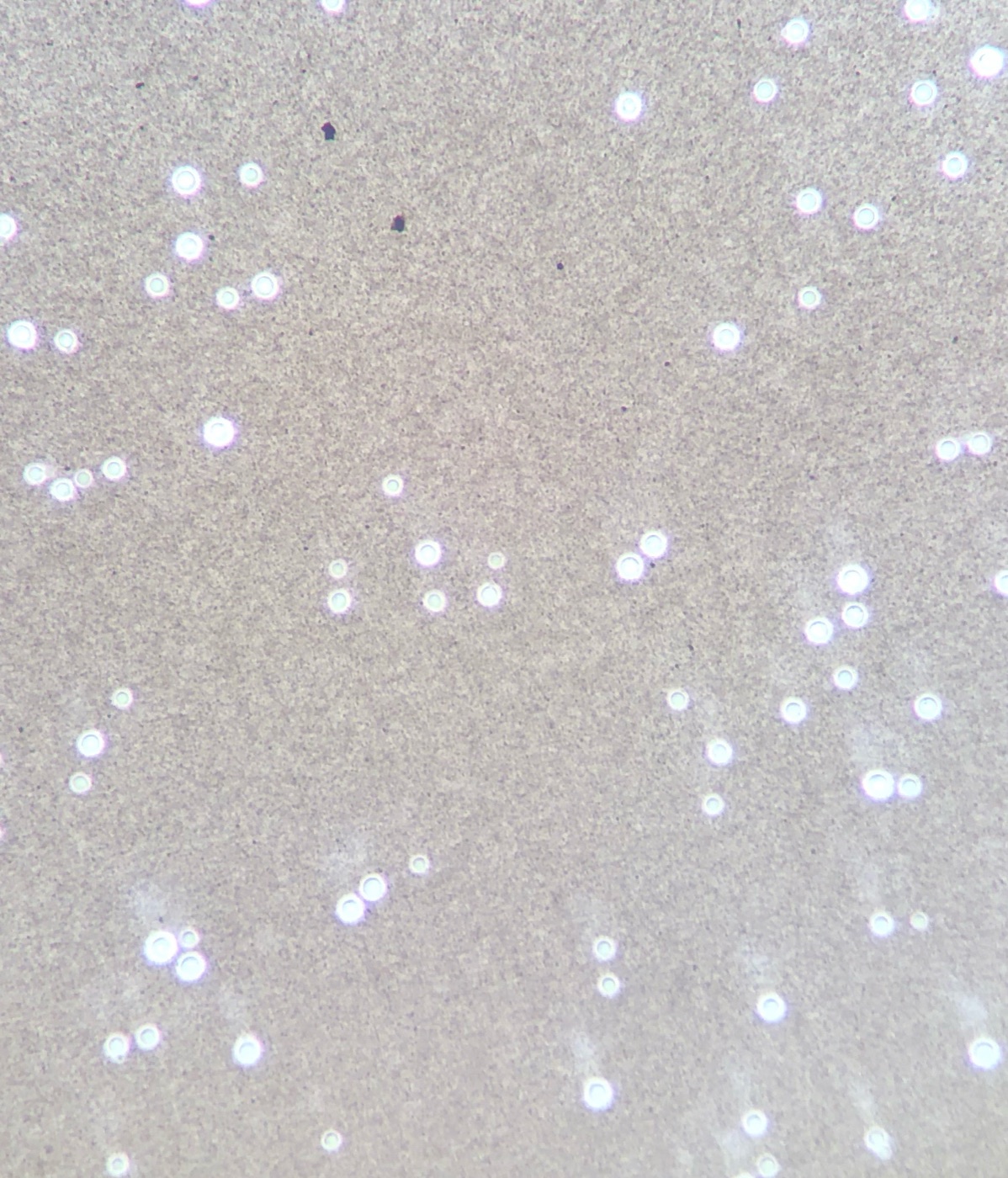
Cryptococcus in cerebrospinal fluid on an India ink preparation: round yeasts outlined by clear halos where the polysaccharide capsule excludes the ink particles. Magnification not stated.Image: Ed Uthman, Wikimedia Commons, CC BY 2.0.
Leukemia, Lymphoma and Plasma Cell Neoplasms in Cerebrospinal Fluid
Definition. Hematolymphoid neoplasms reach the cerebrospinal fluid (CSF) by spread to the leptomeninges, by origin in the central nervous system (CNS) or, during a traumatic lumbar puncture, by contamination with circulating neoplastic cells. CSF examination stages acute lymphoblastic leukemia, detects occult leptomeningeal disease in aggressive B-cell lymphoma, evaluates primary CNS lymphoma and confirms CNS involvement by plasma cell myeloma (Thastrup et al. 2022; Hegde et al. 2005; Abrey et al. 2005; Jurczyszyn et al. 2016). Conventional cytology has a sensitivity below 50% for CNS involvement by hematologic malignancy. Multiparameter flow cytometry detects more cases, particularly when few neoplastic cells are present (Del Principe et al. 2021).
Classification of CNS hematolymphoid neoplasms in WHO-HAEM5 and ICC 2022
Entity
WHO-HAEM5 (2022)
ICC (2022)
Sources
Large B-cell lymphoma arising in the CNS
Primary large B-cell lymphoma of the CNS, one of the large B-cell lymphomas of immune-privileged sites
Primary diffuse large B-cell lymphoma of the CNS, a distinct entity
Alaggio et al. 2022; Campo et al. 2022
Large B-cell lymphoma of the vitreoretina
Primary large B-cell lymphoma of the vitreoretina, within the immune-privileged group
Not listed as a separate entity in the ICC classification of mature lymphoid neoplasms, which lists primary diffuse large B-cell lymphoma of the CNS; the ICC majority judged a subcategorization of extranodal DLBCL by immune-privileged site premature
Alaggio et al. 2022; Campo et al. 2022
Large B-cell lymphoma of the testis
Primary large B-cell lymphoma of the testis, within the immune-privileged group
Primary diffuse large B-cell lymphoma of the testis, a specific entity closely related to the CNS lymphoma
Alaggio et al. 2022; Campo et al. 2022
Change from the revised 4th edition. WHO-HAEM5 introduced the umbrella term large B-cell lymphomas of immune-privileged sites. It combines the former primary diffuse large B-cell lymphoma (DLBCL) of the CNS with DLBCL of the vitreoretina and testis, which were previously included among DLBCL, not otherwise specified (Alaggio et al. 2022).
ICC position. The ICC discussed a broader grouping of extranodal activated B-cell-type lymphomas. The majority considered such a subcategorization premature, so these lymphomas are classified as specific entities (Campo et al. 2022).
Shared features of large B-cell lymphomas of immune-privileged sites
Feature
Description in WHO-HAEM5
Patients
Usually adults older than 60 years; immunocompetent
Spread
Tumors spread to other immune-privileged sites; vitreoretinal tumor can occur with or after CNS tumor
Immunophenotype
Activated B-cell type: usually CD10-negative, MUM1-positive, BCL6-positive; EBV-negative
Mutations
Concomitant MYD88 and CD79B mutations; genetic inactivation of MHC class I and II and B2M
Genomic signature
DLBCL C5/MCD/MYD88 group
Behavior
Aggressive, with generally poor prognosis
Source: Alaggio et al. 2022.
CNS involvement in acute lymphoblastic leukemia
CNS status categories in pediatric acute lymphoblastic leukemia
CNS status
CSF leukocytes/µL
CSF erythrocytes/µL
Leukemic blasts on cytospin
CNS1
5 or fewer
Fewer than 10
Absent
CNS2
5 or fewer
Fewer than 10
Present
CNS3
More than 5
Fewer than 10
Present
Traumatic puncture with blasts (TLP+)
Not applicable
10 or more
Present
Traumatic puncture without blasts (TLP−)
Not applicable
10 or more
Absent
Source: Thastrup et al. 2022, Table 1 (Ponte di Legno review), which gives CNS1 and CNS2 as "≤5" leukocytes/µL and CNS3 as ">5" leukocytes/µL.
CNS status thresholds by source. Thastrup et al. 2022 (Table 1) defines CNS2 as ≤5 leukocytes/µL with blasts and CNS3 as >5 leukocytes/µL with blasts, each with <10 erythrocytes/µL. The Dutch Childhood Oncology Group (protocols ALL-7 and ALL-8) defined CNS2 as blasts with "leukocytes less than 5/microL" and CNS3 as blasts with "leukocytes more than 5/microL" (te Loo et al. 2006).
Clinical criteria for CNS3. CNS3 status is also assigned for clinical or radiologic evidence of CNS leukemia, such as cranial nerve palsy, irrespective of CSF findings (Thastrup et al. 2022).
Frequency of CNS3. Only 2–5% of children are classified as CNS3 at diagnosis. Before universal CNS-directed therapy, up to 75% relapsed in the CNS (Thastrup et al. 2022).
CNS relapse in CNS1 patients. Most CNS relapses occur in children who were CNS1 at diagnosis, so CNS status has limited prognostic value (Thastrup et al. 2022).
Treatment context. Prophylactic cranial irradiation is given to 2–20% of children with acute lymphoblastic leukemia who have an increased risk of CNS relapse. Intensive intrathecal and systemic therapy has largely replaced it, and CNS relapse occurs in 3–8% of patients (Pui and Howard 2008).
Acute myeloid leukemia. About 2% of children with acute myeloid leukemia have overt CNS disease at diagnosis (Pui and Howard 2008).
Traumatic lumbar puncture in acute lymphoblastic leukemia
Study
Patients
Finding
Source
St Jude, 1984–1991
546
Two consecutive CSF samples contaminated with blasts: 5-year event-free survival 46%; hazard ratio 2.39
Gajjar et al. 2000
Dutch Childhood Oncology Group ALL-7 and ALL-8
526
Event-free survival 58% for TLP+ and 82% for TLP−; TLP+ an independent prognostic factor (risk ratio 3.5)
te Loo et al. 2006
Same Dutch cohort
526
Event-free survival 72.6% for CNS1, 70.3% for CNS2 and 66.7% for CNS3, without significant difference
te Loo et al. 2006
Interpretation. Contamination of CSF with circulating blasts during the diagnostic puncture adversely affects outcome and is an indication to intensify intrathecal therapy (Gajjar et al. 2000).
CNS2 status. In trials that gave additional intrathecal therapy to CNS2 patients, CNS2 status had no effect on prognosis. Trials that did not escalate therapy reported an adverse effect (Thastrup et al. 2022).
Limits of cytospin morphology in leukemia staging
Sensitivity and reproducibility. Cytospin cytology has low sensitivity, poor specificity and low reproducibility between laboratories (Thastrup et al. 2022).
Reactive T cells. Leukemic blasts can be difficult to separate from normal or reactive T lymphocytes on cytospins, and false-positive reports can result (Thastrup et al. 2022).
Cell decay. Leukocytes in CSF decay rapidly ex vivo. Delayed processing of unstabilized samples leads to underestimation of CNS leukemia (Thastrup et al. 2022).
Flow cytometry compared with cytospin at diagnosis of acute lymphoblastic leukemia
Patients
Positive by flow cytometry
Positive by cytospin
108 newly diagnosed children
28%
3%
214 newly diagnosed patients
17%
10%
87 newly diagnosed patients
41%
8%
673 newly diagnosed patients
25%
13%
TdT immunostaining study, 1,643 samples
Not tested; 9.1% positive by TdT immunostaining
3.9%
Source: Thastrup et al. 2022, summarizing prospective studies.
CNS involvement in aggressive B-cell lymphoma
Risk of CNS relapse in DLBCL: the CNS International Prognostic Index
CNS-IPI risk group
Share of patients
2-year rate of CNS disease, training set
2-year rate, validation set
Low
46%
0.6%
0.8%
Intermediate
41%
3.4%
3.9%
High
12%
10.2%
12.0%
Components. The CNS-IPI consists of the International Prognostic Index factors plus involvement of the kidneys or adrenal glands (Schmitz et al. 2016).
Population. The model was built in 2,164 patients with aggressive B-cell lymphoma treated with rituximab and CHOP-like chemotherapy and validated in 1,597 patients with DLBCL (Schmitz et al. 2016).
Occult leptomeningeal lymphoma detected by flow cytometry
Study
Patients
Positive by flow cytometry
Positive by cytology
Source
Newly diagnosed aggressive B-cell lymphoma at risk for CNS disease
51
11 (22%)
1
Hegde et al. 2005
Newly diagnosed aggressive B-cell lymphoma at high risk of CNS relapse, stabilized CSF
123
27 (22%)
7 (6%), with 3 more suspicious
Quijano et al. 2009
Analytic sensitivity. Multicolor flow cytometry identified neoplastic clones that made up as little as 0.2% of CSF lymphocytes (Hegde et al. 2005).
Level of infiltration and cytology. Samples positive by both methods typically had more than 20% neoplastic B cells or at least 1 neoplastic B cell/µL. Samples positive only by flow cytometry had lower levels of infiltration (Quijano et al. 2009).
Clinical correlates. The number of extranodal sites was the only factor associated with occult CSF lymphoma in newly diagnosed patients (Hegde et al. 2005).
Routine CSF parameters. CSF chemistry and cell counts were similar in patients with and without CSF lymphoma (Hegde et al. 2005).
Threshold for positivity. The threshold that defines flow cytometric positivity is not universally established (Del Principe et al. 2021).
Secondary CNS involvement by DLBCL
Frequency. In 1,221 Japanese patients with DLBCL treated with R-CHOP without CNS prophylaxis, 82 (6.7%) had a CNS event, and the cumulative 5-year probability of a CNS event was 8.4% (Tomita et al. 2012).
Site-specific risk factors. In that cohort, involvement of the breast (relative risk 10.5), adrenal gland (relative risk 4.6) and bone (relative risk 2.0) were independent risk factors for CNS events. Testicular involvement was already an accepted indication for CNS prophylaxis (Tomita et al. 2012).
Testicular DLBCL. In 134 patients with testicular DLBCL, rituximab reduced the cumulative incidence of progression. Rituximab was not associated with a lower risk of CNS relapse (Kridel et al. 2017).
Timing and outcome. In 291 patients with secondary CNS involvement after first-line immunochemotherapy, CNS involvement was part of the first relapse in 87%, and 39% had concurrent systemic relapse. Median overall survival after CNS involvement was 3.9 months, and the 2-year overall survival rate was 20% (El-Galaly et al. 2018).
Primary CNS lymphoma in CSF
Frequency of meningeal dissemination. In a prospective study of 282 patients, meningeal dissemination was found by cytomorphology in 33 of 205 (16%), by immunoglobulin heavy-chain PCR in 19 of 171 (11%) and by MRI in 8 of 217 (4%). Any method was positive in 17.4% (Fischer et al. 2008).
Discordance between methods. PCR was monoclonal in only 6 of 19 cytologically positive samples and in 10 of 105 cytologically negative samples. The methods are regarded as complementary (Fischer et al. 2008).
Pleocytosis. Detection of meningeal dissemination was more likely with CSF pleocytosis above 5 cells/µL (odds ratio 2.48). CSF protein had no predictive value (Fischer et al. 2008).
Staging and response. International consensus criteria base response assessment in primary CNS lymphoma on neuroimaging, corticosteroid use, ophthalmologic examination and CSF cytology (Abrey et al. 2005).
Ocular involvement. Vitreoretinal lymphoma, which can precede or follow CNS disease, is covered in Vitreous and Ocular Cytology.
MYD88 and other molecular markers in CSF in primary CNS lymphoma
Combined markers. In 54 patients with primary CNS lymphoma tested at diagnosis or relapse, clonal IGH rearrangement, MYD88 L265P or both were detected in 27 (50%) CSF cell pellets and in 24 (44%) cell-free DNA supernatants. Analysis of both fractions detected at least one molecular marker in 36 (66%), and 9 (16%) were positive only in cell-free DNA (Bravetti et al. 2023).
Comparison with cytology and flow cytometry. In the same cohort, cytology was positive in 7 (13.0%) and flow cytometry in 9 (17.3%). A high IL-10 level was found in 36 (66.7%). At least one lymphoma biomarker was detected in CSF in 46 of 54 patients (85%) (Bravetti et al. 2023).
Specificity among CNS tumors.MYD88 L265P has been described as specific to primary CNS lymphoma among CNS tumors. A rapid PCR system detected it in cell-free DNA from the CSF of 2 patients (Yamaguchi et al. 2023).
Biologic basis. Concomitant MYD88 and CD79B mutations are a shared feature of large B-cell lymphomas of immune-privileged sites (Alaggio et al. 2022).
Handling of CSF for flow cytometry
Stabilizing agents. In 99 CSF samples from patients with suspected or proven leptomeningeal hematologic malignancy, leukocyte numbers in TransFix-stabilized CSF were 1.4 times those of native CSF at 30 minutes and 2.3 times at 18 hours. After 18 hours, stabilization improved detection of malignant cells (de Jongste et al. 2014).
Hypocellularity and viability. The typical hypocellularity and low cell viability of CSF are the main technical problems of flow cytometric analysis (Del Principe et al. 2021).
Cytomorphology of hematolymphoid neoplasms and reactive lymphocytes in CSF
Lymphoma cells. Lymphomatous meningitis shows atypical lymphoid cells with enlarged nuclei, coarse chromatin and prominent nucleoli (Nay et al. 2026).
Eosinophils. Eosinophil-dominated pleocytosis is occasionally a reaction to neoplastic meningitis, particularly in CNS involvement by Hodgkin lymphoma (Nay et al. 2026).
Distinction of reactive lymphocytosis from lymphoma or leukemia in CSF
Feature
Reactive lymphocytosis
Lymphoma or leukemia
Sources
Cell population
Mixture of small, intermediate and activated lymphocytes, plasma cells and monocytes
Monotonous atypical lymphoid cells or blasts; intermediate activated forms usually absent
Nay et al. 2026
Mitoses
Can be present in viral meningitis
Can be present
Nay et al. 2026
Flow cytometry
Polytypic B cells and mixed T cells
Clonal population, detectable at 0.2% of lymphocytes
Hegde et al. 2005
Clinical context
Infection, autoimmune disease
Known lymphoma or leukemia; CNS-IPI risk; new cranial neuropathy
Schmitz et al. 2016; Thastrup et al. 2022
Plasma cell myeloma involving the CNS
Frequency. CNS involvement occurs in less than 1% of patients with multiple myeloma (Jurczyszyn et al. 2016).
Timing. In a multi-institutional study of 172 patients, 22% had CNS involvement at the initial myeloma diagnosis and 78% at relapse or progression. The median time from myeloma diagnosis to CNS involvement was 3 years (Jurczyszyn et al. 2016).
Presenting symptoms. The most common symptoms were visual changes (36%), radiculopathy (27%), headache (25%) and confusion (21%) (Jurczyszyn et al. 2016).
CSF findings. CSF cytology showed plasma cells in 86 of 96 patients tested (90%). Flow cytometry showed a monoclonal plasma cell population in 73 of 80 (91%) (Jurczyszyn et al. 2016).
Imaging. MRI showed leptomeningeal involvement in 57% and a cerebral mass in 53%. Imaging alone is insufficient for diagnosis because of false-positive and false-negative results (Jurczyszyn et al. 2016).
Diagnostic evidence. Clonal plasma cells in CSF, or pathologic evidence of soft tissue infiltration, establish CNS involvement (Jurczyszyn et al. 2016).
Immunophenotype and isotype. CD56 was expressed on CSF plasma cells in 70% of patients tested. IgA and IgD myeloma made up 27% and 2% of cases (Jurczyszyn et al. 2016).
Survival. Median overall survival from CNS involvement was 7 months (Jurczyszyn et al. 2016).
Metastatic and Primary Central Nervous System Tumors and Treatment Effects in Cerebrospinal Fluid
Definition. Leptomeningeal metastasis is the spread of a malignant tumor to the leptomeninges and the cerebrospinal fluid (CSF). Malignant cells in CSF, or a positive leptomeningeal biopsy, confirm the diagnosis (Le Rhun et al. 2023). Most solid-tumor leptomeningeal metastases come from breast carcinoma, lung carcinoma and melanoma (Wasserstrom, Glass and Posner 1982; Le Rhun et al. 2023). Primary CNS tumors account for a minority of positive CSF specimens. Embryonal tumors, gliomas, ependymomas and germ cell tumors are the main types (Chhieng et al. 2002). Specimens from shunts, drains and ventricular reservoirs, and specimens taken after intrathecal treatment, contain cells and reactions that mimic tumor (Nay et al. 2026).
Primary tumors that cause leptomeningeal metastasis
Frequency by primary site. In 90 patients with leptomeningeal metastases from solid tumors, the primary tumor was breast carcinoma in 46, lung carcinoma in 23 and melanoma in 11 (Wasserstrom, Glass and Posner 1982).
Clinical suspicion. Leptomeningeal metastasis is considered in patients with breast or lung cancer or melanoma who develop neurologic symptoms or signs (Le Rhun et al. 2023).
Latency. Leptomeningeal symptoms were the presenting sign of cancer in 5 patients and appeared as late as 10 years after the primary diagnosis in 4 (Wasserstrom, Glass and Posner 1982).
Survival. Median survival was 5.8 months after diagnosis in the 1982 series (Wasserstrom, Glass and Posner 1982).
Primary tumor
Concomitant brain metastases in patients with leptomeningeal metastasis
MRI without leptomeningeal findings
Source
Breast carcinoma
36–66%
4% of 318 patients
Le Rhun et al. 2023
Lung carcinoma
66–82%
26% of 171 patients
Le Rhun et al. 2023
Melanoma
69–87%
Not reported
Le Rhun et al. 2023
EANO-ESMO classification of leptomeningeal metastasis
Type
Definition
MRI subtypes and level of evidence
Source
Type I (confirmed)
Positive CSF cytology or positive leptomeningeal biopsy
IA, linear; IB, nodular; IC, linear and nodular; ID, hydrocephalus only or normal MRI
Le Rhun et al. 2023
Type II (probable or possible)
Negative or equivocal cytology; diagnosis based on clinical findings and neuroimaging in a patient with cancer and a reasonable risk of leptomeningeal metastasis
IIA–IIC with typical clinical signs: probable; IIA–IIC without typical signs: possible; IID (hydrocephalus or normal MRI) with typical signs: possible; IID without typical signs: lack of evidence
Le Rhun et al. 2023
Mandatory components. Clinical, imaging and CSF cytologic assessments are all required to classify leptomeningeal metastasis (Le Rhun et al. 2023).
Persistently positive cytology. The role of persisting positive CSF cytology alone in decisions on continuation of treatment remains controversial (Le Rhun et al. 2023).
Sensitivity of CSF cytology for leptomeningeal metastasis
Measure
Value
Source
Cytologically positive first CSF examination
49 of 90 patients
Wasserstrom, Glass and Posner 1982
Positive cytology after repeated sampling
82 of 90 patients
Wasserstrom, Glass and Posner 1982
First-puncture sensitivity across cancers
About 50–60%
Nay et al. 2026
Sensitivity after repeat collections
About 80%
Nay et al. 2026
Measures that raise sensitivity. CSF volume of more than 10 mL (at least 5 mL), processing within 30 minutes and avoidance of hemorrhagic contamination improve sensitivity. A second puncture under these conditions is performed when the first cytologic result is negative or equivocal (Le Rhun et al. 2023).
Cytologic features of malignant cells in CSF
Feature
Description
Source
Cell size
Enlarged; single tumor cells often 20–25 µm and up to 200 µm
Activated lymphocytes, monocytes and granulocytes of an accompanying inflammatory reaction ("neoplastic meningitis")
Nay et al. 2026
Classification by morphology. Morphology alone rarely permits tumor classification. Adenocarcinoma, acute leukemia, some lymphomas and melanoma are exceptions. Immunocytochemistry, flow cytometry or biopsy is used for further classification (Nay et al. 2026).
Immunocytochemistry in equivocal cases. Staining for tumor-specific alterations, such as HER2 in breast carcinoma or BRAF V600E in melanoma, can help in selected equivocal cases (Le Rhun et al. 2023).
Molecular and circulating tumor cell methods in CSF
Circulating tumor cells. Capture of tumor cells with epithelial cell adhesion molecule (EpCAM) antibodies or other antibody-coated magnetic particles has shown promising results. Quantification during follow-up could help define response to treatment (Le Rhun et al. 2023).
Tumor DNA. In leptomeningeal metastasis from lung cancer, a higher sensitivity for EGFR mutations and ALK and ROS1 rearrangements has been reported in CSF than in plasma (Le Rhun et al. 2023).
Status of molecular results. Evidence is insufficient to substitute detection of tumor-specific mutations in CSF for a positive cytologic result in clinical practice (Le Rhun et al. 2023).
Indications for molecular testing. Molecular testing is especially useful when cytology is repeatedly negative, when there is no history of cancer, or when the cause of clinical and imaging findings is uncertain and a treatment decision is required (Le Rhun et al. 2023).
Melanoma in CSF
Pigment. Intracellular or extracellular melanin pigment supports the diagnosis. It is distinguished from hemoglobin-derived pigment such as hemosiderin (Nay et al. 2026).
Distinction of pigmented cells in CSF
Feature
Melanoma cells
Siderophages
Sources
Cell type
Malignant cells with large nuclei, nucleoli and pseudoinclusions
Macrophages
Nay et al. 2026
Pigment
Melanin, intracellular or extracellular
Hemosiderin granules; later hematoidin crystals
Nay et al. 2026
Prussian blue
Negative for melanin
Positive
Nay et al. 2026
Clinical setting
Known melanoma
Prior subarachnoid or intraventricular hemorrhage
Nay et al. 2026
Primary CNS tumors in CSF
Proportion of positive specimens. Two-thirds of CNS tumors are primary, but only 10% of positive CSF specimens come from primary CNS tumors (Chhieng et al. 2002).
Tumor types. In 21 positive specimens from 20 patients, the tumors were 9 medulloblastomas, 7 gliomas (3 glioblastomas, 2 anaplastic astrocytomas and 2 ependymomas), 2 germinomas, 2 large B-cell lymphomas and 1 ganglioneurocytoma. No false-positive reports occurred (Chhieng et al. 2002).
Immunocytochemistry. Placental alkaline phosphatase supported a germinoma, and neuron-specific enolase and synaptophysin supported a ganglioneurocytoma (Chhieng et al. 2002).
Meningioma and other circumscribed tumors. Parenchymal masses with intact pial or ependymal boundaries, such as meningiomas, typically yield negative cytology. Exceptions are CNS WHO grade 2 or 3 histology and overt leptomeningeal or intraventricular seeding (Nay et al. 2026).
Medulloblastoma staging by CSF and MRI
Frequency of leptomeningeal disease. In 106 children with medulloblastoma or a primitive neuroectodermal tumor, 34 (32%) had leptomeningeal disease by CSF cytology, spinal MRI or both (Fouladi et al. 1999).
Discordance. Nine patients (8.5%) had positive MRI with negative cytology. Twelve (11.3%) had positive cytology with negative MRI (Fouladi et al. 1999).
Conclusion of the study. Either method alone would miss leptomeningeal disease in up to 14–18% of patients, so both are used (Fouladi et al. 1999).
Primary CNS tumors that seed CSF in the WHO CNS 5th edition
Tumor
Classification in WHO CNS5 (2021)
Source
Medulloblastoma
Molecularly defined types: medulloblastoma, WNT-activated; SHH-activated and TP53-wildtype; SHH-activated and TP53-mutant; and non-WNT/non-SHH, which comprises the former group 3 and group 4 tumors. Histologically defined: classic, desmoplastic/nodular, extensive nodularity, large cell/anaplastic, described as morphologic patterns
Louis et al. 2021
Ependymoma
Classified by site (supratentorial, posterior fossa, spinal) and molecular group, including ZFTA and YAP1 fusion-positive supratentorial types, posterior fossa groups PFA and PFB, and spinal ependymoma with MYCN amplification; myxopapillary ependymoma is CNS WHO grade 2
Louis et al. 2021
Atypical teratoid/rhabdoid tumor
Three molecular subtypes
Louis et al. 2021
Pineoblastoma
Four molecular subtypes, including MYC/FOXR2-activated and RB1-altered
Medulloblastoma, change from the 2016 classification. Medulloblastoma subgroups are retained. The 2016 histologic types are now described as morphologic patterns in a single section, and WHO CNS5 lists 4 subgroups of SHH-activated and 8 subgroups of non-WNT/non-SHH medulloblastoma (Louis et al. 2021).
Myxopapillary ependymoma, change from the previous edition. Myxopapillary ependymoma is CNS WHO grade 2 in WHO CNS5; it was grade 1 previously (Louis et al. 2021).
Grading of germ cell tumors. The WHO CNS5 summary lists germ cell tumor types without assigning CNS WHO grades. It states that certain malignant tumors, such as medulloblastoma and germinoma, "can be assigned a CNS WHO grade 4 designation", and that grades should not be assigned if a grade could confuse clinical care (Louis et al. 2021).
Ventricular, shunt and reservoir specimens
Ventriculitis. Ventricular CSF pleocytosis in ventriculitis is usually neutrophil-predominant. Prior surgery, intraventricular hemorrhage and intrathecal therapy can reduce it and also raise erythrocyte counts (Nay et al. 2026).
Cell index. A cell index of 4–5 or higher, calculated as the CSF leukocyte-to-erythrocyte ratio divided by the blood leukocyte-to-erythrocyte ratio, has been associated with ventriculitis (Nay et al. 2026).
Reservoir sampling for cytology. Before each intra-CSF injection through a ventricular reservoir, at least the volume to be injected is withdrawn. About 5 mL of nonhemorrhagic CSF is placed in one sterile tube for cytology (Le Rhun et al. 2023).
Eosinophilia in shunted patients
Frequency. CSF eosinophilia has been found in about 30% of patients with intraventricular shunt malfunction (Bezerra et al. 2011).
Causes. Shunt-associated eosinophilia is associated with infection, reactions to foreign material, particles or blood, obstruction of tubing and allergy to silicone. The infections are usually caused by Staphylococcus epidermidis and Cutibacterium (Propionibacterium) acnes (Bezerra et al. 2011; Nay et al. 2026).
Significance. Eosinophils in ventricular CSF correlate with hardware malfunction and CSF extravasation. They prompt evaluation for mechanical problems and targeted microbiology (Nay et al. 2026).
Treatment effects in CSF
Foreign-body macrophages. After intrathecal antisense therapy, such as nusinersen or tofersen, macrophages can contain drug-related basophilic inclusions. These macrophages are distinguished from siderophages (Nay et al. 2026).
Clearance of leukemic cells. In CNS3 leukemia, cells in CSF usually become undetectable after only 1–3 intrathecal treatments, although prolonged therapy is required to reduce relapse (Thastrup et al. 2022).
Differential diagnosis of atypical cells in CSF
Finding
Neoplastic interpretation
Benign mimic
Distinguishing features
Sources
Cohesive epithelial clusters
Metastatic carcinoma
Choroid plexus or ependymal cells in ventricular samples
Uniform round nuclei, delicate chromatin and cilia in benign clusters; malignant nuclear features in carcinoma
Nay et al. 2026
Large vacuolated cells
Signet-ring adenocarcinoma
Activated macrophages and foam cells
Macrophage nuclear features; foamy cytoplasm
Nay et al. 2026
Pigmented cells
Melanoma
Siderophages
Prussian blue positivity of hemosiderin
Nay et al. 2026
Small primitive cells
Medulloblastoma or other embryonal tumor
Germinal matrix cells in infants; marrow contamination
Clinical setting; neuron-specific enolase in germinal matrix cells; trilineage marrow elements
Drug-related macrophages after intrathecal therapy
Treatment history
Nay et al. 2026
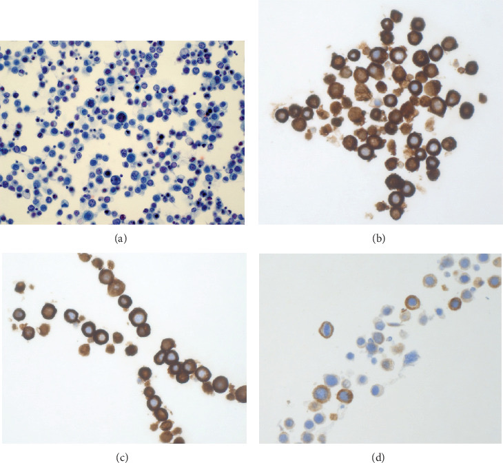
Metastatic high-grade urothelial carcinoma in cerebrospinal fluid. A, dissociated and loosely cohesive malignant epithelial cells, larger than the background inflammatory cells (Papanicolaou stain, 40× objective). B–D, the tumor cells are positive on immunocytochemistry for cytokeratin AE1/AE3 (B), cytokeratin 7 (C) and a urothelial marker (D) (40× objective).Image: Lim Christina et al., PMC open access (Case Rep Oncol Med. 2025 Sep 25;2025:8889550. doi: 10.1155/crom/8889550) Figure 5, CC BY.
Intraoperative Smear Cytology of Central Nervous System Tumors, Including Meningioma
Definition. Intraoperative smear cytology of the central nervous system (CNS) is the examination of squash, smear or touch preparations of neurosurgical tissue during the operation. It is used alone or with frozen sections to establish lesional tissue, a preliminary diagnosis and the need for tissue triage (Sharma and Deb 2011). Smears are particularly useful for small stereotactic biopsies, for the fibrillary background that separates glial from non-glial tumors and for discohesive tumors such as lymphoma and pituitary neuroendocrine tumor (Sharma and Deb 2011). Classification follows the WHO Classification of Tumours of the CNS, 5th edition (WHO CNS5, 2021) (Louis et al. 2021).
Types of preparation. Smear, squash and touch preparations are used. Smears are better suited than frozen sections to tiny stereotactic specimens (Sharma and Deb 2011).
Fixation and staining. In one series, squash or touch smears were immersed in 95% ethanol for 2 minutes and stained with hematoxylin and eosin (Kang et al. 2019). In another, preparations were stained by a quick Papanicolaou method (Mitra et al. 2010).
Air-dried stains. A Diff-Quik stain highlights myxoid material and lymphoglandular bodies (Sharma and Deb 2011).
Firm tissue. Firm lesions, such as schwannomas, metastatic carcinomas and inflammatory lesions, smear poorly. Frozen sections preserve their architecture (Kang et al. 2019).
Correlation with frozen section. Smears and frozen sections are examined together when possible. If the two disagree, the intraoperative diagnosis is based primarily on the frozen section when tissue is adequate (Sharma and Deb 2011).
Necrosis and mitoses on smears. Necrosis adheres poorly to slides, and smearing shears cells, so mitoses and necrosis are underrecognized on smears (Sharma and Deb 2011).
Purposes of the intraoperative diagnosis
Confirmation of lesional tissue. In one study, adequacy of a specimen could be confirmed with 96% sensitivity and 75% specificity (Sharma and Deb 2011).
Triage for infection and lymphoma. When a smear indicates infection or lymphoma, additional tissue is obtained for microbiology or flow cytometry (Sharma and Deb 2011).
Change in surgery. Detection of lymphoma in a ring-enhancing lesion limits surgery. A diagnosis of high-grade glioma leads to maximal safe resection (Sharma and Deb 2011).
Deeper sampling. A nondiagnostic glioma sample can represent the periphery of a high-grade glioma, and the surgeon can obtain a deeper biopsy (Sharma and Deb 2011).
Accuracy of intraoperative CNS cytology
Study
Cases
Result
Source
Squash preparation versus frozen section
96 tumors with a cytologic diagnosis
Cytology 88.5%; frozen section 90.6%; gliomas 84.9% versus 86.8%
Mitra et al. 2010
Frozen cytology with frozen section
454
Accuracy 87.2%; 98.8% after excluding 53 deferred cases
Concordance of intraoperative CNS diagnosis by tumor type
Tumor type
Concordance
Source
Meningeal tumors
97 of 98 (99%)
Kang et al. 2019
Metastatic tumors
51 of 52 (98.1%)
Kang et al. 2019
Pituitary adenomas
86 of 89 (96.6%)
Kang et al. 2019
Schwannomas
45 of 47 (95.8%)
Kang et al. 2019
High-grade astrocytic tumors
47 of 58 (81%)
Kang et al. 2019
Low-grade astrocytic tumors
10 of 13 (76.9%)
Kang et al. 2019
Non-neoplastic lesions
23 of 36 (63.9%)
Kang et al. 2019
Sources of discrepancy in intraoperative CNS diagnosis
Category
Share of 57 discrepancies
Source
Spindle cell lesions, most often schwannoma or meningioma confused with other lesions
21.1%
Plesec and Prayson 2007
Oligodendroglioma versus astrocytoma
21.1%
Plesec and Prayson 2007
CNS lymphoma
15.8%
Plesec and Prayson 2007
Reactive versus neoplastic process, most often gliosis versus glioma
14.0%
Plesec and Prayson 2007
Overgrading of tumors
7.0%
Plesec and Prayson 2007
Other
21.1%
Plesec and Prayson 2007
Cushing disease. In 54 operations (13.4%) with remission, pathologic confirmation of the adenoma came only from intraoperative frozen section or cytology. Addition of intraoperative cytology increased overall pathologic confirmation compared with a historical cohort (80.9% versus 67.1%) (Qiao et al. 2019).
Glands or clusters in carcinoma; whorls in meningioma; focal glial processes in epithelioid glioblastoma
Sharma and Deb 2011
Discohesive monomorphic large cells
Primary CNS lymphoma, germinoma
Lymphoglandular bodies in lymphoma; distinct cell borders and vacuolated cytoplasm in germinoma
Sharma and Deb 2011
Discohesive small cells
Small cell carcinoma, medulloblastoma and other embryonal tumors
Hyperchromatic molded nuclei, mitoses, apoptosis
Sharma and Deb 2011
Macrophage-rich smear
Demyelination, infarct, infection
Foamy, sometimes mitotically active macrophages
Sharma and Deb 2011
Glial tumors on smears
Glioma types in WHO CNS5 relevant to smear diagnosis
Tumor type
WHO CNS5 definition or grading
Source
Glioblastoma, IDH-wildtype
IDH-wildtype diffuse astrocytic glioma in adults with one or more of: microvascular proliferation, necrosis, TERT promoter mutation, EGFR amplification, or +7/−10 chromosome copy-number change
Louis et al. 2021
Astrocytoma, IDH-mutant
CNS WHO grade 2, 3 or 4; homozygous CDKN2A/B deletion confers grade 4
Louis et al. 2021
Oligodendroglioma, IDH-mutant and 1p/19q-codeleted
CNS WHO grade 2 or 3
Louis et al. 2021
Pilocytic astrocytoma
Circumscribed astrocytic glioma; KIAA1549::BRAF fusion, BRAF and NF1 alterations
Louis et al. 2021
Ependymoma
Classified by site and molecular group
Louis et al. 2021
Edition change for glioma grading. WHO CNS5 uses Arabic numerals, grades tumors within types and endorses the term "CNS WHO grade". Previous classifications used Roman numerals and applied grades across different tumor entities. WHO CNS5 also adds molecular grading parameters such as homozygous CDKN2A/B deletion in IDH-mutant astrocytoma (Louis et al. 2021).
Edition change for IDH-mutant astrocytoma. WHO CNS5 treats all IDH-mutant diffuse astrocytic tumors as a single type, astrocytoma, IDH-mutant, graded 2 to 4, and eliminates the term glioblastoma, IDH-mutant. In 2016 the grade IV IDH-mutant tumor was glioblastoma, IDH-mutant (Louis et al. 2021).
Smear features of glial tumors
Pilocytic astrocytoma. Smears show piloid (hairlike) and fibrillary astrocytes with bland elongated nuclei and thin processes, with or without Rosenthal fibers and eosinophilic granular bodies (Sharma and Deb 2011).
Atypical features in pilocytic astrocytoma. Cytologic atypia, smudgy chromatin and microvascular proliferation are acceptable in pilocytic astrocytoma. In a fibrillary-rich sample they can prompt a mistaken high-grade diagnosis (Sharma and Deb 2011).
Reactive gliosis versus astrocytoma. Well-spaced astrocytes with long processes favor reactive gliosis. Astrocytoma shows coarser, more intricately networked processes (Sharma and Deb 2011).
Oligodendroglioma. Nuclei are relatively round, smooth and monomorphic, with bland chromatin, mini-gemistocytes and some glial strands. The perinuclear halo of paraffin sections is a fixation artifact and is absent on smears (Sharma and Deb 2011).
Ependymoma. Smears show monomorphic round cells with salt-and-pepper chromatin and micronucleoli in a fibrillary background. Perivascular pseudorosettes, tumor cells around vessels separated by cell-free fibrillary zones, are essential to the diagnosis (Sharma and Deb 2011).
Ependymoma nuclei. Nuclear grooves and nuclear inclusions separate ependymoma from neurocytoma (Kang et al. 2019).
High-grade astrocytoma and glioblastoma. Smears show pleomorphic or gemistocytic cells with coarse multipolar processes forming a meshwork. Hyperplastic vessels have multilayered cells and complex loops (Sharma and Deb 2011).
Recurrent treated tumors. Recurrent treated tumors are often more anaplastic but have fewer mitoses than primary tumors (Sharma and Deb 2011).
Non-glial tumors on smears
Primary CNS lymphoma. Smears show monotonous large discohesive cells with scant to moderate bluish cytoplasm and prominent nucleoli in a background of lymphoglandular bodies (Sharma and Deb 2011). Lymphoma lacks endovascular proliferation and shows tingible body macrophages (Kang et al. 2019).
Lymphoma with demyelination. Primary CNS lymphoma often has a demyelinating component, which becomes more obvious after corticosteroid-induced tumor regression (Sharma and Deb 2011).
Macrophages mimicking lymphoma. Activated macrophages stripped of cytoplasm during smearing can mimic lymphoma. The differential diagnosis includes demyelination, infarct and infection (Sharma and Deb 2011).
Metastatic carcinoma. Cohesive clusters of malignant epithelial cells, sometimes forming glands, indicate carcinoma. The site of origin is best assessed with immunohistochemistry (Sharma and Deb 2011).
Small cell carcinoma. Small round to spindled hyperchromatic molded nuclei with frequent mitoses and apoptosis suggest small cell carcinoma (Sharma and Deb 2011).
Embryonal tumors. Medulloblastoma and other embryonal tumors are densely cellular, with monomorphic small round cells, molded nuclei, scant cytoplasm and frequent mitoses. Homer Wright rosettes are usually indistinct on smears (Sharma and Deb 2011).
Germinoma. Germinoma cells are large, with distinct borders, vesicular nuclei and prominent nucleoli, with or without cytoplasmic vacuolation (Sharma and Deb 2011).
Atypical teratoid/rhabdoid tumor. Discohesive plasmacytoid cells and characteristic rhabdoid cells support the diagnosis (Sharma and Deb 2011).
Myxopapillary ependymoma and cauda equina neuroendocrine tumor. Both occur in the cauda equina and show perivascular orientation. Myxoid material, highlighted by the Diff-Quik stain, favors myxopapillary ependymoma (Sharma and Deb 2011).
Edition changes for cauda equina tumors. WHO CNS5 grades myxopapillary ependymoma as CNS WHO grade 2 (previously grade 1) and recognizes paraganglioma of the cauda equina and filum terminale as a distinct tumor type (Louis et al. 2021), named cauda equina neuroendocrine tumor (Mittenzwei and Yoda 2024).
Pituitary neuroendocrine tumor terminology. WHO CNS5 adds the term pituitary neuroendocrine tumor (PitNET) to the name pituitary adenoma (Louis et al. 2021).
Pituitary neuroendocrine tumor on smears. Pituitary tissue is recognizable on smears. Separation of hyperplasia from adenoma is easier on frozen sections and best on paraffin sections with a reticulin stain (Sharma and Deb 2011).
Nerve sheath tumor types in WHO CNS5. Cranial and paraspinal nerve tumors comprise schwannoma, neurofibroma, perineurioma, hybrid nerve sheath tumors, malignant melanotic nerve sheath tumor, malignant peripheral nerve sheath tumor and cauda equina neuroendocrine tumor (Mittenzwei and Yoda 2024).
Schwannoma. Schwannomas lack whorls and have elongated, wavy cytoplasm (Kang et al. 2019). Verocay bodies are difficult to recognize on smears (Sharma and Deb 2011).
Meningioma
Classification and grading of meningioma in WHO CNS5
Single type. WHO CNS5 classifies meningioma as a single tumor type with 15 subtypes (Louis et al. 2021).
Molecular grade 3 criteria. A TERT promoter mutation or homozygous CDKN2A/B deletion assigns CNS WHO grade 3, independent of morphology (Louis et al. 2021; Sahm et al. 2025).
Grade 2 subtypes. Chordoid and clear cell meningiomas are CNS WHO grade 2 (Louis et al. 2021).
Brain invasion. Brain invasion is infiltration of tumor cells through the leptomeninges into the underlying brain parenchyma. Extension along Virchow-Robin spaces does not qualify. It has been reported in 4% to one third of meningiomas (Sahm et al. 2025).
Minor histologic criteria. The "soft" criteria are high cellularity, small cells with a high nucleus-to-cytoplasm ratio, prominent nucleoli, sheeting and spontaneous necrosis. cIMPACT-NOW update 8 lists 2 of these 5 criteria with a borderline mitotic count as a scenario for molecular testing (Sahm et al. 2025).
Grading element
WHO CNS5 (2021)
Previous rule
Source
Mitotic threshold, grade 2
2.5 or more mitoses/mm²
4 or more (4–19) mitoses per 10 high-power fields of 0.16 mm²
Sahm et al. 2025
Mitotic threshold, grade 3
12.5 or more mitoses/mm²
20 or more mitoses in 10 high-power fields of 0.16 mm²
Sahm et al. 2025
Rhabdoid and papillary morphology
Graded 1 to 3 by the other histologic criteria
Grade 3 on the basis of rhabdoid or papillary histology alone
Sahm et al. 2025
TERT promoter mutation or homozygous CDKN2A/B deletion
Grade 3
Not a grading criterion
Louis et al. 2021; Sahm et al. 2025
Numerals
Arabic (CNS WHO grade 1, 2, 3)
Roman
Louis et al. 2021
Area for mitotic counts. An area of 1.6 mm² is evaluated, and counts in smaller areas are not extrapolated (Sahm et al. 2025).
cIMPACT-NOW update 8 (not an official WHO classification). A histologically low-grade or borderline meningioma with complete or segmental 1p deletion and concurrent 22q deletion, an NF2 oncogenic variant or both is assigned CNS WHO grade 2, unless markers of grade 3 are present. Testing for 1p status is not recommended for all otherwise low-grade meningiomas. Molecular testing is recommended for borderline grade 1/2 features, brain-invasive otherwise benign tumors, grade 1 tumors with unexpectedly rapid growth or recurrence, chordoid, clear cell, rhabdoid or papillary histology, and meningiomas in children and young adults (Sahm et al. 2025).
Brain-invasive, otherwise benign meningioma. Under cIMPACT-NOW update 8, such a tumor is not graded before molecular data are obtained. Without molecular high-risk features it is CNS WHO grade 1; with 1p deletion plus 22q deletion or an NF2 variant it is grade 2. Without testing it is reported as meningioma, not otherwise specified, without a grade (Sahm et al. 2025).
Markers without recommendations. H3K27me3 status in grade 2 meningioma offers prognostic information but does not inform grading, and data on hemizygous CDKN2A/B deletion are too limited for conclusions (Sahm et al. 2025).
Rhabdoid and papillary meningioma genetics.BAP1 inactivation is frequent in meningiomas with rhabdoid or papillary morphology (Sahm et al. 2025).
Clear cell meningioma.SMARCE1 inactivation is highly useful in establishing the diagnosis and grade of clear cell meningioma, which is CNS WHO grade 2 (Louis et al. 2021; Sahm et al. 2025).
Cytologic features of meningioma on smears
Conventional low-grade meningioma. Smears show syncytial whorls, nuclear inclusions or vesicular nuclear clearing and thick collagenous cytoplasm (Kang et al. 2019).
Fine-needle aspirates of orbital meningioma. Smears showed whorls and syncytial groups with round to oval nuclei and inconspicuous nucleoli. An intranuclear inclusion and psammoma bodies were each seen in one of four cases (Agrawal et al. 2012).
Chordoid meningioma. Imprint smears of five cases showed cohesive cords of bland cells with round nuclei, stippled chromatin, small nucleoli and cytoplasmic vacuolation, with chronic inflammatory cells. Intranuclear inclusions were present in 80% and whorl-like structures in 60%. Necrosis, psammoma bodies and mitoses were absent (Lui et al. 2007).
Rhabdoid and papillary meningioma. These show few whorls and perivascular rosette-like arrangements that have been misdiagnosed as ependymoma. Nuclear inclusions occur in both tumors (Kang et al. 2019).
Rhabdoid features. Eosinophilic hyalinized cytoplasm and hyaline globules of rhabdoid meningioma are distinguished from atypical teratoid/rhabdoid tumor, germinoma and embryonal carcinoma (Kang et al. 2019).
Anaplastic meningioma. Pleomorphic cells can mimic carcinoma or sarcoma (Kang et al. 2019). Focal whorls or typical meningothelial nuclear features are usually still present (Sharma and Deb 2011).
Immunohistochemistry of meningioma
SSTR2A. In 35 meningiomas, 10 schwannomas and 10 solitary fibrous tumors (hemangiopericytomas), SSTR2A was positive in 89% of meningiomas and in none of the other tumors. All grade 1 and 2 meningiomas were positive, and 20% of grade 3 meningiomas were positive (Anis et al. 2018).
Claudin-1. Claudin-1 was positive in 49% of meningiomas and negative in schwannomas and solitary fibrous tumors (Anis et al. 2018).
Solitary fibrous tumor terminology. WHO CNS5 retired the term hemangiopericytoma. The tumor is called solitary fibrous tumor and graded with a three-tiered CNS scheme (Louis et al. 2021).
Differential diagnosis of meningioma on intraoperative smears
Feature
Meningioma
Schwannoma
Ependymoma
Metastatic carcinoma
Architecture
Syncytial clusters and whorls
No whorls
Perivascular pseudorosettes
Cohesive clusters, sometimes glands
Cytoplasm
Thick collagenous cytoplasm
Elongated, wavy cytoplasm
Fibrillary processes
Epithelial
Nuclei
Inclusions, vesicular clearing
Elongated
Round, salt-and-pepper chromatin, grooves and inclusions
Malignant features
Smear quality
Smears well
Firm; frozen section preferred
Smears well
Firm lesions smear poorly
Sources
Kang et al. 2019
Kang et al. 2019; Sharma and Deb 2011
Sharma and Deb 2011; Kang et al. 2019
Sharma and Deb 2011; Kang et al. 2019
Vitreous and Ocular Cytology
Definition. Ocular cytology is the examination of vitreous, aqueous humor and fine-needle aspiration biopsy (FNAB) specimens from intraocular lesions. Its main uses are the diagnosis of vitreoretinal lymphoma in vitrectomy specimens and the diagnosis and molecular prognostication of uveal melanoma by FNAB (Chan et al. 2011; Singh et al. 2016). Biopsy of suspected retinoblastoma is generally avoided because of the risk of extraocular spread (Dimaras et al. 2015; Milman et al. 2023). The WHO Classification of Tumours of the Eye and Orbit, 5th edition, added diagnostic cytopathology for retinal, neuroepithelial and melanocytic uveal tumors and updated the intraocular lymphoma chapter for molecular genetics, cytopathology and diagnostic criteria (Milman et al. 2023).
WHO Eye 5th edition: primary vitreoretinal large cell B-cell lymphoma (PVR-LCBL), the most common high-grade lymphoma arising in the eye. WHO-HAEM5: primary large B-cell lymphoma of the vitreoretina, one of the large B-cell lymphomas of immune-privileged sites
WHO-HAEM5 moved vitreoretinal lymphoma from DLBCL, not otherwise specified, into the immune-privileged group
Milman et al. 2023; Alaggio et al. 2022
Uveal melanoma
WHO Eye 5th edition, uveal tract: iris melanoma (with ocular melanocytosis and melanocytic nevus of the iris); choroidal and ciliary body melanomas, with benign choroidal and ciliary body nevi, uveal melanocytoma and bilateral diffuse uveal melanocytic proliferation
Diagnostic cytopathology added for benign and malignant melanocytic uveal tumors
Milman et al. 2023
Retinoblastoma
WHO Eye 5th edition, retinal tumors, including MYCN-amplified, RB1-wildtype retinoblastoma
Retinocytoma classified as a premalignant tumor
Milman et al. 2023
Vitreoretinal lymphoma
Clinical features of vitreoretinal lymphoma
Patients. Primary vitreoretinal lymphoma most often develops in elderly patients and is usually a diffuse large B-cell lymphoma (Chan et al. 2011).
Presentation. It commonly resembles posterior uveitis. Clinical findings include vitreous cellular infiltration and subretinal tumor infiltration (Chan et al. 2011).
Relation to CNS lymphoma. More than 15% of patients with primary CNS lymphoma develop intraocular lymphoma. Conversely, 65–90% of patients with primary vitreoretinal lymphoma develop CNS lymphoma, usually within 29 months (Chan et al. 2011).
Handling of vitreous specimens
Cell fragility. Lymphoma cells in vitreous easily undergo necrosis, so ocular fluid specimens are processed carefully and promptly (Chan et al. 2011).
Low cellularity. Biopsy is sometimes deferred because the vitreous cells are judged too few for an adequate sample (Chan et al. 2011).
Molecular testing in sparse samples.MYD88 and CD79B hotspot mutation analysis is particularly useful in specimens of low cellularity (Milman et al. 2023).
Cytologic features of vitreoretinal lymphoma
Lymphoma cells. The cells are large atypical lymphoid cells with large irregular nuclei, prominent nucleoli and scant basophilic cytoplasm. They are relatively homogeneous and larger than ordinary inflammatory cells (Chan et al. 2011).
Mitoses. Mitoses can be present, rare or absent (Chan et al. 2011).
Background. The background contains apoptotic and necrotic cells, small T cells and macrophages (Milman et al. 2023).
Reactive T cells. Reactive lymphocytes are often mixed with the lymphoma cells, and CD3-positive T cells can outnumber the malignant cells (Chan et al. 2011).
Immunophenotype. Immunohistochemistry shows CD20-positive cells. Monoclonality is shown as a B-cell population with κ or λ light chain restriction by immunohistochemistry or flow cytometry (Chan et al. 2011).
Ancillary tests for vitreoretinal lymphoma
Test
Pooled sensitivity
Pooled specificity
Studies
Source
IL-10:IL-6 ratio
0.89
0.98
27 studies; 1,370 patients
Lou et al. 2026
IL-10 alone
0.87
0.95
11 studies; 549 patients
Lou et al. 2026
MYD88 mutation
0.73
0.99
12 studies; 382 patients
Lou et al. 2026
IGH clonality
0.85
0.97
12 studies; 649 patients
Lou et al. 2026
CD79B mutation
0.40
1.00
4 studies; 123 patients
Lou et al. 2026
IL-10:IL-6 ratio and MYD88 in parallel
97.1%
97.0%
Combined analysis
Lou et al. 2026
Cytokine ratio. An IL-10:IL-6 ratio greater than 1 is highly suggestive of B-cell vitreoretinal lymphoma but is not diagnostic (Chan et al. 2011).
Aqueous humor. Cytokine testing of aqueous humor gave higher diagnostic odds ratios than vitreous fluid in the 2026 meta-analysis. The certainty of the underlying evidence was low to very low (Lou et al. 2026).
MYD88 mutations.MYD88 mutations were found in 20 of 29 (69%) confirmed vitreoretinal lymphomas. They included 6 cases first diagnosed as reactive or suspicious, so testing improves the diagnostic yield of vitrectomy specimens (Bonzheim et al. 2015).
Clonality analysis.IGH rearrangement analysis of small ocular samples can give false-positive or false-negative results. Microdissection of atypical cells improves the analysis (Chan et al. 2011).
Distinction of vitreoretinal lymphoma from uveitis
Feature
Vitreoretinal lymphoma
Uveitis
Sources
Atypical cells
Large atypical B cells with irregular nuclei and prominent nucleoli
Absent
Chan et al. 2011
Background
Apoptosis, necrosis, small T cells, macrophages
Reactive lymphocytes and macrophages
Milman et al. 2023; Chan et al. 2011
B-cell clonality
κ or λ restriction; clonal IGH
No light chain restriction
Chan et al. 2011
IL-10:IL-6 ratio
Greater than 1 is highly suggestive of lymphoma and is not diagnostic alone
A ratio above 1 is not expected
Chan et al. 2011
MYD88 mutation
69% of confirmed cases in one series; pooled sensitivity 0.73
Rarely detected (pooled specificity 0.99)
Bonzheim et al. 2015; Lou et al. 2026
Uveal melanoma
Classification and cytologic criteria of uveal melanocytic tumors
Entities. The WHO Eye 5th edition lists iris melanocytic tumors (ocular melanocytosis, melanocytic nevus and iris melanoma) separately from choroidal and ciliary body tumors. The malignant choroidal and ciliary body tumor is choroidal and ciliary body melanoma. The benign tumors are nevus, uveal melanocytoma and bilateral diffuse uveal melanocytic proliferation (Milman et al. 2023).
Uveal melanocytoma. Uveal melanocytoma carries solitary GNAQ mutations without additional oncogenic alterations (Milman et al. 2023).
Cytologic criteria in the WHO Eye 5th edition.
Conventional nevus: a paucicellular sample of predominantly small spindle to round cells, with or without pigment, with rare or no Ki-67-positive cells.
Melanocytoma: large ovoid cells with small central nuclei and heavily pigmented cytoplasm; melanin can obscure nuclear detail.
Melanoma: individually dispersed atypical cells or small cohesive groups, in which binucleation and mitoses can be seen (Milman et al. 2023).
Cell type. In the Armed Forces Institute of Pathology modification of the Callender classification, tumors formerly classified as spindle cell type that contained small or rare epithelioid cells were reclassified as mixed cell type, which improved prediction of outcome. Of 52 tumors originally classified as spindle cell type, only 31 of the 100 cases reviewed were reclassified as spindle cell types (2 spindle cell nevi and 29 spindle cell melanomas). Nucleolar size and pleomorphism were identified as important variables (McLean et al. 1983).
Cell type and chromosome 3. In the original monosomy 3 series, histopathologic subtype added no predictive value to monosomy 3, tumor location and tumor diameter (Prescher et al. 1996).
Fine-needle aspiration biopsy of uveal melanoma
Yield and safety. In 150 consecutive patients sampled with a 25-gauge needle, the diagnostic yield was 92% and false-negative results occurred in 8%. Prognostication by fluorescence in situ hybridization for monosomy 3 was possible in 85% (Singh et al. 2016).
Approach and tumor size. Diagnostic yield was 100% by the transcorneal, 96% by the transscleral and 86% by the transvitreal approach. Diagnostic yield was significantly correlated with tumor size, with basal diameter above 5.0 mm and height above 2.5 mm as the size categories (Singh et al. 2016).
Complications. Persistent hemorrhage requiring surgery and rhegmatogenous retinal detachment each occurred in 1%. No endophthalmitis, tumor recurrence or episcleral seeding was seen over a mean follow-up of 37 months (Singh et al. 2016).
Genetic yield. In 119 biopsies, chromosome microarray succeeded more often in post-enucleation samples than in in vivo biopsies (77% versus 50%). Cytologic yield did not differ between FNAB, vitrector and forceps samples (Lin et al. 2022).
Cytologic features of uveal melanoma
Feature
Frequency in 131 adequate FNAB samples
Source
Spindle cells present
98% (63% mixed with epithelioid cells; 35% spindle only)
Medina et al. 2015
Epithelioid cells only
2 samples
Medina et al. 2015
Melanin granules
80%
Medina et al. 2015
Cell shapes. Melanoma cells are spindle-shaped in spindle cell melanoma and ovoid in epithelioid cell melanoma (Milman et al. 2023).
Nuclear grade. Nuclear atypia increased with tumor height. Iris melanomas showed the blandest features (Medina et al. 2015).
Morphology and prognosis in biopsies. In post-enucleation samples, epithelioid cell type correlated with monosomy 3 and BAP1 aberrancy. In in vivo biopsies it did not. Cytomorphology alone is therefore insufficient for prognostication, and immunohistochemistry is a useful surrogate (Lin et al. 2022).
Molecular prognostication. Molecular genetic profiling of cytology material is used for prognostication (Milman et al. 2023).
Prognostic genetic markers in uveal melanoma
Marker
Finding
Source
Monosomy 3
Present in 30 of 54 tumors; 57% of these patients relapsed, and 3-year relapse-free survival was 50%. None of 24 patients with disomy 3 developed metastasis
Prescher et al. 1996
15-gene expression profile
Class 1 in 61.9% and class 2 in 38.1% of 446 classified tumors; metastasis in 1.1% of class 1 and 25.9% of class 2
Onken et al. 2012
Gene expression profile versus chromosome 3
20.8% of tumors were discordant; the expression profile had superior prognostic accuracy, and chromosome 3 added no independent information
Onken et al. 2012
BAP1
Inactivating mutations in 26 of 31 (84%) metastasizing tumors
Harbour et al. 2010
BAP1 in a clinical cohort
Mutated in 45%; associated with class 2 profile; strongest factor for metastasis after the expression class
Decatur et al. 2016
SF3B1
Mutated in 24%; associated with younger age
Decatur et al. 2016
EIF1AX
Mutated in 17%; associated with class 1 profile
Decatur et al. 2016
Assay performance. The 15-gene expression profile assay classified 446 of 459 tumors (97.2%) in a prospective multicenter study (Onken et al. 2012).
Mutual exclusivity.BAP1, SF3B1 and EIF1AX mutations were almost mutually exclusive (Decatur et al. 2016).
Initiating G-protein mutations in uveal melanoma
Gene
Uveal melanoma
Blue nevi
Source
GNAQ Q209
45% of primary tumors
55%
Van Raamsdonk et al. 2010
GNA11 Q209
32% of primary tumors; 57% of metastases
7%
Van Raamsdonk et al. 2010
GNAQ or GNA11, any
83% of uveal melanomas
Not applicable
Van Raamsdonk et al. 2010
Mutual exclusivity.GNAQ and GNA11 mutations are mutually exclusive (Van Raamsdonk et al. 2010; Decatur et al. 2016).
Prognostic value.GNA11 mutations were not associated with any clinicopathologic feature analyzed. GNAQ mutations were associated with absence of ciliary body involvement and greater basal diameter (Decatur et al. 2016).
Retinoblastoma
Epidemiology and genetics of retinoblastoma
Incidence. Retinoblastoma occurs in 1 in 16,000–18,000 live births (Dimaras et al. 2015).
Presentation. Leukocoria is the most common sign (Dimaras et al. 2015).
Age at diagnosis. The median age at diagnosis is 12 months in bilaterally affected patients, who have heritable disease, and 24 months in unilaterally affected patients without family history (Dimaras et al. 2015).
RB1. Retinoblastoma forms when both RB1 alleles are mutated in a susceptible retinal cell, likely a cone photoreceptor precursor (Dimaras et al. 2015).
Retinoma and retinocytoma. The benign retinoma has lost both RB1 alleles (Dimaras et al. 2015). The WHO Eye 5th edition classifies retinocytoma as a premalignant tumor because it carries biallelic RB1 mutations and can dedifferentiate into retinoblastoma (Milman et al. 2023).
MYCN-amplified retinoblastoma. Of 1,068 unilateral non-familial tumors, 29 (2.7%) had no RB1 mutation, and 15 of these had high-level MYCN amplification of 28–121 copies. The median age at diagnosis of children with these tumors was 4.5 months, compared with 24 months for unilateral RB1-mutant tumors (Rushlow et al. 2013).
Laterality of MYCN-amplified tumors.RB1-wildtype, MYCN-amplified tumors are always unilateral and have distinct morphology (Dimaras et al. 2015).
Biopsy and cytology of retinoblastoma
Avoidance of biopsy. Biopsy of retinoblastoma is generally avoided because of the risk of extraocular recurrence and metastasis, and the diagnosis does not rely on histopathologic examination (Dimaras et al. 2015; Milman et al. 2023).
Frequency of diagnostic FNAB. In a survey of 12 high-volume ocular oncology clinics, 8 diagnostic biopsies were performed among 3,651 patients with retinoblastoma. Six were retinoblastoma by cytology, and most were done for a clinical diagnosis of uveitis or endophthalmitis (Karcioglu 2002).
Sampling limitations. Cytologic samples can be scant and can contain retinal neurons (Milman et al. 2023).
Criteria for a cytologic diagnosis. Malignant features such as necrosis, mitoses and nuclear atypia are required for a cytologic diagnosis of retinoblastoma. Consultation with an experienced ophthalmic cytopathologist is recommended when biopsy is considered (Milman et al. 2023).
Cytologic features. In 16 FNAB cases, smears contained undifferentiated cells and more differentiated cells with cytoplasmic processes suggesting early photoreceptor differentiation. Flexner-Wintersteiner rosettes were present in 10 of 16 cases. Rosettes and differentiated cells with processes were the most helpful features (Akhtar et al. 1988).
Metastatic retinoblastoma. Metastasis occurs in about 2% of patients, usually within the first year, and about half of metastases are intracranial. FNA of metastases shows small round blue cells with inconspicuous rosettes, and the differential diagnosis includes other small round cell tumors (Castelino-Prabhu, Stoll and Li 2010).
Aqueous humor as a surrogate biopsy. Cell-free DNA from aqueous humor of eyes with retinoblastoma contained tumor-derived copy number changes and an RB1 mutation matching the tumor (Berry et al. 2017).
Vitreous seeds. Vitreous seeding is the major cause of failure to save an eye (Dimaras et al. 2015).
Small round cell tumors in pediatric ocular and orbital cytology
Tumor
Cytologic features
Source
Retinoblastoma
Flexner-Wintersteiner rosettes; differentiated cells with cytoplasmic processes
Akhtar et al. 1988
Metastatic retinoblastoma
Small round blue cells with inconspicuous rosettes
Castelino-Prabhu, Stoll and Li 2010
Other small round cell tumors
Differential diagnosis of metastatic retinoblastoma: lymphoma, rhabdomyosarcoma, Wilms tumor, Ewing sarcoma and desmoplastic small round cell tumor
Castelino-Prabhu, Stoll and Li 2010
References
Abrey LE, Batchelor TT, Ferreri AJ, et al. Report of an international workshop to standardize baseline evaluation and response criteria for primary CNS lymphoma. J Clin Oncol. 2005;23(22):5034–5043. doi:10.1200/JCO.2005.13.524. PMID 15955902
Agrawal P, Dey P, Saikia UN, et al. Fine-needle aspiration cytology of orbital meningiomas. Diagn Cytopathol. 2012;40(11):967–969. doi:10.1002/dc.21692. PMID 21538953
Akhtar M, Ali MA, Sabbah R, et al. Aspiration cytology of retinoblastoma: light and electron microscopic correlations. Diagn Cytopathol. 1988;4(4):306–311. doi:10.1002/dc.2840040407. PMID 3254808
Alaggio R, Amador C, Anagnostopoulos I, et al. The 5th edition of the World Health Organization Classification of Haematolymphoid Tumours: lymphoid neoplasms. Leukemia. 2022;36(7):1720–1748. doi:10.1038/s41375-022-01620-2. PMID 35732829
Anis SE, Lotfalla M, Zain M, et al. Value of SSTR2A and claudin-1 in differentiating meningioma from schwannoma and hemangiopericytoma. Open Access Maced J Med Sci. 2018;6(2):248–253. doi:10.3889/oamjms.2018.062. PMID 29531582
Berger JR, Aksamit AJ, Clifford DB, et al. PML diagnostic criteria: consensus statement from the AAN Neuroinfectious Disease Section. Neurology. 2013;80(15):1430–1438. doi:10.1212/WNL.0b013e31828c2fa1. PMID 23568998
Berry JL, Xu L, Murphree AL, et al. Potential of aqueous humor as a surrogate tumor biopsy for retinoblastoma. JAMA Ophthalmol. 2017;135(11):1221–1230. doi:10.1001/jamaophthalmol.2017.4097. PMID 29049475
Bezerra S, Frigeri TM, Severo CM, et al. Cerebrospinal fluid eosinophilia associated with intraventricular shunts. Clin Neurol Neurosurg. 2011;113(5):345–349. doi:10.1016/j.clineuro.2011.03.005. PMID 21492998
Bonzheim I, Giese S, Deuter C, et al. High frequency of MYD88 mutations in vitreoretinal B-cell lymphoma: a valuable tool to improve diagnostic yield of vitreous aspirates. Blood. 2015;126(1):76–79. doi:10.1182/blood-2015-01-620518. PMID 25900979
Boulware DR, Rolfes MA, Rajasingham R, et al. Multisite validation of cryptococcal antigen lateral flow assay and quantification by laser thermal contrast. Emerg Infect Dis. 2014;20(1):45–53. doi:10.3201/eid2001.130906. PMID 24378231
Bravetti C, Degaud M, Armand M, et al. Combining MYD88 L265P mutation detection and clonality determination on CSF cellular and cell-free DNA improves diagnosis of primary CNS lymphoma. Br J Haematol. 2023;201(6):1088–1096. doi:10.1111/bjh.18758. PMID 36941788
Campo E, Jaffe ES, Cook JR, et al. The International Consensus Classification of mature lymphoid neoplasms: a report from the Clinical Advisory Committee. Blood. 2022;140(11):1229–1253. doi:10.1182/blood.2022015851. PMID 35653592
Capewell LG, Harris AM, Yoder JS, et al. Diagnosis, clinical course, and treatment of primary amoebic meningoencephalitis in the United States, 1937–2013. J Pediatric Infect Dis Soc. 2015;4(4):e68–e75. doi:10.1093/jpids/piu103. PMID 26582886
Castelino-Prabhu S, Stoll LM, Li QK. Metastatic retinoblastoma presenting as a left shoulder soft tissue mass: FNA findings and review of the literature. Diagn Cytopathol. 2010;38(6):440–446. doi:10.1002/dc.21252. PMID 19937945
Chan CC, Rubenstein JL, Coupland SE, et al. Primary vitreoretinal lymphoma: a report from an International Primary Central Nervous System Lymphoma Collaborative Group symposium. Oncologist. 2011;16(11):1589–1599. doi:10.1634/theoncologist.2011-0210. PMID 22045784
Chan TY, Parwani AV, Levi AW, Ali SZ. Mollaret's meningitis: cytopathologic analysis of fourteen cases. Diagn Cytopathol. 2003;28(5):227–231. doi:10.1002/dc.10261. PMID 12722116
Chhieng DC, Elgert P, Cohen JM, et al. Cytology of primary central nervous system neoplasms in cerebrospinal fluid specimens. Diagn Cytopathol. 2002;26(4):209–212. doi:10.1002/dc.10013. PMID 11933264
Choi J, Kim SH. Liquid-based cytology of the cerebrospinal fluid in a case of cryptococcal meningitis. J Pathol Transl Med. 2018;52(1):61–63. doi:10.4132/jptm.2017.06.13. PMID 29069886
Craver RD, Carson TH. Hematopoietic elements in cerebrospinal fluid in children. Am J Clin Pathol. 1991;95(4):532–535. doi:10.1093/ajcp/95.4.532. PMID 2014779
de Graaf MT, van den Broek PD, Kraan J, et al. Addition of serum-containing medium to cerebrospinal fluid prevents cellular loss over time. J Neurol. 2011;258(8):1507–1512. doi:10.1007/s00415-011-5970-8. PMID 21399987
de Jongste AH, Kraan J, van den Broek PD, et al. Use of TransFix cerebrospinal fluid storage tubes prevents cellular loss and enhances flow cytometric detection of malignant hematological cells after 18 hours of storage. Cytometry B Clin Cytom. 2014;86(4):272–279. doi:10.1002/cyto.b.21097. PMID 23674509
Decatur CL, Ong E, Garg N, et al. Driver mutations in uveal melanoma: associations with gene expression profile and patient outcomes. JAMA Ophthalmol. 2016;134(7):728–733. doi:10.1001/jamaophthalmol.2016.0903. PMID 27123562
Del Principe MI, Gatti A, Johansson U, Buccisano F, Brando B. ESCCA/ISCCA protocol for the analysis of cerebrospinal fluid by multiparametric flow-cytometry in hematological malignancies. Cytometry B Clin Cytom. 2021;100(3):269–281. doi:10.1002/cyto.b.21981. PMID 33369082
El-Galaly TC, Cheah CY, Bendtsen MD, et al. Treatment strategies, outcomes and prognostic factors in 291 patients with secondary CNS involvement by diffuse large B-cell lymphoma. Eur J Cancer. 2018;93:57–68. doi:10.1016/j.ejca.2018.01.073. PMID 29477102
Fischer JR, Davey DD, Gulley ML, Goeken JA. Blast-like cells in cerebrospinal fluid of neonates: possible germinal matrix origin. Am J Clin Pathol. 1989;91(3):255–258. doi:10.1093/ajcp/91.3.255. PMID 2923093
Fischer L, Martus P, Weller M, et al. Meningeal dissemination in primary CNS lymphoma: prospective evaluation of 282 patients. Neurology. 2008;71(14):1102–1108. doi:10.1212/01.wnl.0000326958.52546.f5. PMID 18824675
Fouladi M, Gajjar A, Boyett JM, et al. Comparison of CSF cytology and spinal magnetic resonance imaging in the detection of leptomeningeal disease in pediatric medulloblastoma or primitive neuroectodermal tumor. J Clin Oncol. 1999;17(10):3234–3237. doi:10.1200/JCO.1999.17.10.3234. PMID 10506624
Gajjar A, Harrison PL, Sandlund JT, et al. Traumatic lumbar puncture at diagnosis adversely affects outcome in childhood acute lymphoblastic leukemia. Blood. 2000;96(10):3381–3384. PMID 11071631
Guarner J, Brandt ME. Histopathologic diagnosis of fungal infections in the 21st century. Clin Microbiol Rev. 2011;24(2):247–280. doi:10.1128/CMR.00053-10. PMID 21482725
Harbour JW, Onken MD, Roberson ED, et al. Frequent mutation of BAP1 in metastasizing uveal melanomas. Science. 2010;330(6009):1410–1413. doi:10.1126/science.1194472. PMID 21051595
Hegde U, Filie A, Little RF, et al. High incidence of occult leptomeningeal disease detected by flow cytometry in newly diagnosed aggressive B-cell lymphomas at risk for central nervous system involvement: the role of flow cytometry versus cytology. Blood. 2005;105(2):496–502. doi:10.1182/blood-2004-05-1982. PMID 15358629
Jaffey PB, Varma SK, DeMay RM, McLucas EJ, Campbell GA. Blast-like cells in the cerebrospinal fluid of young infants: further characterization of clinical setting, morphology and origin. Am J Clin Pathol. 1996;105(5):544–547. doi:10.1093/ajcp/105.5.544. PMID 8623760
Joseph P, Calderón MM, Gilman RH, et al. Optimization and evaluation of a PCR assay for detecting toxoplasmic encephalitis in patients with AIDS. J Clin Microbiol. 2002;40(12):4499–4503. doi:10.1128/JCM.40.12.4499-4503.2002. PMID 12454142
Jurczyszyn A, Grzasko N, Gozzetti A, et al. Central nervous system involvement by multiple myeloma: a multi-institutional retrospective study of 172 patients in daily clinical practice. Am J Hematol. 2016;91(6):575–580. doi:10.1002/ajh.24351. PMID 26955792
Kang M, Chung DH, Kim NR, et al. Intraoperative frozen cytology of central nervous system neoplasms: an ancillary tool for frozen diagnosis. J Pathol Transl Med. 2019;53(2):104–111. doi:10.4132/jptm.2018.11.10. PMID 30636391
Kestenbaum LA, Ebberson J, Zorc JJ, Hodinka RL, Shah SS. Defining cerebrospinal fluid white blood cell count reference values in neonates and young infants. Pediatrics. 2010;125(2):257–264. doi:10.1542/peds.2009-1181. PMID 20064869
Kridel R, Telio D, Villa D, et al. Diffuse large B-cell lymphoma with testicular involvement: outcome and risk of CNS relapse in the rituximab era. Br J Haematol. 2017;176(2):210–221. doi:10.1111/bjh.14392. PMID 27739058
Le Rhun E, Weller M, van den Bent M, et al. Leptomeningeal metastasis from solid tumours: EANO-ESMO Clinical Practice Guideline for diagnosis, treatment and follow-up. ESMO Open. 2023;8(5):101624. doi:10.1016/j.esmoop.2023.101624. PMID 37863528
Lin V, Chung IY, Toumi E, et al. Biopsy for molecular risk stratification in uveal melanoma: yields and molecular characteristics in 119 patients. Clin Exp Ophthalmol. 2022;50(1):50–61. doi:10.1111/ceo.14022. PMID 34714583
Lou Y, Ma C, Zhang X, et al. Diagnostic accuracy and complementary value of biomarkers for primary vitreoretinal lymphoma: a systematic review and meta-analysis. Ophthalmology. 2026;133(10):1257–1275. doi:10.1016/j.ophtha.2026.04.034. PMID 42097331
Louis DN, Perry A, Wesseling P, et al. The 2021 WHO Classification of Tumors of the Central Nervous System: a summary. Neuro Oncol. 2021;23(8):1231–1251. doi:10.1093/neuonc/noab106. PMID 34185076
Lui PC, Chau TK, Wong SS, et al. Cytology of chordoid meningioma: a series of five cases with emphasis on differential diagnoses. J Clin Pathol. 2007;60(9):1024–1028. doi:10.1136/jcp.2006.040204. PMID 16837627
McLean IW, Foster WD, Zimmerman LE, Gamel JW. Modifications of Callender's classification of uveal melanoma at the Armed Forces Institute of Pathology. Am J Ophthalmol. 1983;96(4):502–509. doi:10.1016/s0002-9394(14)77914-0. PMID 6624832
Medina CA, Biscotti CV, Singh N, Singh AD. Diagnostic cytologic features of uveal melanoma. Ophthalmology. 2015;122(8):1580–1584. doi:10.1016/j.ophtha.2015.04.013. PMID 26012864
Miller S, Mateen FJ, Aksamit AJ Jr. Herpes simplex virus 2 meningitis: a retrospective cohort study. J Neurovirol. 2013;19(2):166–171. doi:10.1007/s13365-013-0158-x. PMID 23494382
Milman T, Grossniklaus HE, Goldman-Levy G, et al. The 5th edition of the World Health Organization Classification of Tumours of the Eye and Orbit. Ocul Oncol Pathol. 2023;9(3-4):71–95. doi:10.1159/000530730. PMID 37900189
Mitra S, Kumar M, Sharma V, et al. Squash preparation: a reliable diagnostic tool in the intraoperative diagnosis of central nervous system tumors. J Cytol. 2010;27(3):81–85. doi:10.4103/0970-9371.71870. PMID 21187881
Mittenzwei R, Yoda RA. Cytological features of cranial and paraspinal nerve tumours. Cytopathology. 2024;35(5):572–580. doi:10.1111/cyt.13339. PMID 38041220
Nay S, Konen FF, Bachhuber F, et al. CSF cytology: core principles, diagnostic pitfalls and emerging digital tools including artificial intelligence and telecytology. Acta Neuropathol. 2026;152(1):64. doi:10.1007/s00401-026-03101-7. PMID 42961673
Onken MD, Worley LA, Char DH, et al. Collaborative Ocular Oncology Group report number 1: prospective validation of a multi-gene prognostic assay in uveal melanoma. Ophthalmology. 2012;119(8):1596–1603. doi:10.1016/j.ophtha.2012.02.017. PMID 22521086
Plesec TP, Prayson RA. Frozen section discrepancy in the evaluation of central nervous system tumors. Arch Pathol Lab Med. 2007;131(10):1532–1540. doi:10.5858/2007-131-1532-FSDITE. PMID 17922589
Poulikakos PJ, Sergi EE, Margaritis AS, et al. A case of recurrent benign lymphocytic (Mollaret's) meningitis and review of the literature. J Infect Public Health. 2010;3(4):192–195. doi:10.1016/j.jiph.2010.09.006. PMID 21126724
Prescher G, Bornfeld N, Hirche H, et al. Prognostic implications of monosomy 3 in uveal melanoma. Lancet. 1996;347(9010):1222–1225. doi:10.1016/s0140-6736(96)90736-9. PMID 8622452
Procop GW, Yen-Lieberman B, Prayson RA, Gordon SM. Mollaret-like cells in patients with West Nile virus infection. Emerg Infect Dis. 2004;10(4):753–754. doi:10.3201/eid1004.030783. PMID 15211999
Pui CH, Howard SC. Current management and challenges of malignant disease in the CNS in paediatric leukaemia. Lancet Oncol. 2008;9(3):257–268. doi:10.1016/S1470-2045(08)70070-6. PMID 18308251
Qiao N, Swearingen B, Hedley-Whyte ET, Tritos NA. The utility of intraoperative cytological smear and frozen section in the surgical management of patients with Cushing's disease due to pituitary microadenomas. Endocr Pathol. 2019;30(3):180–188. doi:10.1007/s12022-019-09582-5. PMID 31228001
Quijano S, López A, Manuel Sancho J, et al. Identification of leptomeningeal disease in aggressive B-cell non-Hodgkin's lymphoma: improved sensitivity of flow cytometry. J Clin Oncol. 2009;27(9):1462–1469. doi:10.1200/JCO.2008.17.7089. PMID 19224854
Ragland AS, Arsura E, Ismail Y, Johnson R. Eosinophilic pleocytosis in coccidioidal meningitis: frequency and significance. Am J Med. 1993;95(3):254–257. doi:10.1016/0002-9343(93)90276-u. PMID 8368223
Rushlow DE, Mol BM, Kennett JY, et al. Characterisation of retinoblastomas without RB1 mutations: genomic, gene expression, and clinical studies. Lancet Oncol. 2013;14(4):327–334. doi:10.1016/S1470-2045(13)70045-7. PMID 23498719
Sahm F, Aldape KD, Brastianos PK, et al. cIMPACT-NOW update 8: clarifications on molecular risk parameters and recommendations for WHO grading of meningiomas. Neuro Oncol. 2025;27(2):319–330. doi:10.1093/neuonc/noae170. PMID 39212325
Schmitz N, Zeynalova S, Nickelsen M, et al. CNS International Prognostic Index: a risk model for CNS relapse in patients with diffuse large B-cell lymphoma treated with R-CHOP. J Clin Oncol. 2016;34(26):3150–3156. doi:10.1200/JCO.2015.65.6520. PMID 27382100
Sharma S, Deb P. Intraoperative neurocytology of primary central nervous system neoplasia: a simplified and practical diagnostic approach. J Cytol. 2011;28(4):147–158. doi:10.4103/0970-9371.86339. PMID 22090687
Singh AD, Medina CA, Singh N, et al. Fine-needle aspiration biopsy of uveal melanoma: outcomes and complications. Br J Ophthalmol. 2016;100(4):456–462. doi:10.1136/bjophthalmol-2015-306921. PMID 26231747
te Loo DM, Kamps WA, van der Does-van den Berg A, van Wering ER, de Graaf SS. Prognostic significance of blasts in the cerebrospinal fluid without pleiocytosis or a traumatic lumbar puncture in children with acute lymphoblastic leukemia: experience of the Dutch Childhood Oncology Group. J Clin Oncol. 2006;24(15):2332–2336. doi:10.1200/JCO.2005.03.9727. PMID 16710032
Thastrup M, Duguid A, Mirian C, et al. Central nervous system involvement in childhood acute lymphoblastic leukemia: challenges and solutions. Leukemia. 2022;36(12):2751–2768. doi:10.1038/s41375-022-01714-x. PMID 36266325
Tomita N, Yokoyama M, Yamamoto W, et al. Central nervous system event in patients with diffuse large B-cell lymphoma in the rituximab era. Cancer Sci. 2012;103(2):245–251. doi:10.1111/j.1349-7006.2011.02139.x. PMID 22044593
Tunkel AR, Glaser CA, Bloch KC, et al. The management of encephalitis: clinical practice guidelines by the Infectious Diseases Society of America. Clin Infect Dis. 2008;47(3):303–327. doi:10.1086/589747. PMID 18582201
Tunkel AR, Hartman BJ, Kaplan SL, et al. Practice guidelines for the management of bacterial meningitis. Clin Infect Dis. 2004;39(9):1267–1284. doi:10.1086/425368. PMID 15494903
van de Beek D, de Gans J, Spanjaard L, et al. Clinical features and prognostic factors in adults with bacterial meningitis. N Engl J Med. 2004;351(18):1849–1859. doi:10.1056/NEJMoa040845. PMID 15509818
Van Raamsdonk CD, Griewank KG, Crosby MB, et al. Mutations in GNA11 in uveal melanoma. N Engl J Med. 2010;363(23):2191–2199. doi:10.1056/NEJMoa1000584. PMID 21083380
Yamaguchi J, Ohka F, Kitano Y, et al. Rapid detection of the MYD88 L265P mutation for pre- and intra-operative diagnosis of primary central nervous system lymphoma. Cancer Sci. 2023;114(6):2544–2551. doi:10.1111/cas.15762. PMID 36859777
Yildiz Aktas IZ, Chakraborty B, Zhang JM, et al. Application of the International System for Serous Fluid Cytopathology in cerebrospinal fluid cytology: overall survival analysis associated with diagnostic categories. Cancer Cytopathol. 2026;134(7):e70127. doi:10.1002/cncy.70127. PMID 42324785
Yoon H, Chen CV, Krishnan V, et al. Utility and performance of cell blocks in cerebrospinal fluid cytology: experience at two teaching hospitals. Cancer Cytopathol. 2024;132(10):621–628. doi:10.1002/cncy.22836. PMID 38812401
Image credits
Cryptococcus in cerebrospinal fluid on an India ink preparation. Image: Ed Uthman, Wikimedia Commons, CC BY 2.0.
Metastatic high-grade urothelial carcinoma in cerebrospinal fluid. A, dissociated and loosely cohesive malignant epithelial cells, larger than the background inflammatory cells (Papanicolaou stain, 40× objective). B–D, the tumor cells are positive on immunocytochemistry for cytokeratin AE1/AE3 (B), cytokeratin 7 (C) and a urothelial marker (D) (40× objective). Image: Lim Christina et al., PMC open access (Case Rep Oncol Med. 2025 Sep 25;2025:8889550. doi: 10.1155/crom/8889550) Figure 5, CC BY.