Library › Kidney and adrenal fine-needle aspiration cytology
Kidney and adrenal fine-needle aspiration cytology
Last reviewed 2026-11-17 by a board-certified pathologist. 76 references.
Kidney Fine-Needle Aspiration: Indications, Normal Elements and Benign Lesions
Definition. Renal mass biopsy (RMB) is the percutaneous sampling of a renal mass by core needle biopsy (CB), fine-needle aspiration (FNA) or both. Its purpose is to establish whether a mass is malignant, to subtype renal tumors and to guide the choice between surveillance, ablation, surgery and systemic therapy (Choy, Nayar and Lin 2023). Benign lesions sampled by renal FNA include inflammatory processes such as abscess and xanthogranulomatous pyelonephritis, cysts, renal oncocytoma and angiomyolipoma (Truong et al. 1999; Renshaw and Gould 2018). No WHO reporting system for renal cytology was in use in 2026. A WHO Reporting System for Kidney and Adrenal Cytopathology, developed by the International Academy of Cytology (IAC), the International Agency for Research on Cancer (IARC) and the WHO, was described as nearing completion (Field et al. 2026).
Related page: Renal Cell Carcinoma Subtypes on FNA (renal cell carcinoma subtypes on FNA) Related page: Urothelial Carcinoma of the Renal Pelvis, Wilms Tumor, Metastases and Lymphoma in Kidney FNA (urothelial carcinoma, Wilms tumor, metastases and lymphoma) Related page: Adrenal FNA: Indications, Normal Cortex, Cortical Adenoma, Cortical Carcinoma and Myelolipoma (adrenal FNA)
Indications for renal mass biopsy
The frequency of detection of renal masses has increased. Early surgery for every small mass contributed to overtreatment, and this led to wider use of preoperative RMB (Choy, Nayar and Lin 2023). The European Association of Urology (EAU) guideline sets out the following indications and limits (EAU Guidelines on Renal Cell Carcinoma 2025).
| Clinical setting | EAU position | Strength |
|---|---|---|
| Before ablative therapy without previous pathology | Biopsy is performed | Strong |
| Before systemic therapy without previous pathology | Biopsy is performed | Strong |
| Patient considering active surveillance | Biopsy is performed in selected patients | Weak |
| Radiologically indeterminate renal mass | Biopsy can establish the histology | Text statement |
| Comorbid or frail patient managed conservatively | Biopsy is not indicated | Text statement |
| Surgery planned regardless of the result | Biopsy is not indicated | Text statement |
| Cystic renal mass | Biopsy is not performed unless a significant solid component is visible on imaging | Strong |
- Effect on benign resections. Benign findings in resection specimens were less frequent at centers that perform biopsy (5% versus 16%). In one series, 50 of 182 patients (27.5%) avoided active treatment after a benign biopsy result (EAU Guidelines on Renal Cell Carcinoma 2025).
- Choice of technique. The EAU recommends core biopsy over FNA for histologic characterization of solid renal tumors (strong recommendation). Core biopsy combined with FNA may improve accuracy in complex cystic lesions (EAU Guidelines on Renal Cell Carcinoma 2025).
- Core technique. At least two good-quality cores are obtained and necrotic areas are avoided. Peripheral sampling is preferred in larger tumors. Eighteen-gauge needles give low morbidity and sufficient tissue (EAU Guidelines on Renal Cell Carcinoma 2025).
- Role of cytology with core biopsy. Rapid on-site evaluation (ROSE) of touch preparations of cores or of FNA smears gives an immediate assessment of adequacy and triage. ROSE also secures material for immunohistochemistry and molecular studies (Choy, Nayar and Lin 2023).
Diagnostic performance of renal FNA and core biopsy
A meta-analysis of 57 studies and 5,228 patients compared CB and FNA of renal tumors (Marconi et al. 2016).
| Measure | Core biopsy | FNA | Source |
|---|---|---|---|
| Sensitivity for malignancy | 99.1% | 93.2% | Marconi et al. 2016 |
| Specificity for malignancy | 99.7% | 89.8% | Marconi et al. 2016 |
- Pooled measures for all biopsy types. The overall median diagnostic rate of renal tumor biopsy was 92%. Agreement with the resection specimen was good for histologic subtype (κ 0.683) and fair for Fuhrman grade (κ 0.34) (Marconi et al. 2016).
- Non-diagnostic core biopsies. Between 0% and 22.6% of core biopsies are non-diagnostic, with 8% in the meta-analysis. Repeat biopsies were diagnostic in 83–100% of cases (EAU Guidelines on Renal Cell Carcinoma 2025).
- Grading on biopsy. Grading accuracy was 62.5% with the full nuclear grading system and 87% with a two-tier (high versus low) system in a pooled analysis (Marconi et al. 2016, cited in EAU Guidelines on Renal Cell Carcinoma 2025).
- Performance of FNA alone. A systematic review of 23 studies with 2,231 renal FNAs reported an accuracy of 91–95%, a sensitivity of 90.9–96.7% and a specificity of 82–92%, depending on how the indeterminate categories were counted (Katipoglu et al. 2024).
- Ancillary studies. Most renal aspirates are diagnosed on morphology alone and do not require ancillary studies. Immunohistochemistry improves subtyping and accuracy in a minority of cases (Renshaw and Gould 2018).
Complications of renal mass biopsy and needle tract seeding
- Bleeding. Spontaneously resolving hematomas occurred in 4.3% of biopsies. Clinically significant bleeding occurred in 0.7% (range 0–1.4%). Hilar masses had a similar diagnostic yield and a higher rate of post-procedural bleeding (EAU Guidelines on Renal Cell Carcinoma 2025).
- Overall morbidity. The meta-analysis reported a very low rate of Clavien grade 2 or higher complications (Marconi et al. 2016).
- Needle tract seeding. A literature review found 16 reported cases of seeding after renal mass biopsy. Seeding occurred almost exclusively with papillary renal cell carcinoma (RCC). It was associated with multiple punctures, core needles of 20 gauge or larger and the absence of a coaxial sheath (Renshaw et al. 2019).
- Seeding with FNA. FNA had a significantly lower rate of seeding than large core needle biopsy (Renshaw et al. 2019).
- Coaxial technique. The EAU recommends a coaxial technique for every renal tumor biopsy (strong recommendation). In one series seeding was found in seven resection specimens, six of them papillary RCC (EAU Guidelines on Renal Cell Carcinoma 2025).
Normal renal elements and adequacy of renal FNA
- Normal parenchymal elements. Renal aspirates can contain glomeruli and benign tubular epithelial cells from the parenchyma traversed by the needle (Truong et al. 1999).
- Unsatisfactory aspirates. In a series of 108 adult renal FNAs, 17 aspirates contained only blood, soft tissue, necrotic material, glomeruli or tubular cells. These were classified as unsatisfactory because they did not sample the lesion (Truong et al. 1999).
- Risk of malignancy in non-diagnostic aspirates. In the pooled literature, 6.36% of renal FNAs were non-diagnostic, and 65.4% of these lesions proved malignant on follow-up (Katipoglu et al. 2024). A non-diagnostic aspirate therefore does not indicate a benign lesion.
Proposed reporting categories for renal FNA
A systematic review recategorized 2,231 published renal FNAs into a proposed seven-tier system and calculated the risk of malignancy (ROM) of each category (Katipoglu et al. 2024).
| Proposed category | Share of aspirates | ROM |
|---|---|---|
| Nondiagnostic | 6.36% | 65.4% |
| Nonneoplastic | 12.1% | 18.1% |
| Benign neoplasm | 12.14% | 16.6% |
| Renal neoplasm with unknown malignant potential, oncocytic type | 2.91% | 16.9% |
| Atypia of undetermined significance | 1.12% | 60% |
| Suspicious for malignancy | 2.68% | 73.3% |
| Malignant | 62.66% | 96.9% |
- Purpose of the proposal. The authors concluded that standardized reporting would improve sensitivity and accuracy, reduce indeterminate diagnoses and support management decisions (Katipoglu et al. 2024).
- Terminology for benign and low-risk tumors. Renshaw and Pitman (2024) proposed diagnostic terms for biopsies of benign and low-risk renal tumors, grouped by level of risk.
- Status of the WHO system. Each WHO reporting system links standardized categories to a hierarchical ROM and to management recommendations (Field et al. 2026).
Renal abscess and cystic lesions on FNA
- Renal abscess. Aspirates from four renal abscesses showed abundant clusters of neutrophils (Truong et al. 1999).
- Benign cysts. Thirty aspirates were interpreted as benign cysts. Twenty-eight were confirmed. The other two were an acquired cystic kidney and a cystic RCC (Truong et al. 1999).
- Suspicious cystic patterns. Eleven aspirates were classified as suspicious. Final diagnoses included cystic papillary RCC (three), multilocular cystic nephroma (two), adult polycystic kidney (two), angiomyolipoma, simple cyst, acquired cystic kidney and solid RCC (Truong et al. 1999).
- Correlation with imaging. Cytologic-radiologic correlation is required in cystic lesions to avoid misinterpretation (Truong et al. 1999). Biopsy of cystic renal masses has a lower diagnostic yield and accuracy (EAU Guidelines on Renal Cell Carcinoma 2025).
Xanthogranulomatous pyelonephritis on FNA
Xanthogranulomatous pyelonephritis (XGP) is a destructive chronic inflammatory process that forms a mass and can mimic a renal neoplasm on imaging.
- Cytologic findings. Aspirates showed foamy cells and gland-like groups of degenerated tubular cells. Imprint smears showed multinucleated giant cells and cells with pale yellowish cytoplasm (Sugie et al. 1991).
- Differential diagnosis. The cytologic differential diagnosis includes well-differentiated RCC and renal oncocytoma (Sugie et al. 1991).
- Immunohistochemistry. CD68 and keratin separate XGP from RCC. The foamy cells of XGP are CD68-positive histiocytes. RCC cells express keratin (Renshaw and Gould 2018). All RCCs express cytokeratin, sometimes in limited amounts. Broad-spectrum keratin antibodies are therefore used (Truong and Shen 2011).
Renal oncocytoma on FNA
Renal oncocytoma (RO) is a benign oncocytic tumor. Its main cytologic differential diagnosis is chromophobe RCC (ChRCC). The distinction affects management because most patients with RO choose active surveillance (Zhu, Rohan and Lin 2020).
Cytomorphology of oncocytoma compared with chromophobe RCC
| Feature | Renal oncocytoma | Chromophobe RCC | Source |
|---|---|---|---|
| Cell size and shape | Large cells, uniform in size and shape | Round to polygonal, moderately pleomorphic | Wiatrowska and Zakowski 1999; Zhu, Rohan and Lin 2020 |
| Cytoplasm | Homogeneous, uniformly granular | Variegated: dense, flocculent or vacuolated, with prominent membranes | Wiatrowska and Zakowski 1999 |
| Nuclear membranes | Minimal or no irregularity | Irregular, with grooves at least focally | Wiatrowska and Zakowski 1999 |
| Nucleoli | Tiny | Small; rarely prominent | Wiatrowska and Zakowski 1999 |
| Binucleation and nuclear atypia | Occasional large hyperchromatic nucleus | Frequent binucleation | Wiatrowska and Zakowski 1999 |
| Architecture | Large sheets with transverse vessels less often | Large sheets with transverse vessels more often | Zhu, Rohan and Lin 2020 |
Ancillary studies in oncocytoma and chromophobe RCC
| Study | Renal oncocytoma | Chromophobe RCC | Source |
|---|---|---|---|
| CK7 | Negative or focal; diffuse in 4% | Diffusely positive; diffuse in 54% in one cytology series | Renshaw and Gould 2018; Zhu, Rohan and Lin 2020 |
| CD117 | Positive | Positive | Renshaw and Gould 2018 |
| HNF1β (moderate to strong nuclear) | 73% | 21% | Conner, Hirsch and Jo 2015 |
| S100A1 (moderate to strong) | 80% | 8% | Conner, Hirsch and Jo 2015 |
| Hale colloidal iron | Apical or perinuclear (73%) or negative (27%) | Diffuse or focal cytoplasmic | Liu and Fanning 2001 |
| Vimentin | Negative | Negative | Liu and Fanning 2001 |
| Electron microscopy | Cytoplasm densely packed with mitochondria | Not characteristic | Liu and Fanning 2001 |
- Combined markers. No ChRCC in the Conner series was positive for both HNF1β and S100A1 (Conner, Hirsch and Jo 2015).
- CD117 and clear cell RCC. CD117 separates ChRCC and RO, which are positive, from the granular variant of clear cell RCC, which is negative (Renshaw and Gould 2018).
- No single defining marker. No immunomarker or morphologic finding is specific by itself. A combination of morphology and immunohistochemistry is the most reliable basis for distinguishing ChRCC, hybrid oncocytic tumor and RO on biopsy (Zhu, Rohan and Lin 2020).
- Reporting as oncocytic neoplasm. Eight of ten RO aspirates in one series were originally reported as oncocytic neoplasm (Liu and Fanning 2001). For tumors with features between RO and ChRCC, the Genitourinary Pathology Society (GUPS) proposed the term "oncocytic renal neoplasm of low malignant potential, not further classified" (Trpkov, Hes et al. 2021).
- Management after subtyping. Active surveillance was chosen by 88% of patients with RO, 29% with hybrid oncocytic tumor and 12% with ChRCC (Zhu, Rohan and Lin 2020).
WHO 5th edition changes affecting renal oncocytic tumors
| Topic | WHO 5th edition (2022) | Previous rule (WHO 4th edition, 2016) | Source |
|---|---|---|---|
| Biopsy diagnosis of oncocytoma | A definitive diagnosis of oncocytoma on needle core biopsy is precluded | No such restriction stated in the 2016 WHO summary | EAU Guidelines on Renal Cell Carcinoma 2025; Moch et al. 2016 |
| Other oncocytic tumors | Category of "other oncocytic tumours" with oncocytoma or ChRCC-like features, including eosinophilic vacuolated tumor and low-grade oncocytic tumor | Oncocytoma and ChRCC only | Lobo et al. 2022; Trpkov, Williamson et al. 2021 |
- Biopsy reports of oncocytic tumors. The EAU states that the 2022 WHO classification categorically precludes a definitive diagnosis of oncocytoma on needle core biopsy (EAU Guidelines on Renal Cell Carcinoma 2025). In one FNA series, most oncocytoma aspirates were reported as oncocytic neoplasm (Liu and Fanning 2001).
- Other oncocytic tumors. Most of these emerging entities carry TSC/mTOR pathway alterations (Lobo et al. 2022). They are part of the differential diagnosis of an oncocytic aspirate.
Angiomyolipoma on FNA
Angiomyolipoma (AML) is a mesenchymal renal tumor of the perivascular epithelioid cell (PEC) family. A preoperative diagnosis allows conservative management of small asymptomatic tumors (Bonzanini et al. 1994).
- Discordance with imaging. In a series of eight aspirates, radiology and cytology suggested carcinoma in three cases, and these patients underwent surgery (Bonzanini et al. 1994).
- Immunocytochemistry. A panel of keratin, vimentin, actin and HMB-45 indicated AML in seven of eight aspirates, including two of the three misdiagnosed as carcinoma (Bonzanini et al. 1994).
- HMB-45-positive perivascular epithelioid cells. These cells were the most significant finding in the aspirates (Bonzanini et al. 1994). HMB-45 is useful for recognizing scant aspirates of AML (Renshaw and Gould 2018).
Differential diagnosis of benign renal lesions on FNA
| Lesion | Main cytologic findings | Discriminating ancillary result | Main malignant mimic | Source |
|---|---|---|---|---|
| Renal abscess | Abundant clusters of neutrophils | Not reported in the series | Not reported in the series | Truong et al. 1999 |
| Xanthogranulomatous pyelonephritis | Foamy cells, multinucleated giant cells, degenerated tubular cells | CD68-positive, keratin-negative foamy cells | Well-differentiated RCC | Sugie et al. 1991; Renshaw and Gould 2018 |
| Renal oncocytoma | Uniform cells with homogeneous granular cytoplasm | CK7 negative or focal; HNF1β and S100A1 positive | Chromophobe RCC | Wiatrowska and Zakowski 1999; Conner, Hirsch and Jo 2015 |
| Angiomyolipoma | Perivascular epithelioid and spindle cells | HMB-45 positive | Carcinoma | Bonzanini et al. 1994 |
| Benign cyst | Scant cyst contents | Imaging correlation | Cystic RCC | Truong et al. 1999 |
Renal Cell Carcinoma Subtypes on Fine-Needle Aspiration
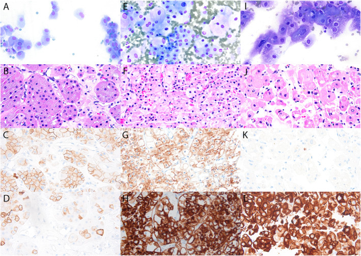
Definition. Renal cell carcinoma (RCC) is a group of malignant epithelial tumors of the renal tubules. The WHO Classification of Tumours of the Urinary and Male Genital Organs, 5th edition (2022), lists renal tumors hierarchically by site, category, family and type. It gives essential and desirable diagnostic criteria that combine morphology with immunohistochemistry and molecular tests, and it introduces a group of molecularly defined RCCs (Moch et al. 2022). On fine-needle aspiration (FNA), clear cell RCC, papillary RCC and chromophobe RCC are subtyped mainly on morphology. Immunohistochemistry on cell block is used to classify high-grade, eosinophilic and unusual tumors (Renshaw and Gould 2018; Kim et al. 2020).
Related page: Kidney FNA: Indications, Normal Elements and Benign Lesions (indications, adequacy, oncocytoma and angiomyolipoma) Related page: Urothelial Carcinoma of the Renal Pelvis, Wilms Tumor, Metastases and Lymphoma in Kidney FNA (urothelial carcinoma, Wilms tumor, metastases and lymphoma) Related page: Pheochromocytoma, Metastases and Lymphoma in Adrenal FNA (metastatic RCC in the adrenal gland)
WHO 5th edition classification of renal cell carcinoma and changes from the 4th edition
The 5th edition retains the main morphologic types and adds molecularly defined tumors (Moch et al. 2022).
| Topic | WHO 5th edition (2022) | Previous rule (WHO 4th edition, 2016, unless stated) | Source |
|---|---|---|---|
| Molecularly defined RCC | Group of seven molecularly defined RCCs: TFE3-rearranged, TFEB-altered, ELOC-mutated, fumarate hydratase-deficient, succinate dehydrogenase-deficient, ALK-rearranged and SMARCB1-deficient medullary RCC; SMARCB1-deficient medullary, TFEB-altered, ALK-rearranged and ELOC-mutated RCC are named as novel | No separate molecular group; HLRCC syndrome-associated RCC and SDH-deficient RCC were newly recognized types listed with the other RCCs | Moch et al. 2022; Goswami et al. 2024; Moch et al. 2016 |
| Translocation RCC | TFE3-rearranged RCC and TFEB-altered RCC listed separately | MiT family translocation RCC category | Goswami et al. 2024 |
| Eosinophilic solid and cystic RCC | Accepted as a new, independent entity | Not a listed entity | Moch et al. 2022; Lobo et al. 2022 |
| Papillary RCC | Type 1 and type 2 subcategories eliminated | Divided into type 1 and type 2 | Lobo et al. 2022 |
| Oncocytic tumors | New category of "other oncocytic tumours", including eosinophilic vacuolated tumor and low-grade oncocytic tumor | Oncocytoma and chromophobe RCC only | Lobo et al. 2022 |
| Clear cell papillary tumor | Clear cell papillary renal cell tumor | Clear cell papillary RCC, a newly recognized type in 2016 | Moch et al. 2016; Goswami et al. 2024; Sagan et al. 2026 |
| Fumarate hydratase-deficient RCC | Fumarate hydratase-deficient RCC | Hereditary leiomyomatosis and RCC syndrome-associated RCC | Moch et al. 2016; Goswami et al. 2024; Trpkov, Hes et al. 2021 |
| Medullary carcinoma | SMARCB1-deficient medullary RCC | Renal medullary carcinoma | Moch et al. 2022; Goswami et al. 2024 |
- Reason for dropping papillary subtypes. Mixed type 1 and type 2 phenotypes are frequent, and the former type 2 category contained entities with different molecular backgrounds (Lobo et al. 2022). WHO/ISUP grade and tumor architecture predict outcome better than the type (Trpkov, Hes et al. 2021).
- Papillary RCC spectrum. Biphasic squamoid alveolar RCC, biphasic hyalinizing psammomatous RCC, papillary renal neoplasm with reverse polarity and Warthin-like papillary RCC are included in the papillary RCC spectrum (Lobo et al. 2022).
Immunohistochemical panel for renal cell carcinoma subtypes
A study of 214 renal tumors found a CK7, carbonic anhydrase IX (CAIX) and α-methylacyl-CoA racemase (AMACR) panel to be the best primary screen for subtyping. CD10, epithelial membrane antigen, cathepsin K, CD117, HNF1β and E-cadherin form an adjunctive panel (Kim et al. 2020).
| Tumor | CK7 | CAIX | AMACR | CD117 | Other discriminating markers | Source |
|---|---|---|---|---|---|---|
| Clear cell RCC | Negative or focal (diffuse in 0%) | Diffuse, box pattern (100%) | Focal (70%) or diffuse (26%) | Negative | CD10 with saw-tooth membranous pattern | Kim et al. 2020; Renshaw and Gould 2018 |
| Papillary RCC (former type 1 series) | Diffuse (94%) | Negative (100%) | Diffuse (93%) | Negative | HNF1β positive | Kim et al. 2020 |
| Chromophobe RCC | Diffuse (85%) | Negative (100%) | Negative (65%) or focal | Positive | HNF1β lost in 80%; E-cadherin positive | Kim et al. 2020 |
| Oncocytoma | Negative (80%) or focal | Negative | Negative | Positive | HNF1β and S100A1 positive | Kim et al. 2020; Conner, Hirsch and Jo 2015 |
| Clear cell papillary renal cell tumor | Diffuse (100%) | Cup-like (100%) | Positive in 35–50% | Negative | CD10 reverse cup-like in about half | Lin 2017; Griffin and Lin 2021 |
| TFE3-rearranged or TFEB-altered RCC (MiT family in Kim 2020) | Negative (100%) | Negative (100%) | Negative or focal | Negative | Cathepsin K positive; TFE3 with or without FISH | Kim et al. 2020; Jin et al. 2020 |
| Collecting duct carcinoma | Diffuse (100%) | Negative (100%) | Focal or diffuse | Negative | E-cadherin lost or broken | Kim et al. 2020 |
- Renal lineage. PAX2 and PAX8 are sensitive and relatively specific markers of renal neoplasms of every subtype (Truong and Shen 2011). In cytology cell blocks, PAX8 stained 88% and PAX2 83% of RCCs, and either marker stained 92% (Knoepp, Kunju and Roh 2012).
- Proximal and distal nephron markers. CD10 and RCC marker are sensitive for tumors derived from the proximal tubule, including clear cell and papillary RCC. Kidney-specific cadherin, parvalbumin, claudin-7 and claudin-8 mark tumors of the distal nephron, including chromophobe RCC and oncocytoma (Truong and Shen 2011).
- Metastatic setting. Markers are expressed less frequently and less diffusely in metastatic RCC than in primary RCC. A panel of at least CD10, RCC marker, PAX2 and PAX8 is advised (Truong and Shen 2011).
Clear cell renal cell carcinoma on FNA
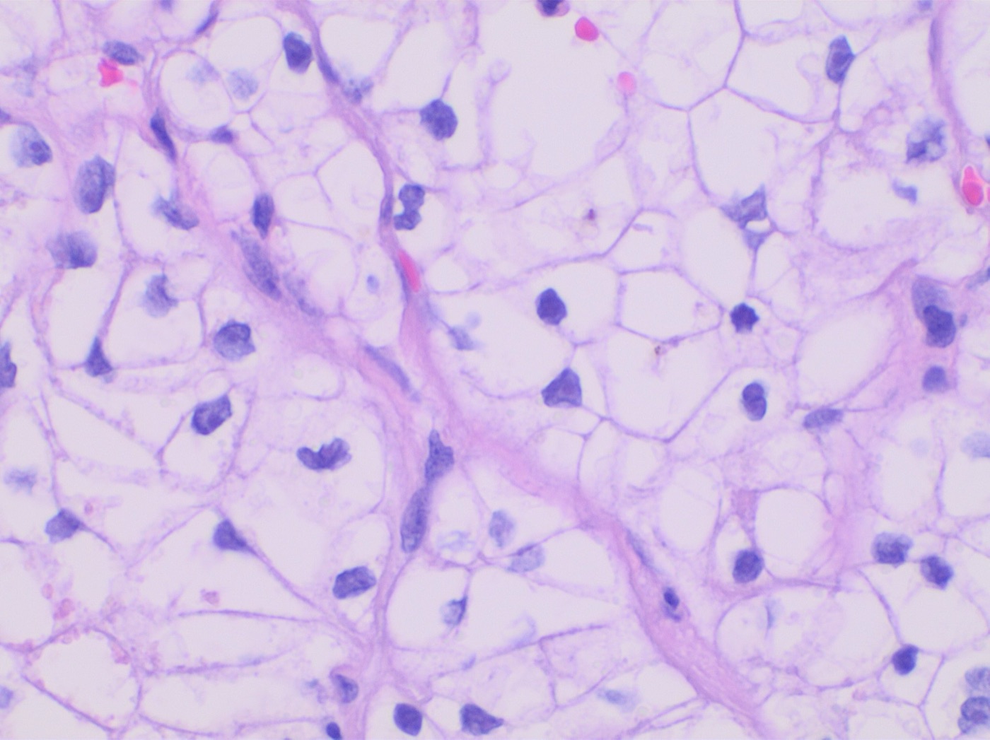
- Cytomorphology. In a comparison of 20 clear cell RCC aspirates with clear cell papillary tumors, clear cell RCC showed sheets in 70%, naked nuclei in 100% and prominent nucleoli in 65%. Vessels traversed the cell groups, and the cells had large amounts of cytoplasm. Three-dimensional clusters and papillae were absent (Griffin and Lin 2021).
- Granular variant. Clear cell RCC with granular cytoplasm is part of the differential diagnosis of oncocytic tumors. CD117 is negative in the granular variant of clear cell RCC and positive in chromophobe RCC and oncocytoma (Renshaw and Gould 2018).
- Immunoprofile. CAIX stained every clear cell RCC diffusely and intensely, in a box pattern around each cell border. CK7 was negative or focal (Kim et al. 2020). CK7 negativity separates clear cell RCC from papillary RCC, clear cell papillary tumor and multilocular cystic tumors (Renshaw and Gould 2018).
- Prognostic features. The prognostic features of clear cell RCC are WHO/ISUP grade, AJCC pTNM stage, coagulative necrosis and rhabdoid or sarcomatoid differentiation (Trpkov, Hes et al. 2021).
Papillary renal cell carcinoma on FNA
- Cytomorphology. In 18 papillary RCC aspirates, sheets were present in 61%, naked nuclei in 67%, nuclear grooves in 67%, intranuclear inclusions in 67% and pigmented cytoplasm in 83% (Griffin and Lin 2021).
- Immunoprofile. CK7 and AMACR are sensitive markers of papillary RCC (Truong and Shen 2011). In the former type 1 group, CK7 and AMACR were diffuse in more than 90% of tumors, and CAIX was negative in all (Kim et al. 2020).
- Needle tract seeding. Seeding after renal mass biopsy has been reported almost exclusively in papillary RCC (Renshaw et al. 2019).
Chromophobe renal cell carcinoma on FNA
- Cytomorphology. Six chromophobe RCC aspirates showed round, oval or polygonal, moderately pleomorphic large cells, single and in small clusters. The abundant cytoplasm was variegated, ranging from dense to flocculent to vacuolated, with prominent cytoplasmic membranes. Nuclei were large and hyperchromatic with membrane irregularities and grooves, and binucleation was frequent (Wiatrowska and Zakowski 1999).
- Architecture. Large sheets with transverse vessels and higher nuclear grade were more frequent in chromophobe RCC than in oncocytoma (Zhu, Rohan and Lin 2020).
- Ancillary studies. Chromophobe RCC is diffusely CK7-positive and CD117-positive (Renshaw and Gould 2018). Hale colloidal iron shows diffuse or focal cytoplasmic staining (Liu and Fanning 2001). HNF1β and S100A1 are usually negative (Conner, Hirsch and Jo 2015).
- Grading. The International Society of Urological Pathology (ISUP) agreed that chromophobe RCC should not be graded (Delahunt et al. 2013). A reproducible, clinically relevant grading system for chromophobe RCC was still missing in the WHO 5th edition (Lobo et al. 2022).
- Biopsy diagnosis of oncocytoma. The 2022 WHO classification precludes a definitive diagnosis of oncocytoma on needle core biopsy (EAU Guidelines on Renal Cell Carcinoma 2025). This affects the distinction from chromophobe RCC on biopsy (Kidney FNA: Indications, Normal Elements and Benign Lesions).
Clear cell papillary renal cell tumor on FNA
Clear cell papillary renal cell tumor (CCPRCT), formerly clear cell papillary RCC, is a low-grade renal neoplasm with CK7 and cup-like CAIX positivity and without von Hippel-Lindau (VHL) abnormalities. Some tumors with typical features carry monoallelic VHL alterations (Sagan et al. 2026). GUPS had proposed reclassifying it as a tumor of low malignant potential (Trpkov, Hes et al. 2021).
- Cytomorphology. In seven aspirates and touch preparations, cells formed small nests (100%), three-dimensional clusters (71%), papillary, tubular or acinar arrays (43%) and single cells (57%). The cells were columnar with eccentric, small, round to oval nuclei, smooth membranes and fine chromatin, corresponding to Fuhrman grade 1 to 2. The cytoplasm was delicate or clear with small vacuoles (Lin 2017).
- Features separating CCPRCT from clear cell RCC. Three-dimensional clusters (72% versus 0%), papillae (50% versus 0%), sheets (22% versus 70%), naked nuclei (17% versus 100%) and prominent nucleoli (0% versus 65%) differ between the two. Vessels lie within papillae in CCPRCT and traverse cell groups in clear cell RCC (Griffin and Lin 2021).
- Features separating CCPRCT from papillary RCC. Sheets (22% versus 61%), naked nuclei (17% versus 67%), nuclear grooves (5% versus 67%), intranuclear inclusions (17% versus 67%) and pigmented cytoplasm (17% versus 83%) are less frequent in CCPRCT (Griffin and Lin 2021).
- Immunoprofile. CK7 was positive in 100%, CAIX in 100% with a cup-like pattern, CD10 in 53% with a reverse cup-like pattern and AMACR in 35%. CD117 was negative (Griffin and Lin 2021). In the earlier series, CD10 was positive in 43% and AMACR in 50% (Lin 2017).
- Behavior and management. In a prospective cohort of 36 patients with a median tumor size of 1.2 cm, no recurrence or cancer-specific death occurred during follow-up (Kim et al. 2023). Of 18 patients with a cytologic diagnosis, 50% underwent nephrectomy, 28% cryoablation and 22% surveillance (Griffin and Lin 2021).
Molecularly defined and other renal cell carcinomas on FNA
| Tumor | Key finding on cytology or biopsy | Confirmatory test | Source |
|---|---|---|---|
| TFE3-rearranged RCC and TFEB-altered RCC (formerly MiT family translocation RCC) | Large cells with voluminous vacuolated cytoplasm; "tigroid" strips of detached cytoplasm; features overlap other RCC types | TFE3 immunohistochemistry with or without TFE3 FISH; cathepsin K | Jin et al. 2020; Kim et al. 2020 |
| Fumarate hydratase-deficient RCC | Difficult-to-classify RCC, particularly eosinophilic tumors in younger patients | Loss of FH and positive 2SC on immunohistochemistry | Trpkov, Hes et al. 2021 |
| Succinate dehydrogenase-deficient RCC | Difficult-to-classify RCC, particularly eosinophilic tumors | Loss of SDHB on immunohistochemistry | Trpkov, Hes et al. 2021; Renshaw and Gould 2018 |
| SMARCB1-deficient medullary RCC | Defined by immunohistochemistry and clinical context | Loss of SMARCB1 (INI1) together with sickle cell trait or disease | Trpkov, Hes et al. 2021; Moch et al. 2022 |
| Collecting duct carcinoma | Separated from urothelial carcinoma by immunohistochemistry | Diffuse CK7; PAX2 and PAX8 with high-molecular-weight keratin separate it from urothelial carcinoma | Kim et al. 2020; Truong and Shen 2011 |
| Mucinous tubular and spindle cell carcinoma | Morphology overlaps papillary RCC | Losses of chromosomes 1, 4, 6, 8, 9, 13, 14, 15 and 22 | Trpkov, Hes et al. 2021 |
| Eosinophilic solid and cystic RCC | Morphologically defined entity, more frequent in females | CK20 positive; CK7 and CD117 typically negative; association with tuberous sclerosis complex | Goswami et al. 2024; Moch et al. 2022 |
- Translocation RCC in adults and children. Xp11 translocation RCC constitutes a large percentage of primary renal neoplasms in children and less than 4% of adult cases (Jin et al. 2020).
- Threshold for ancillary testing. GUPS recommends a low threshold for FH, 2SC and SDHB immunohistochemistry in difficult-to-classify RCC, especially eosinophilic tumors in younger patients (Trpkov, Hes et al. 2021). Succinate dehydrogenase B, fumarate hydratase, INI1 and OCT3/4 immunostains are used to identify less common subtypes on FNA (Renshaw and Gould 2018).
- Collecting duct carcinoma and GATA3. GATA3 was positive in 88% of upper tract urothelial carcinomas and 6% of collecting duct carcinomas, and PAX8 in 12% and 100%. A GATA3-positive or p63-positive, PAX8-negative profile had a specificity of 100% for urothelial carcinoma (Gonzalez-Roibon et al. 2013). GATA3 is used in a panel with PAX8 and p63.
Grading of renal cell carcinoma on biopsy
- WHO/ISUP grading. Nucleolar prominence defines grades 1 to 3 of clear cell and papillary RCC. Extreme nuclear pleomorphism or sarcomatoid or rhabdoid differentiation defines grade 4 (Delahunt et al. 2013).
- Sarcomatoid differentiation. No minimum proportion of sarcomatoid tumor is required for the diagnosis. The underlying subtype is reported, and a sarcomatoid tumor without an identifiable subtype is classified as a grade 4 unclassified carcinoma with a sarcomatoid component (Delahunt et al. 2013).
- Accuracy of grading on biopsy. Agreement between biopsy and resection was fair for Fuhrman grade (κ 0.34) (Marconi et al. 2016). Accuracy rose from 62.5% with full nuclear grading to 87% with a two-tier high-grade versus low-grade system (EAU Guidelines on Renal Cell Carcinoma 2025).
Differential diagnosis of renal cell carcinoma subtypes on FNA
| Pattern on FNA | Leading diagnoses | Discriminating findings | Source |
|---|---|---|---|
| Clear cells in sheets with naked nuclei | Clear cell RCC; xanthogranulomatous pyelonephritis; adrenal cortical cells | CAIX box pattern and keratin in RCC; CD68 in XGP; SF-1 and melan-A in adrenal cortex | Kim et al. 2020; Renshaw and Gould 2018; Sbiera et al. 2010; Ghorab et al. 2003 |
| Clear cells in three-dimensional clusters and papillae, low grade | Clear cell papillary renal cell tumor; clear cell RCC | Diffuse CK7 and cup-like CAIX in CCPRCT | Griffin and Lin 2021 |
| Papillae with pigmented cytoplasm and grooves | Papillary RCC; CCPRCT | Diffuse CK7 and AMACR, CAIX negative in papillary RCC | Griffin and Lin 2021; Kim et al. 2020 |
| Eosinophilic or granular cells | Chromophobe RCC; oncocytoma; granular clear cell RCC; FH- or SDH-deficient RCC | CK7, CD117, HNF1β, S100A1, FH, SDHB | Renshaw and Gould 2018; Conner, Hirsch and Jo 2015; Trpkov, Hes et al. 2021 |
| Carcinoma of uncertain renal or urothelial origin | Collecting duct carcinoma; urothelial carcinoma; SMARCB1-deficient medullary RCC | PAX8, p63, GATA3, high-molecular-weight keratin, SMARCB1 | Truong and Shen 2011; Gonzalez-Roibon et al. 2013; Trpkov, Hes et al. 2021 |
Reporting of renal cell carcinoma on FNA
- Subtype. Biopsy agreement with the resection subtype was good (κ 0.683) (Marconi et al. 2016). The WHO 5th edition subtype name is used.
- Grade. A WHO/ISUP grade is reported for clear cell and papillary RCC when the sample allows. Biopsy grading is more accurate with a two-tier system (EAU Guidelines on Renal Cell Carcinoma 2025).
- Sarcomatoid or rhabdoid features. Their presence is reported because they define grade 4 (Delahunt et al. 2013).
- Risk of malignancy. Aspirates placed in a malignant category had a pooled ROM of 96.9% (Katipoglu et al. 2024).
CAP cancer protocols for kidney biopsy and resection
- Protocols in use. The College of American Pathologists (CAP) lists a Kidney, Biopsy protocol and a Kidney, Resection protocol, both version 4.3.0.0 (June 2026) (College of American Pathologists 2026a).
- Scope of the biopsy protocol. The biopsy protocol applies to core needle, endoscopic and other biopsies of renal cell carcinomas of all subtypes. Urothelial tumors, nephroblastic tumors, hematopoietic neoplasms and sarcomas are reported with other protocols (College of American Pathologists 2026b).
- Biopsy reporting elements. The biopsy case summary records procedure, laterality, histologic type, WHO/ISUP grade, rhabdoid features, sarcomatoid features and lymphatic or vascular invasion. Tumor site and necrosis are optional elements (College of American Pathologists 2026b).
- Magnifications in the protocol grade definitions. G1 has nucleoli absent or inconspicuous at 400x. G2 has nucleoli visible at 400x and not prominent at 100x. G3 has nucleoli conspicuous at 100x (College of American Pathologists 2026b).
- Renal cell carcinoma, NOS. The protocol lists "renal cell carcinoma, NOS (unclassified)" among the WHO 5th edition types. The category is reserved for carcinomas that cannot be placed in a morphologically or molecularly defined type and is usually high grade. Low-grade oncocytic tumors that are difficult to classify are placed in the "other oncocytic tumors of the kidney" group (College of American Pathologists 2026b).
- Former names. TFE3-rearranged RCC was formerly called Xp11 translocation RCC. SMARCB1-deficient renal medullary carcinoma was formerly called renal medullary carcinoma (College of American Pathologists 2026b).
- SDH-deficient RCC. Its bland, bubbly eosinophilic cells overlap with oncocytic tumors. Unlike most other oncocytic tumors, it is typically KIT-negative and entirely keratin 7-negative (College of American Pathologists 2026b).
renal epithelial tumor immunoprofile lookup
Urothelial Carcinoma of the Renal Pelvis, Wilms Tumor, Metastases and Lymphoma in Kidney Fine-Needle Aspiration
Definition. Malignant renal masses other than renal cell carcinoma (RCC) include urothelial carcinoma of the renal pelvis, nephroblastoma (Wilms tumor) in children, metastatic tumors and lymphoma. In a series of 108 adult renal fine-needle aspirations (FNAs), the 46 malignant aspirates comprised 27 RCCs, five urothelial (transitional cell) carcinomas, four lymphomas, one small cell carcinoma and nine metastatic carcinomas (Truong et al. 1999). FNA recognition of a metastasis avoids unnecessary surgery (Gattuso et al. 1999), and FNA has an essential role in treatment planning for renal lymphoma (Truong et al. 2001).
Related page: Kidney FNA: Indications, Normal Elements and Benign Lesions (indications, adequacy and benign lesions) Related page: Renal Cell Carcinoma Subtypes on FNA (renal cell carcinoma subtypes) Related page: Pheochromocytoma, Metastases and Lymphoma in Adrenal FNA (metastases and lymphoma in the adrenal gland)
Urothelial carcinoma of the renal pelvis on FNA
- Clinical presentation. In a series of seven urothelial carcinomas of the renal pelvis diagnosed by FNA, most patients presented with pain and hematuria of short duration. Imaging showed a renal mass in three patients and a filling defect in the collecting system in the others (Santamaría et al. 1995).
- Cytomorphology. The cytologic pattern was similar to that of urothelial carcinoma of the bladder. The resemblance was closest in well-differentiated papillary carcinomas (Santamaría et al. 1995).
- Role of FNA. FNA contributed both to the preoperative diagnosis and to follow-up (Santamaría et al. 1995).
- Urothelial markers. Thrombomodulin, uroplakin III, p63 and S100P are useful markers of urothelial carcinoma (Truong and Shen 2011).
- Distinction from collecting duct carcinoma. Urothelial markers, high-molecular-weight cytokeratins, PAX2 and PAX8 together separate renal pelvic urothelial carcinoma from collecting duct carcinoma (Truong and Shen 2011). PAX2 and PAX8 mark renal neoplasms of all subtypes (Truong and Shen 2011).
- GATA3 in non-urothelial tumors. GATA3 labels urothelial carcinoma, breast carcinoma and lymphoma, and it labeled 78.7% of paragangliomas (So and Epstein 2013). In a direct comparison, GATA3 was positive in 88% of upper tract urothelial carcinomas and 6% of collecting duct carcinomas, and p63 in 92% and 11%. A GATA3-positive or p63-positive, PAX8-negative profile had a specificity of 100% for upper tract urothelial carcinoma (Gonzalez-Roibon et al. 2013). GATA3 alone does not establish urothelial origin.
- Lymphoma mimicking urothelial carcinoma. One renal lymphoma in an FNA series was misdiagnosed as transitional cell carcinoma on smears. The diagnosis was revised on tissue sections (Truong et al. 2001).
| Marker | Urothelial carcinoma | Collecting duct carcinoma | Clear cell or papillary RCC | Source |
|---|---|---|---|---|
| PAX8 | Positive in 12% | Positive in 100% | Positive in 88% of RCCs in cytology specimens | Gonzalez-Roibon et al. 2013; Knoepp, Kunju and Roh 2012 |
| p63 | Positive in 92% | Positive in 11% | Used to exclude urothelial carcinoma | Gonzalez-Roibon et al. 2013; Truong and Shen 2011 |
| Uroplakin III, thrombomodulin, S100P | Useful positive markers | Used to exclude urothelial carcinoma | Used to exclude urothelial carcinoma | Truong and Shen 2011 |
| GATA3 | Positive in 88% of upper tract urothelial carcinomas | Rarely positive (1 of 18, 6%) | Negative in metastatic clear cell RCC | Gonzalez-Roibon et al. 2013; Perrino et al. 2017 |
| CK7 | Not used to separate | Diffuse (100%) | Clear cell negative or focal; papillary diffuse | Kim et al. 2020 |
Urine and upper tract cytology in urothelial carcinoma of the renal pelvis
- Place in the work-up. Urothelial carcinomas of the upper urinary tract are uncommon. Cytology of voided urine or of washings from the upper tract has been part of the standard work-up, and its value remains controversial (Zhang, VandenBussche et al. 2021).
- Effect of standardized reporting. The Paris System for Reporting Urinary Cytology, published in 2016, introduced diagnostic categories with specific cytomorphologic criteria. Most studies of its effect concern lower urinary tract specimens, and few have analyzed upper tract specimens (Zhang, Miki et al. 2021).
- Predictive features in upper tract specimens. It remains uncertain which cytologic features best predict high-grade urothelial carcinoma in upper tract specimens (Zhang, Miki et al. 2021).
Related page: The Paris System for Reporting Urinary Cytology (The Paris System for Reporting Urinary Cytology)
Wilms tumor (nephroblastoma) on FNA
Wilms tumor is the most common kidney tumor of childhood (Portugal and Barroca 2008). Its classical components are blastema, epithelium and stroma.
- Diagnostic accuracy. In a series of 121 pediatric renal neoplasms, 109 of them Wilms tumors, FNA with cell block was adequate in 99.18% and gave a definitive diagnosis in 96.7%. Sensitivity for Wilms tumor was 97.4% and specificity 98.7% (Gupta et al. 2021).
- Components on cytology. Among 68 histologically proven Wilms tumors, 58.8% were triphasic, 35.3% biphasic and four were composed of blastema only on cytology (Gupta et al. 2021).
- Cell blocks. Cell blocks added information over smears in 33.8% of cases, including recognition of epithelial or mesenchymal elements and rhabdomyoblastic differentiation (Gupta et al. 2021).
- Smears of blastemal tumors. Smears of blastemal tumors are highly cellular. The similarity of blastemal cells across neuroblastic tumor, nephroblastoma and other blastemal tumors requires clinical and radiologic correlation (Gajdzis and Klijanienko 2026).
- WT1 immunostaining. Blastemal nuclear WT1 staining was present in 44% of nephroblastomas and correlated with high stage. Clear cell sarcoma of the kidney and neuroblastoma showed no nuclear WT1 staining (Chen et al. 2004).
- Predominance of one component. When one classical component predominates on smears, the differential diagnosis extends to clear cell sarcoma of the kidney, mesoblastic nephroma of the cellular type and metanephric adenoma (Portugal and Barroca 2008).
Biopsy of pediatric renal tumors under SIOP and COG approaches
- Two treatment approaches. The International Society of Paediatric Oncology Renal Tumour Study Group (SIOP-RTSG) treats Wilms tumor with preoperative chemotherapy. The Children's Oncology Group (COG) advocates primary nephrectomy without biopsy when feasible (Jackson et al. 2022).
- SIOP biopsy indications. Successive SIOP protocols recommended pretreatment biopsy of unilateral tumors only when features suggested a higher probability of a non-Wilms tumor that would change management (Jackson et al. 2022).
- Biopsy of all children. In the UK experience within the SIOP WT 2001 trial, biopsy of every child with a renal mass rarely changed management and could result in misdiagnosis, particularly in the typical Wilms tumor age range (Jackson et al. 2022).
- UMBRELLA protocol. The SIOP-RTSG biopsy working group recommendations are incorporated in the UMBRELLA SIOP-RTSG 2016 study (Jackson et al. 2022; van den Heuvel-Eibrink et al. 2017).
- Practice where FNA is routine. One center performs FNA with cell block before neoadjuvant chemotherapy for all pediatric malignant renal neoplasms (Gupta et al. 2021).
Differential diagnosis of pediatric renal tumors on FNA
| Tumor | Distinguishing cytologic finding | Immunohistochemistry | Source |
|---|---|---|---|
| Nephroblastoma (Wilms tumor) | Blastema, epithelial tubules and stroma in triphasic or biphasic combinations | Blastemal nuclear WT1 in a subset | Gupta et al. 2021; Chen et al. 2004 |
| Clear cell sarcoma of the kidney | Moderate amount of pale blue cytoplasm | Dot-like vimentin; no nuclear WT1; negative for the markers usually positive in Wilms tumor | Portugal and Barroca 2008; Chen et al. 2004 |
| Mesoblastic nephroma (cellular type) | Tightly cohesive, bland spindle cells | Not discriminating in the reported case | Portugal and Barroca 2008 |
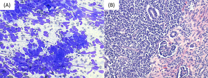
| Metanephric adenoma | Small, well-differentiated tubules, sometimes with psammoma bodies; microfollicular and papillary groups of small uniform cells with scant cytoplasm, fine chromatin and absent nucleoli; no atypia, necrosis or mitoses | BRAF mutations are shared by the metanephric family | Portugal and Barroca 2008; Blanco et al. 2013; Trpkov, Hes et al. 2021 |
| Neuroblastoma | Blastemal-type small round cells | No nuclear WT1 | Chen et al. 2004; Gajdzis and Klijanienko 2026 |
- Metanephric adenoma as a mimic. Metanephric adenoma shares cytologic features with Wilms tumor and papillary RCC. Recognition of its features avoids a diagnosis of malignancy (Blanco et al. 2013).
Metastatic tumors to the kidney on FNA
- Frequency. Of 261 radiologically guided renal FNAs, 136 were malignant, and 28 of these (21%) were metastatic tumors. Metastases accounted for 11% of all renal FNAs (Gattuso et al. 1999).
- Primary sites. Of the 28 patients, 89% had a known primary malignancy. The primaries were lung carcinoma (11 cases, 39%), lymphoma (8 cases, 29%), hepatocellular carcinoma (3 cases, 11%) and one case each of breast, pancreatic and cervical cancer. In three patients the primary remained unknown (Gattuso et al. 1999).
- Demographics and outcome. Twenty-three of the 28 patients were men, the mean age was 58 years and the mean survival after FNA was 9.8 months (Gattuso et al. 1999).
- Excluding RCC. PAX8 or PAX2 stained 92% of RCCs in cytology specimens (Knoepp, Kunju and Roh 2012).
- Melanoma. SOX10 had a sensitivity of 1.0 and a specificity of 0.93 for melanoma. Ovarian, endometrial, lung and colon carcinomas were SOX10-negative (Mohamed et al. 2013).
| Suspected primary | First-line markers on the cell block | Source |
|---|---|---|
| Renal cell carcinoma (primary) | PAX8, PAX2; CAIX for clear cell RCC | Knoepp, Kunju and Roh 2012; Kim et al. 2020 |
| Urothelial carcinoma | p63, uroplakin III, thrombomodulin, S100P, GATA3 | Truong and Shen 2011; So and Epstein 2013 |
| Melanoma | SOX10 | Mohamed et al. 2013 |
| Lymphoma | Immunophenotyping by immunostains or flow cytometry | Truong et al. 2001 |
Lymphoma of the kidney on FNA
- Series. Nineteen patients with renal lymphoma had FNA: ten with secondary renal lymphoma, eight with primary renal lymphoma and one with lymphoma in a transplanted kidney. Eighteen lymphomas were B-cell and one was T-cell (Truong et al. 2001).
- Initial cytologic reports. Of 20 initial specimens, 14 were reported as positive for lymphoma, three as suggestive of lymphoma, one as transitional cell carcinoma and two as unsatisfactory (Truong et al. 2001).
- Role of tissue and phenotyping. Tissue correlation confirmed seven positive diagnoses, resolved three inconclusive cases and corrected the transitional cell carcinoma report. Immunophenotyping by immunostains or flow cytometry established B or T lineage in every tested case. Gene rearrangement studies showed light-chain restriction in both tested cases (Truong et al. 2001).
- Primary renal lymphoma. A high index of suspicion and awareness of atypical or misleading cytomorphology are needed, especially for primary renal lymphoma (Truong et al. 2001).
- Triage at the time of sampling. When ROSE suggests lymphoma, a dedicated pass for flow cytometry allows lineage and clonality to be established (Truong et al. 2001).
Specimen triage for non-RCC malignancies in kidney FNA
- Rapid on-site evaluation. ROSE of FNA smears or touch preparations gives an immediate assessment of adequacy and allows triage of material for immunohistochemistry and molecular studies (Choy, Nayar and Lin 2023).
- Cell block. Immunostains on cell block sections confirm renal, urothelial, melanocytic or lymphoid lineage (Knoepp, Kunju and Roh 2012; Truong and Shen 2011; Mohamed et al. 2013; Truong et al. 2001).
- Ancillary studies for unusual RCC. TFE3 immunohistochemistry with or without TFE3 FISH is needed to diagnose translocation RCC (Jin et al. 2020).
Diagnostic errors in non-RCC malignancies of the kidney on FNA
| Situation | Error | Correct approach | Source |
|---|---|---|---|
| GATA3-positive high-grade carcinoma | Diagnosis of urothelial carcinoma on GATA3 alone | PAX8, p63 and high-molecular-weight keratin; GATA3 is occasionally positive in collecting duct carcinoma and usually positive in paraganglioma | Gonzalez-Roibon et al. 2013; So and Epstein 2013; Truong and Shen 2011 |
| Discohesive malignant cells in a renal mass | Diagnosis of carcinoma | Immunophenotyping for lymphoma | Truong et al. 2001 |
| Cellular aspirate of small uniform cells in tubules in a child | Diagnosis of Wilms tumor | Consideration of metanephric adenoma; absence of atypia, necrosis and mitoses | Blanco et al. 2013 |
| Renal mass in a patient with a known primary | Diagnosis of primary RCC | Comparison with the known primary; PAX8 | Gattuso et al. 1999; Knoepp, Kunju and Roh 2012 |
CAP cancer protocols for renal pelvic urothelial carcinoma
- Protocols in use. The College of American Pathologists lists Ureter, Renal Pelvis, Biopsy and Ureter, Renal Pelvis, Resection protocols, both version 2.4.0.0 (March 2025) (College of American Pathologists 2026a).
- Separation from the kidney protocol. The kidney biopsy protocol excludes urothelial tumors, which are reported with the ureter and renal pelvis protocol. Nephroblastic tumors and hematopoietic neoplasms are also excluded (College of American Pathologists 2026b).
Adrenal Fine-Needle Aspiration: Indications, Normal Cortex, Cortical Adenoma, Cortical Carcinoma and Myelolipoma
Definition. Adrenal fine-needle aspiration (FNA) samples an adrenal mass percutaneously or by endoscopic ultrasound (EUS). Adrenal lesions are present in 1% to 5% of patients and are most often found incidentally on abdominal imaging. The most common reason for adrenal FNA is to distinguish a benign cortical lesion, such as adrenal cortical adenoma, from metastatic malignancy, especially in patients with a known cancer (Cantley 2022). Most adrenal incidentalomas are nonfunctioning adrenal cortical adenomas (Fassnacht et al. 2023). The adrenal cortical lesions sampled by FNA are cortical nodules and adenomas, adrenal cortical carcinoma (ACC) and myelolipoma. A WHO Reporting System for Kidney and Adrenal Cytopathology was nearing completion in 2026 (Field et al. 2026).
Related page: Pheochromocytoma, Metastases and Lymphoma in Adrenal FNA (pheochromocytoma, metastases and lymphoma) Related page: Kidney FNA: Indications, Normal Elements and Benign Lesions (renal FNA) Related page: Renal Cell Carcinoma Subtypes on FNA (renal cell carcinoma, a mimic of adrenal cortical cells)
Indications for adrenal biopsy
The European Society of Endocrinology (ESE) and the European Network for the Study of Adrenal Tumors (ENSAT) guideline restricts adrenal biopsy to selected patients (Fassnacht et al. 2023).
| Recommendation | Content | Source |
|---|---|---|
| R.2.7 | Adrenal biopsy is not used in the diagnostic work-up of adrenal masses, unless there is a history of extra-adrenal malignancy | Fassnacht et al. 2023 |
| R.6.3.4 | In a patient with extra-adrenal malignancy and an indeterminate adrenal lesion, when management would change if the lesion were malignant, FDG-PET/CT, surgical resection or biopsy is suggested | Fassnacht et al. 2023 |
| R.6.3.5 | Three criteria are fulfilled before biopsy: the lesion is hormonally inactive, in particular pheochromocytoma has been excluded; it has not been characterized as benign by imaging; and the histology would change management | Fassnacht et al. 2023 |
| R.3.9 | Pheochromocytoma is excluded by plasma free metanephrines or urinary fractionated metanephrines in all patients with adrenal lesions with features not typical for a benign adenoma | Fassnacht et al. 2023 |
- Imaging thresholds. A homogeneous lesion with an unenhanced CT attenuation of ≤10 Hounsfield units (HU) is benign and needs no further imaging, regardless of size. Lesions ≥4 cm that are inhomogeneous or have an attenuation >20 HU carry a risk of malignancy high enough that surgery is the usual management (Fassnacht et al. 2023).
- Endocrine work-up. Every patient has a 1-mg overnight dexamethasone suppression test, with a cortisol cutoff of ≤50 nmol/L (≤1.8 µg/dL). Plasma or urinary metanephrines are measured when the lesion has features not typical for a benign adenoma (R.3.9) (Fassnacht et al. 2023).
- Biopsy and catecholamine-producing tumors. Adrenal biopsy is performed only if the findings are likely to alter management and only after biochemical exclusion of a catecholamine-producing tumor, to prevent life-threatening complications (Bancos et al. 2016).
- Suspected adrenocortical carcinoma. For suspected ACC, the guideline recommends measurement of sex steroids and steroid precursors, ideally by multisteroid profiling with tandem mass spectrometry (R.2.8). It gives no recommendation to biopsy (Fassnacht et al. 2023).
Imaging work-up of adrenal incidentalomas
- First imaging test. The guideline recommends imaging of every adrenal incidentaloma to determine whether the mass is homogeneous and lipid-rich. Noncontrast CT is the first modality (R.2.2) (Fassnacht et al. 2023).
- Indeterminate unenhanced CT. For a homogeneous mass of 11–20 HU and <4 cm without significant hormone excess, the guideline suggests immediate additional imaging. Interval noncontrast CT or MRI at 12 months is the alternative (R.2.4) (Fassnacht et al. 2023).
- High-risk imaging. A mass ≥4 cm that is heterogeneous or has an unenhanced attenuation >20 HU is discussed in a multidisciplinary team meeting. Before surgery the patient is staged with at least thoracic CT or FDG-PET/CT (R.2.5) (Fassnacht et al. 2023).
- Contrast washout. Malignant adrenal lesions usually enhance rapidly and show slow washout of contrast medium. Relative and absolute washout are calculated from attenuation measured before contrast, at 60 seconds and at 10–15 minutes (Fassnacht et al. 2023).
- Chemical-shift MRI. Adenomas rich in intracellular lipid lose signal on out-of-phase images. Malignant lesions, pheochromocytomas and some lipid-poor adenomas lack intracellular lipid and retain signal (Fassnacht et al. 2023).
- FDG-PET/CT. FDG-PET/CT is mainly used to detect malignant disease. FDG uptake reflects increased glucose metabolism and also increases with conditions such as active infection (Fassnacht et al. 2023).
Diagnostic performance and complications of adrenal biopsy
A systematic review included 32 studies with 2,174 patients and 2,190 adrenal biopsies (Bancos et al. 2016).
| Measure | Result | Source |
|---|---|---|
| Pooled non-diagnostic rate | 8.7% (95% CI 6–11%) | Bancos et al. 2016 |
| Pooled complication rate | 2.5% (95% CI 1.5–3.4%) | Bancos et al. 2016 |
| Sensitivity and specificity for malignancy | 87% and 100% | Bancos et al. 2016 |
| Sensitivity and specificity for ACC | 70% and 98% | Bancos et al. 2016 |
| Sensitivity and specificity for metastasis | 87% and 96% | Bancos et al. 2016 |
- Lesions sampled. Of 1,621 lesions with pathology in the review, 689 were metastases, 464 adenomas, 68 ACCs, 36 pheochromocytomas, 64 other malignancies, 226 other benign lesions and 74 others (Bancos et al. 2016).
- Most useful setting. Adrenal biopsy was most useful for diagnosing metastasis in patients with a history of extra-adrenal malignancy. The evidence came from small samples with a moderate risk of bias (Bancos et al. 2016).
- Earlier FNA series. In 188 FNAs, the diagnostic rate was 86%. Primary adrenal lesions and metastases each made up 43% of lesions (Saboorian, Katz and Charnsangavej 1995). In another series of 50 FNAs, 12% were insufficient, and FNA was diagnostic in all 29 malignant cases with follow-up, without false-positive diagnoses (Wadih, Nance and Silverman 1992).
- Endoscopic ultrasound. EUS-guided FNA sampled almost exclusively the left adrenal gland (23 of 24 procedures), mainly for staging of other malignancies. Diagnostic material was obtained in every case, and 19 of 24 aspirates showed cortical tissue consistent with cortical adenoma (Stelow et al. 2005).
Reporting categories for adrenal FNA
A multi-institutional study of 473 adrenal cytology specimens proposed a seven-tier reporting scheme (Trabzonlu et al. 2022).
| Category | Definition | ROM with histologic follow-up |
|---|---|---|
| Nondiagnostic | Insufficient diagnostic material, such as no adrenal cortical cells or abnormal elements | About 30% (10 cases) |
| Nonneoplastic | Inflammation, infection, pseudocyst and similar elements | 0% (1 case) |
| Benign adrenal cortical elements | Bland adrenal cortical cells; no mitoses; no necrosis | 0% (21 cases) |
| Primary neoplasm of noncortical origin | Features suggesting a primary noncortical neoplasm | 0% (6 cases: 3 pheochromocytomas, 3 schwannomas) |
| Atypia of undetermined significance | Atypia not meeting criteria for another category | 47.6% (21 cases) |
| Suspicious for malignancy | Findings highly suggestive of malignancy without quantitative sufficiency | 100% (6 cases) |
| Malignant | High cellularity, marked atypia, frequent mitoses and necrosis | 98.9% (88 cases) |
Source: Trabzonlu et al. 2022.
- Category frequency. Benign adrenal cortical elements made up 21.8% of cases and the malignant category 57.5% (Trabzonlu et al. 2022).
- Single discordant malignant case. The only malignant cytology report with benign follow-up was an adrenal cortical adenoma with marked atypia on resection (Trabzonlu et al. 2022).
- WHO categories. A meta-analysis of 15 studies recategorized adrenal FNAs into the proposed WHO categories. Pooled ROMs were 18% for inadequate, 3.7% for benign, 46.2% for atypical, 76.5% for suspicious and 99.6% for malignant. Pooled sensitivity was 92.2% and specificity 99.5% (Nigam et al. 2026).
Normal adrenal cortex and benign cortical nodules on FNA
- Cytomorphology. In 162 adrenal FNAs, the three features most often seen in benign adrenal cortical nodules were a bubbly, vacuolated lipid background; large cohesive tissue fragments with a syncytial nesting arrangement admixed with sinusoidal endothelial cells; and abundant oval to round bare nuclei (Wu et al. 1998).
- Specificity of the combination. All three features were present in 89% of benign cortical nodules. None of the primary or metastatic malignancies showed the combination, except four metastatic carcinomas with coexisting cortical hyperplasia (Wu et al. 1998).
- Size. Benign cortical nodules measured a mean of 2.5 cm (range 1–5 cm). The combined features in a nodule <3.5 cm were highly specific for a benign cortical nodule. The four metastases with coexisting hyperplasia had a mean size of 4.8 cm (Wu et al. 1998).
- Size in other series. Benign cortical nodules and adenomas averaged 2.4 cm (range 1–4 cm). Cortical neoplasms and carcinomas measured 4–12 cm (Saboorian, Katz and Charnsangavej 1995).
Immunohistochemistry of adrenal cortical cells
| Marker | Adrenal cortical lesions | Specificity and limitations | Source |
|---|---|---|---|
| SF-1 (steroidogenic factor-1) | 98% of ACCs; all normal and benign steroidogenic tissues | Absent in all nonsteroidogenic tumors tested | Sbiera et al. 2010 |
| Melan-A (A103) | Strong, diffuse granular cytoplasmic staining in 31 of 32 cortical neoplasms | Positive in melanoma; one clear cell RCC was positive | Ghorab et al. 2003; Busam et al. 1998 |
| Alpha-inhibin | Panel marker | Does not separate a cortical lesion from pheochromocytoma | Mete, Erickson et al. 2022 |
| Calretinin | Panel marker | Non-specific | Mete, Erickson et al. 2022 |
| Synaptophysin | Panel marker | Non-specific; also a neuroendocrine marker | Mete, Erickson et al. 2022 |
| Cytokeratins | Rarely detected in fixed tissue: one of 31 fixed ACCs | Normal cortex stains variably; fixation reduces detection | Gaffey et al. 1992 |
- Most reliable marker. SF-1 is the most reliable and specific biomarker of adrenal cortical origin. Melan-A, synaptophysin, alpha-inhibin and calretinin are non-specific and are used only in a panel (Mete, Erickson et al. 2022).
- Melan-A and carcinomas. In one study, 111 carcinomas, 40 germ cell tumors and 33 other non-melanocytic epithelioid tumors were all A103-negative. Once melanoma is excluded, A103 positivity makes another carcinoma in the differential diagnosis unlikely (Busam et al. 1998).
- Melan-A and benign versus malignant. Staining patterns did not differ between adenomas and carcinomas (Ghorab et al. 2003).
- Prognostic value of SF-1. Strong SF-1 expression in ACC correlated with poor outcome, with a stage-adjusted hazard ratio for death of 2.46 (Sbiera et al. 2010).
Immunohistochemistry separating adrenal cortical neoplasms from renal cell and hepatocellular carcinoma
Adrenal cortical neoplasms, renal cell carcinoma and hepatocellular carcinoma form one histologic differential diagnosis that occasionally requires immunohistochemistry (Ghorab et al. 2003; Gaffey et al. 1992).
| Marker | Adrenal cortical neoplasms | Renal cell carcinoma | Hepatocellular carcinoma | Source |
|---|---|---|---|---|
| Melan-A (A103) | Positive in 31 of 32 | Negative in 85 of 86; one clear cell RCC positive | Negative in 57 of 57 | Ghorab et al. 2003 |
| Keratin (fixed tissue) | Positive in 1 of 31 carcinomas | Positive in 34 of 37 | Positive in 33 of 33 | Gaffey et al. 1992 |
WHO 5th edition classification of adrenal cortical lesions and changes from the 4th edition
The WHO Classification of Endocrine and Neuroendocrine Tumours, 5th edition (2022), revised adrenal cortical terminology (Mete, Erickson et al. 2022).
| Topic | WHO 5th edition (2022) | Previous rule or usage | Source |
|---|---|---|---|
| Sporadic non-functional nodules | Sporadic nodular adrenocortical disease; "nodular adrenal cortical hyperplasia" no longer endorsed | "Nodular adrenal cortical hyperplasia" | Mete, Erickson et al. 2022 |
| Bilateral nodular disease | Bilateral micronodular or macronodular adrenal cortical disease | Primary bilateral micronodular or macronodular adrenocortical hyperplasia | Mete, Erickson et al. 2022 |
| Hyperplasia | Restricted to bilateral diffuse adrenal cortical hyperplasia | Used more broadly | Mete, Erickson et al. 2022 |
| Oncocytic cortical tumors | "Oncocytoma" no longer recommended; oncocytic adrenal cortical neoplasms are assessed with the Lin-Weiss-Bisceglia system, reticulin algorithm or Helsinki score | "Oncocytoma" used for oncocytic adrenal cortical neoplasms | Mete, Erickson et al. 2022 |
| ACC grade | Low grade ≤20 and high grade >20 mitoses per 10 mm², adopting the mitotic grade introduced by Weiss | The original Weiss grade counted ≤20 versus >20 mitoses per 50 high-power fields | Mete, Erickson et al. 2022; Weiss, Medeiros and Vickery 1989 |
| New chapters | Adrenal cysts, adrenal ectopia and myelolipoma as separate chapters | No separate chapters | Mete, Erickson et al. 2022 |
Adrenal cortical carcinoma on FNA
- Cytomorphology. ACC shows nuclear overlap, hyperchromasia and nuclear pleomorphism (Trabzonlu et al. 2022).
- Separation from adenoma. Benign adrenal cortical elements are defined by bland cells without mitoses or necrosis (Trabzonlu et al. 2022). The histologic scoring systems below require architecture, capsule and vessels and are applied to resection specimens.
- Separation from other tumors. SF-1 confirms adrenal cortical origin (Sbiera et al. 2010). GATA3 was negative in 90% of ACCs and positive in 71% of pheochromocytomas (Perrino et al. 2017).
| Histologic system | Criterion for malignancy | Source |
|---|---|---|
| Weiss criteria | 3 of 9 histologic parameters | Mete, Erickson et al. 2022 |
| Modified Weiss score | Score ≥3 | Mete, Erickson et al. 2022 |
| Lin-Weiss-Bisceglia (oncocytic tumors) | At least one major criterion | Mete, Erickson et al. 2022 |
| Reticulin algorithm | Altered reticulin network with mitoses (>5 per 10 mm²), necrosis or angioinvasion | Mete, Erickson et al. 2022 |
| Helsinki score | Score >8.5 | Mete, Erickson et al. 2022 |
| Pediatric tumors | Wieneke criteria preferred | Mete, Erickson et al. 2022 |
- Grading. The WHO 5th edition grades ACC as low grade with ≤20 mitoses per 10 mm² and high grade with >20 mitoses per 10 mm² (Mete, Erickson et al. 2022). Weiss and colleagues introduced the cut-off as 20 mitoses per 50 high-power fields; patients above it had a median survival of 14 months and those at or below it 58 months (Weiss, Medeiros and Vickery 1989).
- Ki-67. Most ACCs have a Ki-67 labeling index above 5%. Ki-67-based grading is not endorsed in the WHO 5th edition (Mete, Erickson et al. 2022).
- Grading cut-off. A cut-off of 10 mitoses per 10 mm² performed better in two series, and further validation is required (Mete, Erickson et al. 2022).
- Variants. Oncocytic ACC is composed of more than 90% oncocytic cells. Myxoid ACC shows prominent extracellular mucin, and myxoid change alone does not indicate malignancy. Sarcomatoid ACC often shows adrenal cortical differentiation, and sarcomatoid tumors without another ACC subtype are distinguished from sarcomas (Mete, Erickson et al. 2022).
- Molecular and hereditary findings. High-risk ACCs carry frequent Wnt/P53 pathway mutations. Activating CTNNB1 and deleterious ZNRF3 mutations activate the Wnt pathway. Li-Fraumeni syndrome increases the risk of ACC, and about 3% of adults with ACC have Lynch syndrome. Mismatch repair immunohistochemistry identifies patients who need genetic counseling (Mete, Erickson et al. 2022).
Adrenal myelolipoma on FNA
- Definition. Adrenal myelolipoma is a nonfunctioning tumor composed of adipose tissue and extramedullary hematopoietic elements (Settakorn et al. 1999). The WHO 5th edition gives it a separate chapter (Mete, Erickson et al. 2022).
- Cytomorphology. Aspirates contain adipose tissue and extramedullary hematopoietic elements (Settakorn et al. 1999). In a presacral (extra-adrenal) myelolipoma, smears showed hematopoietic cells of all lineages and fragments of collapsed stroma in a background of fat droplets, and megakaryocytic, myeloid and eosinophilic lineages were demonstrated with factor VIII immunostain, chloroacetate esterase and Luna stains (Yang et al. 1992).
- Clinical setting. In one report a myelolipoma was found at staging for lung carcinoma and was clinically suspected to be a metastasis. FNA established the benign diagnosis (Settakorn et al. 1999).
- Association with congenital adrenal hyperplasia. Almost 40% of patients with congenital adrenal hyperplasia may exhibit adrenal myelolipoma (Mete, Erickson et al. 2022).
- Frequency in FNA series. Myelolipoma was one of 81 primary adrenal lesions in one series (Saboorian, Katz and Charnsangavej 1995) and one of 15 nonneoplastic or benign lesions in another (Kumar and Dey 2016).
Nonneoplastic adrenal lesions on FNA
- Infections. Fifteen nonmalignant adrenal FNAs comprised five tuberculosis, five Histoplasma, two acute inflammation, two granulomatous inflammation and one myelolipoma. Six patients had bilateral lesions. Acid-fast bacilli were shown with Ziehl-Neelsen stain, and the capsule of Histoplasma stained bright pink with periodic acid-Schiff (Kumar and Dey 2016).
- Histoplasmosis in FNA series. Two adrenal histoplasmoses were found among 188 adrenal FNAs (Saboorian, Katz and Charnsangavej 1995).
Differential diagnosis of adrenal cortical lesions on FNA
| Lesion | Cytologic findings | Immunohistochemistry | Source |
|---|---|---|---|
| Benign cortical nodule or adenoma | Bubbly lipid background, cohesive fragments with endothelium, bare nuclei; usually <3.5 cm | SF-1 and melan-A positive | Wu et al. 1998; Sbiera et al. 2010; Ghorab et al. 2003 |
| Adrenal cortical carcinoma | Overlapping, hyperchromatic, pleomorphic nuclei; mitoses; necrosis; usually >4 cm | SF-1 positive; GATA3 usually negative | Trabzonlu et al. 2022; Sbiera et al. 2010; Perrino et al. 2017 |
| Myelolipoma | Fat droplets and trilineage hematopoiesis | Lineage stains for megakaryocytes and myeloid cells (shown in a presacral myelolipoma) | Settakorn et al. 1999; Yang et al. 1992 |
| Pheochromocytoma | Discohesive plasmacytoid cells, pleomorphism, intranuclear pseudoinclusions | Chromogranin, synaptophysin, GATA3; keratin negative | Jiménez-Heffernan et al. 2006; Mete, Asa et al. 2022 |
| Metastatic clear cell RCC | Clear cells, prominent nucleoli, bloody background | PAX8 positive; SF-1 and melan-A negative | Knoepp, Kunju and Roh 2012; Sbiera et al. 2010; Ghorab et al. 2003 |
| Metastatic melanoma | Variable | Melan-A and SOX10 positive; SF-1 negative | Busam et al. 1998; Mohamed et al. 2013; Sbiera et al. 2010 |
CAP cancer protocols for adrenal and paraganglionic tumors
- Protocols in use. The College of American Pathologists lists an Adrenal protocol, version 4.3.1.0 (March 2023), and a Paraganglioma Pheochromocytoma protocol, version 1.0.0.0 (March 2025) (College of American Pathologists 2026a).
Pheochromocytoma, Metastases and Lymphoma in Adrenal Fine-Needle Aspiration
Definition. Adrenal lesions of noncortical origin sampled by fine-needle aspiration (FNA) include pheochromocytoma, metastatic tumors and lymphoma. Pheochromocytoma is the intra-adrenal paraganglioma, the classical sympathetic form of paraganglioma (Mete, Asa et al. 2022). Metastases are the largest group of adrenal lesions sampled by biopsy (Bancos et al. 2016). Primary adrenal lymphoma is rare, with fewer than 200 cases reported by 2013 (Rashidi and Fisher 2013).
Related page: Adrenal FNA: Indications, Normal Cortex, Cortical Adenoma, Cortical Carcinoma and Myelolipoma (indications, benign cortex, adrenal cortical carcinoma and myelolipoma) Related page: Renal Cell Carcinoma Subtypes on FNA (renal cell carcinoma) Related page: Urothelial Carcinoma of the Renal Pelvis, Wilms Tumor, Metastases and Lymphoma in Kidney FNA (metastases and lymphoma in the kidney)
Pheochromocytoma in the WHO 5th edition classification
- Terminology. Paragangliomas are sympathetic or parasympathetic. The term pheochromocytoma is reserved for intra-adrenal paragangliomas of the classical sympathetic type. The WHO 5th edition classifies paragangliomas as non-epithelial neuroendocrine neoplasms (Mete, Asa et al. 2022).
- No benign or malignant categories. Since the WHO 4th edition (2017), paragangliomas have not been classified as benign or malignant, because any tumor can metastasize and no feature reliably predicts metastasis. The 5th edition (2022) keeps this rule. Some tumors are lethal through local invasion of critical structures without metastasis (Mete, Asa et al. 2022).
- Scoring systems. The WHO 5th edition reviews scoring systems for aggressive behavior. It neither endorses nor discourages their use (Mete, Asa et al. 2022).
- Staging. The staging system for pheochromocytoma and extra-adrenal sympathetic paraganglioma introduced in the AJCC 8th edition is included (Mete, Asa et al. 2022).
- Genetic predisposition. Paragangliomas have an exceptionally high rate of germline predisposition. Immunohistochemistry identifies associations with SDHx, VHL, FH, MAX and MEN1 mutations and with pseudohypoxia-related pathogenesis (Mete, Asa et al. 2022).
- Multifocal disease. In patients with germline syndromes, multiple lesions can be multifocal primary tumors. A paraganglioma in an unusual site such as lung or liver is not by itself proof of metastasis (Mete, Asa et al. 2022).
Biopsy of pheochromocytoma
- Recognized hazards. Percutaneous biopsy of pheochromocytomas and paragangliomas has been associated with life-threatening hemorrhage, hypertensive crisis, capsular disruption with tumor implantation and death (Vanderveen et al. 2009).
- Complications in one series. Of 20 patients biopsied before referral, 90% had no biochemical testing before biopsy. Fourteen (70%) had a complication: increased difficulty of the definitive operation in 7 of 17 operated cases, hematoma (30%), incorrect or inadequate biopsy (25%), severe pain (25%), severe hypertension (15%) and delay in surgery (15%) (Vanderveen et al. 2009).
- Hypertensive episode during FNA. In a cytology series of 13 tumors, one patient developed a hypertensive episode during FNA that was controlled medically. The authors performed FNA only in a setting equipped to treat a pheochromocytoma crisis (Jiménez-Heffernan et al. 2006).
- Guideline requirement. Pheochromocytoma is excluded biochemically before any adrenal biopsy (Fassnacht et al. 2023; Bancos et al. 2016).
- Frequency in biopsy series. Pheochromocytomas made up 36 of 1,621 biopsied adrenal lesions with pathology (Bancos et al. 2016) and 5 of 473 adrenal cytology cases (about 1.1%) (Trabzonlu et al. 2022).
Cytomorphology of pheochromocytoma on FNA
In a study of 15 cytologic specimens from 13 pheochromocytomas and extra-adrenal paragangliomas (Jiménez-Heffernan et al. 2006):
- Cellularity. Two aspirates were hypocellular, and the others were cellular.
- Arrangement. Cells lay singly or in discohesive groups.
- Cytoplasm. When present, the cytoplasm was abundant and ill defined.
- Nuclei. Most cells had an eccentric nucleus and a plasmacytoid appearance. Nuclear pleomorphism, binucleation, multinucleation, naked nuclei and intranuclear pseudoinclusions were common.
- Background. A lipid background was seen focally in two cases. This finding overlaps with the background of adrenal cortical cells (Wu et al. 1998).
- Scant material. Scant cellularity was a source of diagnostic error.
- Reporting category. In a proposed adrenal reporting scheme, pheochromocytoma is placed in the category "primary neoplasm of noncortical origin". None of the three pheochromocytomas in that category with histology had proved malignant (Trabzonlu et al. 2022).
Immunohistochemistry of pheochromocytoma
| Marker | Pheochromocytoma | Relevance | Source |
|---|---|---|---|
| Chromogranin, synaptophysin, INSM1 | Positive | General neuroendocrine markers; synaptophysin or chromogranin stained all 10 aspirates tested | Mete, Asa et al. 2022; Jiménez-Heffernan et al. 2006 |
| Keratins | Typically negative | Separates pheochromocytoma from epithelial neuroendocrine neoplasms and carcinomas | Mete, Asa et al. 2022 |
| GATA3 | Positive in 71% of pheochromocytomas; 78.7% of paragangliomas | Shared with urothelial and breast carcinoma; separates pheochromocytoma from ACC (GATA3-negative in 90%) | Perrino et al. 2017; So and Epstein 2013 |
| Tyrosine hydroxylase | Positive | Rate-limiting enzyme of catecholamine synthesis | Mete, Asa et al. 2022 |
| Dopamine β-hydroxylase | Positive in norepinephrine-producing cells | Catecholamine enzyme | Mete, Asa et al. 2022 |
| Phenylethanolamine N-methyltransferase | Positive in epinephrine-producing tumors | Identifies epinephrine production | Mete, Asa et al. 2022 |
| S100 and SOX10 | Sustentacular cells only; tumor cells negative | Sustentacular cells also occur in epithelial neuroendocrine neoplasms | Mete, Asa et al. 2022; Nonaka, Chiriboga and Rubin 2008 |
| SDHB and other molecular immunostains | Loss indicates SDHx-related disease | Identifies germline predisposition | Mete, Asa et al. 2022 |
- Alpha-inhibin in adrenal cortical lesions and pheochromocytoma. Alpha-inhibin cannot distinguish an adrenal cortical lesion from pheochromocytoma (Mete, Erickson et al. 2022).
- ACC and pheochromocytoma. Confusion between adrenal cortical carcinoma and pheochromocytoma is the most common source of error in diagnosing ACC. GATA3 narrows this differential diagnosis as part of a panel (Perrino et al. 2017).
- GATA3 in metastatic carcinoma. Metastatic lung adenocarcinoma in the adrenal gland expressed GATA3 in 36% of cases. Metastatic clear cell renal cell carcinoma (RCC) did not express GATA3 (Perrino et al. 2017).
Sustentacular cells in pheochromocytoma and paraganglioma
- Markers. Paragangliomas have a well-formed network of sustentacular cells that express SOX10 and S100 (Mete, Asa et al. 2022).
- SOX10 in tumor cells. SOX10 stained only the sustentacular cells of pheochromocytomas and paragangliomas. The tumor cells were negative (Nonaka, Chiriboga and Rubin 2008).
- Specificity. Epithelial neuroendocrine neoplasms also contain sustentacular cells, so their presence does not establish paraganglioma. Misinterpretation of these cells led to the former terms cauda equina paraganglioma and gangliocytic paraganglioma. The 2022 WHO terms are cauda equina neuroendocrine tumor and composite gangliocytoma/neuroma and neuroendocrine tumor (Mete, Asa et al. 2022).
Neuroblastic tumors and other primary medullary tumors in adrenal FNA
- Neuroblastoma in adrenal FNA series. Three neuroblastomas were among six primary adrenal malignancies in a series of 50 adrenal FNAs from patients aged 12 months to 79 years (Wadih, Nance and Silverman 1992).
- Cytology of blastemal tumors. Neuroblastic tumors share the round to oval blastemal cell morphology of nephroblastoma and other blastemal tumors. Clinical and radiologic information is required for an accurate diagnosis, and the cellular smears provide material for prognostic ancillary studies (Gajdzis and Klijanienko 2026).
- WT1 in neuroblastoma. Neuroblastomas showed no nuclear WT1 staining, which separates them from blastemal Wilms tumor (Chen et al. 2004).
- Schwannoma. Three schwannomas were found among the primary noncortical neoplasms in a multi-institutional adrenal cytology series (Trabzonlu et al. 2022). Schwannomas are diffusely SOX10-positive (Nonaka, Chiriboga and Rubin 2008).
Metastatic tumors to the adrenal gland
| Series | Setting | Most common primary | Other primaries | Source |
|---|---|---|---|---|
| Lam and Lo 2002 | 464 patients over 30 years; autopsy, adrenalectomy and FNA | Lung (35%) | Stomach (14%), esophagus (12%), liver and bile ducts (10%) | Lam and Lo 2002 |
| Saboorian et al. 1995 | 188 adrenal FNAs | Lung (55 cases; 47 adenocarcinomas) | Melanoma (5), RCC (7), mixed müllerian tumor (1) | Saboorian, Katz and Charnsangavej 1995 |
| Wadih et al. 1992 | 50 adrenal FNAs | Lung (60% of metastases) | Melanoma and RCC (8.6% each) | Wadih, Nance and Silverman 1992 |
| Trabzonlu et al. 2022 | 473 adrenal cytology cases, seven centers | Lung (83 cases) | Genitourinary (44), gastrointestinal and hepatobiliary (25) | Trabzonlu et al. 2022 |
- Frequency. Adrenal metastases were present in 3.1% of autopsies, 7.5% of adrenalectomies and 33% of adrenal FNAs (Lam and Lo 2002).
- Histologic type. Ninety percent of adrenal metastases were carcinomas, and 56% of these were adenocarcinomas (Lam and Lo 2002).
- Bilaterality and size. Metastases were bilateral in 49% of patients. The mean diameter was 2 cm (Lam and Lo 2002). In an FNA series, metastases averaged 5.1 cm (range 1.5–10 cm) (Saboorian, Katz and Charnsangavej 1995).
- Clinical course. Most adrenal metastases were asymptomatic and appeared a mean of 7 months after detection of the primary tumor. Five patients presented with adrenal insufficiency (Lam and Lo 2002).
- Hepatocellular carcinoma. Four of 15 extrahepatic metastases of hepatocellular carcinoma diagnosed by FNA were adrenal. A trabecular pattern was present in nine of the 15 cases, and the FNA was the first diagnosis of hepatocellular carcinoma in two patients (Dusenbery and Carr 1996).
- Melanoma. Melanoma made up 5 of 80 metastases in one adrenal FNA series (Saboorian, Katz and Charnsangavej 1995). SOX10 was expressed in 97% of melanomas (Nonaka, Chiriboga and Rubin 2008).
- Symptomatic metastases. Twenty of 464 patients (4%) had symptomatic adrenal metastases. These lesions were larger and occurred in younger patients, and the median survival after their detection was 3 months (Lam and Lo 2002).
- Accuracy of biopsy. Adrenal biopsy had a sensitivity of 87% and a specificity of 96% for metastasis (Bancos et al. 2016).
- Coexisting cortical hyperplasia. Four metastatic carcinomas in one series also contained benign cortical cells from coexisting hyperplasia (Wu et al. 1998). Benign cortical cells in an aspirate therefore do not exclude a metastasis in a large mass.
Immunohistochemical approach to adrenal metastases
The most frequently used immunostains in one multi-institutional adrenal series were TTF-1 (60 cases), inhibin (56 cases) and CK7 (46 cases) (Trabzonlu et al. 2022).
| Question | Marker and result | Source |
|---|---|---|
| Adrenal cortical origin | SF-1 positive (98% of ACCs; absent in nonsteroidogenic tumors) | Sbiera et al. 2010 |
| Adrenal cortex versus RCC or hepatocellular carcinoma | Melan-A positive in cortical neoplasms; negative in RCC except one case and in all hepatocellular carcinomas | Ghorab et al. 2003 |
| Renal origin | PAX8 positive in 88% and PAX8 or PAX2 in 92% of RCCs in cytology specimens | Knoepp, Kunju and Roh 2012 |
| Melanoma | SOX10 sensitivity 1.0 and specificity 0.93; lung, colon, ovarian and endometrial carcinomas negative | Mohamed et al. 2013 |
| Pheochromocytoma | Chromogranin and GATA3 positive; keratin negative | Mete, Asa et al. 2022; Perrino et al. 2017 |
| Lymphoma | B- or T-lineage immunophenotype | Truong et al. 2001 |
- Melan-A in adrenal aspirates. Melan-A stains adrenal cortical neoplasms (Busam et al. 1998). A melan-A-positive adrenal aspirate is classified as melanoma only with SOX10 positivity and a negative SF-1 result (Mohamed et al. 2013; Sbiera et al. 2010).
- Clear cells. Clear cell RCC and adrenal cortical cells both have clear cytoplasm. PAX8, SF-1 and melan-A separate them (Knoepp, Kunju and Roh 2012; Sbiera et al. 2010; Ghorab et al. 2003).
Primary adrenal lymphoma
- Frequency. A systematic review found 187 cases of primary adrenal lymphoma (PAL) reported in the English literature by June 2013 (Rashidi and Fisher 2013).
- Clinical features. PAL is typically a highly symptomatic, aggressive, high-grade lymphoma that mainly affects elderly males and presents with large bilateral adrenal masses. Most patients have adrenal insufficiency, B symptoms and elevated lactate dehydrogenase (Rashidi and Fisher 2013).
- Imaging. PAL is metabolically hyperactive, hypovascular and hypoechoic, and it is hypodense with slight to moderate enhancement on CT (Rashidi and Fisher 2013).
- Subtypes. Diffuse large B-cell lymphoma made up 78% of cases and peripheral T-cell lymphoma 7% (WHO 2008 subtypes). Epstein-Barr virus was positive in more than half of cases (Rashidi and Fisher 2013).
- Uncommon findings. Hepatosplenomegaly, lymphadenopathy, immune dysregulation and bone marrow involvement were uncommon. Disease was disseminated at presentation in 18% (Rashidi and Fisher 2013).
- Prognosis. Prognosis improved with rituximab-containing chemotherapy. Chemotherapy and adrenal insufficiency were independent predictors of outcome (Rashidi and Fisher 2013).
- FNA series. Three large cell lymphomas were found among 188 adrenal FNAs (Saboorian, Katz and Charnsangavej 1995).
- Cytologic confirmation. Immunophenotyping by immunostains or flow cytometry establishes B or T lineage in aspirates of lymphoma (Truong et al. 2001).
Differential diagnosis of noncortical adrenal lesions on FNA
| Lesion | Cytologic findings | Key ancillary results | Source |
|---|---|---|---|
| Pheochromocytoma | Discohesive plasmacytoid cells, pleomorphism, binucleation, intranuclear pseudoinclusions | Chromogranin, synaptophysin, GATA3 positive; keratin negative | Jiménez-Heffernan et al. 2006; Mete, Asa et al. 2022 |
| Metastatic adenocarcinoma | Malignant epithelial cells (mean mass size 5.1 cm in one series) | Site-specific markers; TTF-1 was the most used stain in one series | Saboorian, Katz and Charnsangavej 1995; Trabzonlu et al. 2022 |
| Metastatic clear cell RCC | Clear cells; bloody background | PAX8 positive; SF-1 and melan-A negative; GATA3 negative | Knoepp, Kunju and Roh 2012; Perrino et al. 2017 |
| Metastatic melanoma | Variable | SOX10 and melan-A positive; SF-1 negative | Mohamed et al. 2013; Sbiera et al. 2010 |
| Primary adrenal lymphoma | Large bilateral masses on imaging; lineage established by immunophenotyping | B-cell markers in 78% (diffuse large B-cell lymphoma) | Rashidi and Fisher 2013; Truong et al. 2001 |
| Adrenal cortical carcinoma | Overlapping, hyperchromatic, pleomorphic nuclei; necrosis; mitoses | SF-1 positive; GATA3 negative in 90% | Trabzonlu et al. 2022; Sbiera et al. 2010; Perrino et al. 2017 |
References
- Bancos I, Tamhane S, Shah M, et al. Diagnosis of endocrine disease: the diagnostic performance of adrenal biopsy: a systematic review and meta-analysis. Eur J Endocrinol. 2016;175(2):R65–R80. doi:10.1530/EJE-16-0297. PMID 27257146
- Bex A, Ghanem YA, Albiges L, et al. European Association of Urology Guidelines on Renal Cell Carcinoma: the 2025 update. Eur Urol. 2025;87(6):683–696. doi:10.1016/j.eururo.2025.02.020. PMID 40118739. Full recommendations: EAU Guidelines on Renal Cell Carcinoma, chapter "Diagnostic evaluation", https://uroweb.org/guidelines/renal-cell-carcinoma (accessed 2026-11-04). Cited in the text as EAU Guidelines on Renal Cell Carcinoma 2025.
- Blanco LZ Jr, Schein CO, Patel T, et al. Fine-needle aspiration of metanephric adenoma of the kidney with clinical, radiographic and histopathologic correlation: a review. Diagn Cytopathol. 2013;41(8):742–751. doi:10.1002/dc.22962. PMID 23447142
- Bonzanini M, Pea M, Martignoni G, et al. Preoperative diagnosis of renal angiomyolipoma: fine needle aspiration cytology and immunocytochemical characterization. Pathology. 1994;26(2):170–175. doi:10.1080/00313029400169421. PMID 8090589
- Busam KJ, Iversen K, Coplan KA, et al. Immunoreactivity for A103, an antibody to melan-A (Mart-1), in adrenocortical and other steroid tumors. Am J Surg Pathol. 1998;22(1):57–63. doi:10.1097/00000478-199801000-00007. PMID 9422316
- Cantley RL. Approach to fine needle aspiration of adrenal gland lesions. Adv Anat Pathol. 2022;29(6):373–379. doi:10.1097/PAP.0000000000000356. PMID 35878423
- Chen BF, Tzen CY, Liang DC, Liu HC, Huang YW, Fan CC. Immunohistochemical expression of Wilms' tumor 1 protein in nephroblastoma. J Chin Med Assoc. 2004;67(10):506–510. PMID 15648285
- Choy B, Nayar R, Lin X. Role of renal mass biopsy for diagnosis and management: review of current trends and future directions. Cancer Cytopathol. 2023;131(8):480–494. doi:10.1002/cncy.22697. PMID 36973920
- College of American Pathologists. Cancer protocol templates (Adrenal, v4.3.1.0, March 2023; Paraganglioma Pheochromocytoma, v1.0.0.0, March 2025). https://www.cap.org/protocols-and-guidelines/cancer-reporting-tools/cancer-protocol-templates (accessed 2026-11-04). No DOI or PMID. Cited as College of American Pathologists 2026a.
- College of American Pathologists. Cancer protocol templates (Kidney, Biopsy and Kidney, Resection, v4.3.0.0, June 2026). https://www.cap.org/protocols-and-guidelines/cancer-reporting-tools/cancer-protocol-templates (accessed 2026-11-04). No DOI or PMID. Cited as College of American Pathologists 2026a.
- College of American Pathologists. Cancer protocol templates (Ureter, Renal Pelvis, Biopsy and Resection, v2.4.0.0, March 2025). https://www.cap.org/protocols-and-guidelines/cancer-reporting-tools/cancer-protocol-templates (accessed 2026-11-04). No DOI or PMID. Cited as College of American Pathologists 2026a.
- College of American Pathologists. Protocol for the examination of biopsy specimens from patients with renal cell carcinoma, version 4.3.0.0, June 2026. https://www.cap.org/wp-content/uploads/2026/06/Kidney.Bx_4.3.0.0.REL_CAPCP.pdf (accessed 2026-11-04). No DOI or PMID. Cited as College of American Pathologists 2026b.
- Conner JR, Hirsch MS, Jo VY. HNF1β and S100A1 are useful biomarkers for distinguishing renal oncocytoma and chromophobe renal cell carcinoma in FNA and core needle biopsies. Cancer Cytopathol. 2015;123(5):298–305. doi:10.1002/cncy.21530. PMID 25739652
- Delahunt B, Cheville JC, Martignoni G, et al. The International Society of Urological Pathology (ISUP) grading system for renal cell carcinoma and other prognostic parameters. Am J Surg Pathol. 2013;37(10):1490–1504. doi:10.1097/PAS.0b013e318299f0fb. PMID 24025520
- Dusenbery D, Carr BI. Fine needle aspiration diagnosis of hepatocellular carcinoma in metastatic sites. Acta Cytol. 1996;40(3):443–449. doi:10.1159/000333896. PMID 8669176
- Fassnacht M, Tsagarakis S, Terzolo M, et al. European Society of Endocrinology clinical practice guidelines on the management of adrenal incidentalomas, in collaboration with the European Network for the Study of Adrenal Tumors. Eur J Endocrinol. 2023;189(1):G1–G42. doi:10.1093/ejendo/lvad066. PMID 37318239
- Fatima N, Cohen C, Lawson D, Siddiqui MT. TTF-1 and napsin A double stain: a useful marker for diagnosing lung adenocarcinoma on fine-needle aspiration cell blocks. Cancer Cytopathol. 2011;119(2):127–133. doi:10.1002/cncy.20135. PMID 21287692
- Field AS, Pitman MB, Rekhi B, Cree IA, Lokuhetty D, Schmitt F. WHO Reporting Systems in cytopathology: a review of current status and the future. Acta Cytol. 2026 (online ahead of print). doi:10.1159/acy/abjag010. PMID 42814652
- Gaffey MJ, Traweek ST, Mills SE, et al. Cytokeratin expression in adrenocortical neoplasia: an immunohistochemical and biochemical study with implications for the differential diagnosis of adrenocortical, hepatocellular, and renal cell carcinoma. Hum Pathol. 1992;23(2):144–153. doi:10.1016/0046-8177(92)90235-u. PMID 1371262
- Gajdzis P, Klijanienko J. Blastemal tumours: cytological features and the usefulness of fine needle aspiration. Acta Cytol. 2026;70(3):310–319. doi:10.1159/000549899. PMID 41348649
- Gattuso P, Ramzy I, Truong LD, et al. Utilization of fine-needle aspiration in the diagnosis of metastatic tumors to the kidney. Diagn Cytopathol. 1999;21(1):35–38. doi:10.1002/(sici)1097-0339(199907)21:1<35::aid-dc10>3.0.co;2-#. PMID 10405806
- Ghorab Z, Jorda M, Ganjei P, Nadji M. Melan A (A103) is expressed in adrenocortical neoplasms but not in renal cell and hepatocellular carcinomas. Appl Immunohistochem Mol Morphol. 2003;11(4):330–333. doi:10.1097/00129039-200312000-00009. PMID 14663359
- Gonzalez-Roibon N, Albadine R, Sharma R, et al. The role of GATA binding protein 3 in the differential diagnosis of collecting duct and upper tract urothelial carcinomas. Hum Pathol. 2013;44(12):2651–2657. doi:10.1016/j.humpath.2013.07.006. PMID 24071011
- Goswami PR, Singh G, Patel T, Dave R. The WHO 2022 classification of renal neoplasms (5th edition): salient updates. Cureus. 2024;16(4):e58470. doi:10.7759/cureus.58470. PMID 38765391
- Griffin BB, Lin X. Cytomorphologic analysis of clear cell papillary renal cell carcinoma: distinguishing diagnostic features. Cancer Cytopathol. 2021;129(3):192–203. doi:10.1002/cncy.22355. PMID 33036062
- Gupta S, Srinivasan R, Trehan A, et al. Diagnostic accuracy and cytomorphological spectrum of Wilms tumour in fine needle aspiration biopsy cytology samples supplemented with cell blocks. Pediatr Blood Cancer. 2021;68(7):e28996. doi:10.1002/pbc.28996. PMID 33745231
- Jackson TJ, Brisse HJ, Pritchard-Jones K, et al. How we approach paediatric renal tumour core needle biopsy in the setting of preoperative chemotherapy: a review from the SIOP Renal Tumour Study Group. Pediatr Blood Cancer. 2022;69(9):e29702. doi:10.1002/pbc.29702. PMID 35587187
- Jiménez-Heffernan JA, Vicandi B, López-Ferrer P, et al. Cytologic features of pheochromocytoma and retroperitoneal paraganglioma: a morphologic and immunohistochemical study of 13 cases. Acta Cytol. 2006;50(4):372–378. doi:10.1159/000325975. PMID 16900997
- Jin M, Parwani A, Li Z, Wakely PE Jr. Cytopathology of Xp11 translocation renal cell carcinoma: a report of 5 cases. J Am Soc Cytopathol. 2020;9(2):95–102. doi:10.1016/j.jasc.2019.10.005. PMID 32029406
- Katipoglu K, Kilic I, Kurtulan O, Akdas Y, Barkan GA. A systematic review on performance characteristics of FNA of renal lesions: it is time for a standardized classification system. Cancer Cytopathol. 2024;132(8):510–524. doi:10.1002/cncy.22826. PMID 38713617
- Kim M, Joo JW, Lee SJ, Cho YA, Park CK, Cho NH. Comprehensive immunoprofiles of renal cell carcinoma subtypes. Cancers (Basel). 2020;12(3):602. doi:10.3390/cancers12030602. PMID 32150988
- Kim SH, Han JH, Jeong SH, et al. Clinical features and surgical outcome of clear cell papillary renal cell tumor: result from a prospective cohort. BMC Urol. 2023;23(1):41. doi:10.1186/s12894-023-01216-7. PMID 36944962
- Knoepp SM, Kunju LP, Roh MH. Utility of PAX8 and PAX2 immunohistochemistry in the identification of renal cell carcinoma in diagnostic cytology. Diagn Cytopathol. 2012;40(8):667–672. doi:10.1002/dc.21590. PMID 22807381
- Kumar R, Dey P. Fine-needle aspiration cytology of non-neoplastic adrenal pathology. Diagn Cytopathol. 2016;44(6):472–476. doi:10.1002/dc.23467. PMID 26956774
- Lam KY, Lo CY. Metastatic tumours of the adrenal glands: a 30-year experience in a teaching hospital. Clin Endocrinol (Oxf). 2002;56(1):95–101. doi:10.1046/j.0300-0664.2001.01435.x. PMID 11849252
- Lin X. Cytomorphology of clear cell papillary renal cell carcinoma. Cancer Cytopathol. 2017;125(1):48–54. doi:10.1002/cncy.21779. PMID 27636377
- Liu J, Fanning CV. Can renal oncocytomas be distinguished from renal cell carcinoma on fine-needle aspiration specimens? A study of conventional smears in conjunction with ancillary studies. Cancer. 2001;93(6):390–397. doi:10.1002/cncr.10141. PMID 11748579
- Lobo J, Ohashi R, Amin MB, et al. WHO 2022 landscape of papillary and chromophobe renal cell carcinoma. Histopathology. 2022;81(4):426–438. doi:10.1111/his.14700. PMID 35596618
- Marconi L, Dabestani S, Lam TB, et al. Systematic review and meta-analysis of diagnostic accuracy of percutaneous renal tumour biopsy. Eur Urol. 2016;69(4):660–673. doi:10.1016/j.eururo.2015.07.072. PMID 26323946
- Mete O, Asa SL, Gill AJ, Kimura N, de Krijger RR, Tischler A. Overview of the 2022 WHO classification of paragangliomas and pheochromocytomas. Endocr Pathol. 2022;33(1):90–114. doi:10.1007/s12022-022-09704-6. PMID 35285002
- Mete O, Erickson LA, Juhlin CC, et al. Overview of the 2022 WHO classification of adrenal cortical tumors. Endocr Pathol. 2022;33(1):155–196. doi:10.1007/s12022-022-09710-8. PMID 35288842
- Moch H, Amin MB, Berney DM, et al. The 2022 World Health Organization Classification of Tumours of the Urinary System and Male Genital Organs—Part A: renal, penile, and testicular tumours. Eur Urol. 2022;82(5):458–468. doi:10.1016/j.eururo.2022.06.016. PMID 35853783
- Moch H, Cubilla AL, Humphrey PA, Reuter VE, Ulbright TM. The 2016 WHO Classification of Tumours of the Urinary System and Male Genital Organs—Part A: renal, penile, and testicular tumours. Eur Urol. 2016;70(1):93–105. doi:10.1016/j.eururo.2016.02.029. PMID 26935559
- Mohamed A, Gonzalez RS, Lawson D, Wang J, Cohen C. SOX10 expression in malignant melanoma, carcinoma, and normal tissues. Appl Immunohistochem Mol Morphol. 2013;21(6):506–510. doi:10.1097/PAI.0b013e318279bc0a. PMID 23197006
- Nigam JS, Bharti JN, Pradeep I, Rath A. Diagnostic performance of adrenal gland cytology following the World Health Organization standardized categories: systematic review and meta-analysis. Acta Cytol. 2026;70(4):450–460. doi:10.1159/000549667. PMID 41264540
- Nonaka D, Chiriboga L, Rubin BP. Sox10: a pan-schwannian and melanocytic marker. Am J Surg Pathol. 2008;32(9):1291–1298. doi:10.1097/PAS.0b013e3181658c14. PMID 18636017
- Perrino CM, Ho A, Dall CP, Zynger DL. Utility of GATA3 in the differential diagnosis of pheochromocytoma. Histopathology. 2017;71(3):475–479. doi:10.1111/his.13229. PMID 28374498
- Portugal R, Barroca H. Clear cell sarcoma, cellular mesoblastic nephroma and metanephric adenoma: cytological features and differential diagnosis with Wilms tumour. Cytopathology. 2008;19(2):80–85. doi:10.1111/j.1365-2303.2007.00460.x. PMID 17681013
- Rashidi A, Fisher SI. Primary adrenal lymphoma: a systematic review. Ann Hematol. 2013;92(12):1583–1593. doi:10.1007/s00277-013-1812-3. PMID 23771429
- Renshaw AA, Gould EW. Ancillary studies in fine needle aspiration of the kidney. Cancer Cytopathol. 2018;126(Suppl 8):711–723. doi:10.1002/cncy.22029. PMID 30156770
- Renshaw AA, Pitman MB. Diagnostic terminology for benign/low-risk tumors on renal cytology. Cancer Cytopathol. 2024;132(5):270–273. doi:10.1002/cncy.22776. PMID 37950489
- Renshaw AA, Powell A, Caso J, Gould EW. Needle track seeding in renal mass biopsies. Cancer Cytopathol. 2019;127(6):358–361. doi:10.1002/cncy.22147. PMID 31116493
- Saboorian MH, Katz RL, Charnsangavej C. Fine needle aspiration cytology of primary and metastatic lesions of the adrenal gland: a series of 188 biopsies with radiologic correlation. Acta Cytol. 1995;39(5):843–851. PMID 7571958
- Sagan K, Kojima F, Matsuzaki I, et al. Clear cell papillary renal cell tumor revisited: comparison of histologic and immunohistochemical profiles in relation to VHL allelic status. Am J Surg Pathol. 2026;50(7):758–769. doi:10.1097/PAS.0000000000002535. PMID 41797440
- Santamaría M, Jauregui I, Urtasun F, Bertol A. Fine needle aspiration biopsy in urothelial carcinoma of the renal pelvis. Acta Cytol. 1995;39(3):443–448. PMID 7762330
- Sbiera S, Schmull S, Assie G, et al. High diagnostic and prognostic value of steroidogenic factor-1 expression in adrenal tumors. J Clin Endocrinol Metab. 2010;95(10):E161–E171. doi:10.1210/jc.2010-0653. PMID 20660055
- Settakorn J, Sirivanichai C, Rangdaeng S, Chaiwun B. Fine-needle aspiration cytology of adrenal myelolipoma: case report and review of the literature. Diagn Cytopathol. 1999;21(6):409–412. doi:10.1002/(sici)1097-0339(199912)21:6<409::aid-dc9>3.0.co;2-a. PMID 10572274
- So JS, Epstein JI. GATA3 expression in paragangliomas: a pitfall potentially leading to misdiagnosis of urothelial carcinoma. Mod Pathol. 2013;26(10):1365–1370. doi:10.1038/modpathol.2013.76. PMID 23599157
- Stelow EB, Debol SM, Stanley MW, Mallery S, Lai R, Bardales RH. Sampling of the adrenal glands by endoscopic ultrasound-guided fine-needle aspiration. Diagn Cytopathol. 2005;33(1):26–30. doi:10.1002/dc.20273. PMID 15945088
- Sugie S, Tanaka T, Nishikawa A, et al. Fine-needle aspiration cytology of xanthogranulomatous pyelonephritis. Urology. 1991;37(4):376–379. doi:10.1016/0090-4295(91)80272-9. PMID 2014608
- Trabzonlu L, Jager L, Tabibi S, et al. Adrenal gland cytology reporting: a multi-institutional proposal for a standardized reporting system. Cancer Cytopathol. 2022;130(6):423–432. doi:10.1002/cncy.22564. PMID 35244974
- Trpkov K, Hes O, Williamson SR, et al. New developments in existing WHO entities and evolving molecular concepts: the Genitourinary Pathology Society (GUPS) update on renal neoplasia. Mod Pathol. 2021;34(7):1392–1424. doi:10.1038/s41379-021-00779-w. PMID 33664427
- Trpkov K, Williamson SR, Gill AJ, et al. Novel, emerging and provisional renal entities: the Genitourinary Pathology Society (GUPS) update on renal neoplasia. Mod Pathol. 2021;34(6):1167–1184. doi:10.1038/s41379-021-00737-6. PMID 33526874
- Truong LD, Caraway N, Ngo T, Laucirica R, Katz R, Ramzy I. Renal lymphoma: the diagnostic and therapeutic roles of fine-needle aspiration. Am J Clin Pathol. 2001;115(1):18–31. doi:10.1309/25PQ-9TWL-1P9U-J31M. PMID 11190804
- Truong LD, Shen SS. Immunohistochemical diagnosis of renal neoplasms. Arch Pathol Lab Med. 2011;135(1):92–109. doi:10.5858/2010-0478-RAR.1. PMID 21204715
- Truong LD, Todd TD, Dhurandhar B, Ramzy I. Fine-needle aspiration of renal masses in adults: analysis of results and diagnostic problems in 108 cases. Diagn Cytopathol. 1999;20(6):339–349. doi:10.1002/(sici)1097-0339(199906)20:6<339::aid-dc4>3.0.co;2-7. PMID 10352906
- van den Heuvel-Eibrink MM, Hol JA, Pritchard-Jones K, et al. Position paper: rationale for the treatment of Wilms tumour in the UMBRELLA SIOP-RTSG 2016 protocol. Nat Rev Urol. 2017;14(12):743–752. doi:10.1038/nrurol.2017.163. PMID 29089605
- Vanderveen KA, Thompson SM, Callstrom MR, et al. Biopsy of pheochromocytomas and paragangliomas: potential for disaster. Surgery. 2009;146(6):1158–1166. doi:10.1016/j.surg.2009.09.013. PMID 19958944
- Wadih GE, Nance KV, Silverman JF. Fine-needle aspiration cytology of the adrenal gland: fifty biopsies in 48 patients. Arch Pathol Lab Med. 1992;116(8):841–846. PMID 1497466
- Weiss LM, Medeiros LJ, Vickery AL Jr. Pathologic features of prognostic significance in adrenocortical carcinoma. Am J Surg Pathol. 1989;13(3):202–206. doi:10.1097/00000478-198903000-00004. PMID 2919718
- Wiatrowska BA, Zakowski MF. Fine-needle aspiration biopsy of chromophobe renal cell carcinoma and oncocytoma: comparison of cytomorphologic features. Cancer. 1999;87(3):161–167. doi:10.1002/(sici)1097-0142(19990625)87:3<161::aid-cncr10>3.0.co;2-i. PMID 10385448
- Wu HH, Cramer HM, Kho J, Elsheikh TM. Fine needle aspiration cytology of benign adrenal cortical nodules: a comparison of cytologic findings with those of primary and metastatic adrenal malignancies. Acta Cytol. 1998;42(6):1352–1358. doi:10.1159/000332167. PMID 9850642
- Yang GC, Coleman B, Daly JM, Gupta PK. Presacral myelolipoma: report of a case with fine needle aspiration cytology and immunohistochemical and histochemical studies. Acta Cytol. 1992;36(6):932–936. PMID 1449031
- Zhang ML, Miki Y, Hang JF, et al. A review of upper urinary tract cytology performance before and after the implementation of The Paris System. Cancer Cytopathol. 2021;129(4):264–274. doi:10.1002/cncy.22343. PMID 32897658
- Zhang ML, VandenBussche CJ, Hang JF, et al. A review of urinary cytology in the setting of upper tract urothelial carcinoma. J Am Soc Cytopathol. 2021;10(1):29–35. doi:10.1016/j.jasc.2020.06.011. PMID 32792229
- Zhu B, Rohan SM, Lin X. Cytomorphology, immunoprofile, and management of renal oncocytic neoplasms. Cancer Cytopathol. 2020;128(12):962–970. doi:10.1002/cncy.22330. PMID 32697415
Image credits
- Renal oncocytic tumors compared, one column each. Image: Lin Xiaoqi, PMC open access (Cancer Cytopathol. 2025 Dec 16;134(1):e70064. doi: 10.1002/cncy.70064) FIGURE 1, CC BY.
- Clear cell renal cell carcinoma, histology for correlation with the aspirate. Image: Mikael Häggström, M.D., Wikimedia Commons, CC0.
- Wilms tumor without anaplasia. A, FNA. Image: Severseike Benjamin O. et al., PMC open access (J Clin Lab Anal. 2023 Feb 26;37(4):e24854. doi: 10.1002/jcla.24854) FIGURE 2, CC BY.
