Library › Lymph node cytology and the Sydney and WHO systems
Lymph node cytology and the Sydney and WHO systems
Last reviewed 2026-11-04 by a board-certified pathologist. 92 references.
Sydney System and WHO Reporting System for Lymph Node, Spleen and Thymus Cytopathology
Definition. The Sydney system (2020) is a proposal for the performance, classification and reporting of lymph node fine-needle aspiration cytology (FNAC). It was developed by an international committee of cytopathologists and endorsed by the International Academy of Cytology (IAC) and the European Federation of Cytology Societies (Al-Abbadi 2020). The WHO Reporting System for Lymph Node, Spleen and Thymus Cytopathology (WHO system) is volume 3 of the IAC-IARC-WHO Cytopathology Reporting Systems series. It was published by the International Agency for Research on Cancer (IARC) in 2024 and is aligned with the WHO Classification of Haematolymphoid Tumours, 5th edition (Yuil Valdes 2026). The WHO system retains the five diagnostic categories of the Sydney system. It adds cytomorphologic diagnostic criteria, standardized terminology, category-specific risks of malignancy (ROM) and considerations for pediatric lymphadenopathy (Cozzolino 2026c). Related topics: Lymph Node FNA Triage for Flow Cytometry, Cultures and Molecular Testing, Normal and Reactive Lymph Node Cytology and Classic Hodgkin Lymphoma (Cytology).
Clinical use of the Sydney System and WHO Reporting System for Lymph Node Cytopathology
- Indications: fine-needle aspiration biopsy (FNAB) of lymph nodes has a rapid turnaround time, low cost and minimal morbidity. It is useful for deeply located nodes and for patients with significant comorbidities (Cozzolino 2026a).
- Integration: both systems combine clinical, imaging and serologic data with cytomorphology and ancillary tests (Al-Abbadi 2020; Cozzolino 2026c).
- Rapid on-site evaluation (ROSE): the WHO system emphasizes ROSE to improve diagnostic accuracy and reduce additional procedures (Cozzolino 2026a). ROSE is endorsed to reduce the nondiagnostic rate and to triage material for ancillary tests (Yuil Valdes 2026).
- Scope: the WHO system applies equally to lymph node, spleen and thymus. It is intended for high-income and low- and middle-income countries (Cozzolino 2026a).
- Entities outside the system: some WHO 5th edition entities are not included because cytology plays no role in their diagnosis (Yuil Valdes 2026).
Two diagnostic levels
- First level: one of five categories. Each category has specific recommendations (Al-Abbadi 2020).
- Second level: when achievable, the identification of a specific benign or malignant entity with additional information from ancillary tests (Al-Abbadi 2020). The findings of both levels are given in one integrated cytopathology report (Al-Abbadi 2020).
Diagnostic categories of the Sydney System and WHO Reporting System for Lymph Node Cytopathology
| Sydney category | WHO category | Definition (WHO system) | Estimated ROM (WHO system) |
|---|---|---|---|
| L1 Inadequate/insufficient | Inadequate/insufficient/non-diagnostic | Material insufficient in quantity or quality for a reliable diagnosis | 17–67% (34%) |
| L2 Benign | Benign | Unequivocal cytopathologic features, with or without a specific benign process or neoplasm | 2–16% (5%) |
| L3 Atypical lymphoid cells of undetermined/uncertain significance (ALUS/AUS) | Atypical | Mainly benign features with minimal features that raise the possibility of malignancy; insufficient in number or quality for a benign or malignant diagnosis | 29–77% (65%) |
| L4 Suspicious | Suspicious for malignancy | Some features suggestive of malignancy; insufficient in number or quality for an unequivocal diagnosis | 88–100% (93%) |
| L5 Malignant | Malignant | Unequivocal cytopathologic features of malignancy | 98–100% (100%) |
Source: Yuil Valdes 2026, Table 1 and text (WHO system definitions and estimated ROM, typical value in parentheses).
- ROM varies by setting. The values derive mainly from retrospective series and differ across cohorts and practices (Yuil Valdes 2026).
- Review data for the two indeterminate categories: published ROM for atypical ranges from 28.6% to 76.9% (mean about 65%). Published series report a ROM of 82–100% (mean about 95%) for suspicious for malignancy. The WHO system estimate is 88–100% (typical 93%) (Cozzolino 2026b; Yuil Valdes 2026).
- Sydney system validation: in 6983 lymph node FNACs from one institution, the categories were L1 4.1%, L2 48.6%, L3 0.5%, L4 1.4% and L5 45.4%. With histologic follow-up in 618 cases, the ROM was 27.5% (L1), 11.5% (L2), 66.7% (L3), 88% (L4) and 99.6% (L5). Overall diagnostic accuracy was 89.3% (Gupta 2021).
Category details and management
Inadequate/insufficient/non-diagnostic
- Sydney examples: scant cellularity, extensive necrosis or technical limitations (Al-Abbadi 2020).
- Management (Sydney): repeat FNAC, core needle biopsy (CNB) or excision biopsy, chosen by the clinical context (Al-Abbadi 2020).
Benign
- Sydney examples: suppurative and granulomatous inflammation, specific infections, and other cases with a heterogeneous lymphoid population in which small lymphocytes predominate (Al-Abbadi 2020).
- Management (Sydney): clinical follow-up when the FNAC findings agree with the clinical presentation and ultrasound features. Discrepant or suspicious clinical or ultrasound features require repeat FNAC with immunophenotyping, preferably by flow cytometry (Al-Abbadi 2020).
Atypical
- Sydney examples (ALUS/AUS): features that suggest a reactive process in which follicular lymphoma cannot be excluded, an excess of large cells (centroblasts or immunoblasts) or immature small lymphoid cells, and atypical cells that are not lymphoid (AUS) (Al-Abbadi 2020).
- WHO examples: a monotonous small lymphoid population in which the differential includes a resting lymph node and a small cell lymphoma, a mixed population of centrocytes and centroblasts in which the differential includes follicular hyperplasia and follicular lymphoma, technically limited material, and a few atypical cells of possible nonlymphoid origin (Cozzolino 2026b).
- Use: the category is used sparingly and only when the report can state a specific reason. The report states the reason for the atypical categorization (Cozzolino 2026b).
- Management (Sydney): repeat FNAC, preferably with flow cytometry and cytogenetics, or CNB or excision biopsy, regardless of the clinical and ultrasound findings (Al-Abbadi 2020).
Suspicious for malignancy
- Sydney examples: small or medium-sized monomorphic atypical lymphoid cells in which cytomorphology alone is insufficient, polymorphous smears with a few Reed-Sternberg-like cells, scantly cellular large cell or Burkitt lymphoma, and atypical cells suspicious for metastasis that are too scant for diagnosis (Chong 2023).
- WHO examples: single markedly atypical cells that resemble Hodgkin cells, or features strongly concerning for metastasis, without sufficient ancillary evidence (Cozzolino 2026b).
- Reporting: the report names the malignancy suspected (Cozzolino 2026b).
- Management: repeat FNAC to obtain a diagnostic cell block, CNB or excision biopsy (Al-Abbadi 2020). A suspicious result is followed by prompt tissue biopsy (Cozzolino 2026b).
Malignant
- Sydney criteria: small to medium-sized non-Hodgkin lymphoma supported by evidence of clonality, large cell non-Hodgkin lymphoma identifiable by cytology, Hodgkin lymphoma with an appropriate cellular background and diagnostic Hodgkin and Reed-Sternberg cells, and metastatic neoplasms (Al-Abbadi 2020).
- WHO patterns: the malignant chapter uses 7 cell types and patterns: mixed lymphoid; predominantly small/intermediate; predominantly intermediate/large/pleomorphic/blastic; single very large atypical cell; histiocytic/dendritic cell neoplasms; stroma-derived neoplasms of lymphoid tissue; and metastases (Yuil Valdes 2026).
- Histologic confirmation (Sydney): recommended for the primary diagnosis of Hodgkin lymphoma, except in specific clinical situations or anatomic locations. For chronic lymphocytic leukemia/small lymphocytic lymphoma (CLL/SLL), FNAC with flow cytometry is usually sufficient without histologic confirmation (Al-Abbadi 2020).
Reproducibility
- DELYCYUS study: 15 cytopathologists from 12 institutions reviewed 85 digitized lymph node FNAC cases. Overall interobserver concordance was moderate (Fleiss κ = 0.476) (Caputo 2023).
- By category: agreement was substantial for inadequate (κ = 0.794) and malignant (κ = 0.729), moderate for benign (κ = 0.490), and very slight for suspicious (κ = 0.104) and atypical (κ = 0.075) (Caputo 2023).
Ancillary testing within the system
- Role: ancillary tests guide the choice between the atypical and suspicious categories and the second diagnostic level (Cozzolino 2026b).
- Immunocytochemistry without flow cytometry (B-cell processes): CD20, CD3, CD5, CD10, CD23, kappa/lambda, cyclin D1 and BCL2 (Cozzolino 2026b).
- Pitfall: small monoclonal B-cell populations occur in reactive conditions such as Hashimoto thyroiditis and AIDS-related lymphadenopathy. Overinterpretation of these populations results in a suspicious or malignant report (Cozzolino 2026b).
- Specimen handling is covered in Lymph Node FNA Triage for Flow Cytometry, Cultures and Molecular Testing.
Differential diagnosis of common categorization errors
| Situation | Correct category | Key point |
|---|---|---|
| Mixed centrocytes and centroblasts, follicular lymphoma not excluded | Atypical | Report states the reason; flow cytometry or repeat sampling |
| Monotonous small lymphocytes without flow cytometry | Atypical | Differential includes a resting node and small cell lymphoma |
| A few Hodgkin-like cells in a polymorphous background | Suspicious for malignancy | Report names Hodgkin lymphoma as the suspected malignancy |
| Benign cytology with suspicious ultrasound features | Benign | Repeat FNAC with flow cytometry (Sydney) |
| Granulomatous or suppurative inflammation | Benign | Material for microbiology |
Sources: Al-Abbadi 2020; Cozzolino 2026b; Chong 2023.
Sydney System and WHO Reporting System for Lymph Node Cytopathology category lookup
Normal and Reactive Lymph Node on FNA
Definition. A benign lymph node aspirate shows a heterogeneous lymphoid population in which small lymphocytes predominate. Germinal center elements, including dendritic cells and tingible-body macrophages, are present (Al-Abbadi 2020). Reactive lymphoid hyperplasia without a specific cause is the most common pattern in lymph node FNA (Chong 2023). In the Sydney system and the WHO Reporting System for Lymph Node, Spleen and Thymus Cytopathology, these aspirates are reported in the Benign category (Al-Abbadi 2020; Yuil Valdes 2026). Related topics: Sydney System and WHO Reporting System for Lymph Node Cytopathology, Lymph Node FNA Triage for Flow Cytometry, Cultures and Molecular Testing and Granulomatous Lymphadenitis (Cytology).
Clinical features of normal and reactive lymph node cytology
- Frequency: benign lymphadenopathy accounts for about 90% of lymphadenopathy. Reactive hyperplasia accounts for 60% and infectious or inflammatory lymphadenitis for 30% (Chong 2023).
- Malignant causes: malignant lymphadenopathy accounts for about 10%, with primary lymphoma about 3% and metastatic carcinoma about 7% (Chong 2023).
- Clinical data used in interpretation: age, site, node size, B symptoms, prior malignancy, infectious signs, autoimmune disease, and drug or chemical exposure (Chong 2023).
- Ultrasound of benign nodes: relatively small, oval and hypoechoic, with a regular, well-demarcated border and a visible hilum. Necrosis and calcification are absent (Chong 2023).
- Ultrasound of metastatic nodes: usually large and round, with ill-defined borders and loss of the hilum. Cystic necrosis and calcifications can occur (Chong 2023).
- Ultrasound of lymphoma: enlarged or conglomerated hypoechoic nodes with cortical thickening and no visible hilum (Chong 2023).
Cytomorphology of normal and reactive lymph node cytology
- Low-power pattern: the reactive pattern is an evenly distributed smear of normal cellularity with some lymphoid clusters (Chong 2023).
- Cell population: small lymphocytes, centrocytes, centroblasts, immunoblasts, tingible-body macrophages, follicular dendritic cells and histiocytes in follicular hyperplasia (Yuil Valdes 2026).
- Two-cell population pattern: reactive hyperplasia shows small and large plasmacytoid lymphocytes. Tingible-body macrophages, dendritic-lymphocytic aggregates (intact follicles), capillaries, eosinophils and mast cells accompany them (Chong 2023).
- Lymphoglandular bodies: round, pale, basophilic fragments of cytoplasm with smooth borders. One series found them in >90% of non-Hodgkin lymphomas, 86% of reactive lymphadenitis and 66% of Hodgkin lymphomas (Chong 2023). They therefore indicate lymphoid tissue and do not separate reactive from neoplastic processes.
- Disease-specific clues in benign nodes (Chong 2023):
- granulomas with caseous necrosis in tuberculosis
- C-shaped macrophages in Kikuchi-Fujimoto disease
- emperipolesis in Rosai-Dorfman disease
- melanin-containing macrophages in dermatopathic lymphadenopathy
Ancillary testing in normal and reactive lymph node cytology
- Flow cytometry: some low-grade lymphomas closely resemble reactive proliferations. Cell block preparation and flow cytometric immunophenotyping are often required for the distinction (Reagans 2026).
- Small B-cell clones in reactive nodes: small monoclonal B-cell populations occur in reactive conditions such as Hashimoto thyroiditis and AIDS-related lymphadenopathy. Overinterpretation of these populations is a pitfall (Cozzolino 2026).
- Example: a polymorphous node with eosinophils showed a lambda-restricted B-cell population of about 10% within a polytypic background. The case was reported as atypical, and the node resolved at 2 months. The authors note that minor B-cell clones in reactive conditions can indicate monoclonal B-cell lymphocytosis (Cozzolino 2026).
- IGH clonality: detection of a B-cell clone is not synonymous with neoplasia. Germinal centers can contain one or more expanded clones (Yuil Valdes 2026).
Differential diagnosis of normal and reactive lymph node cytology
| Entity | Distinguishing features |
|---|---|
| Reactive follicular hyperplasia | Polymorphous population; many tingible-body macrophages; dendritic-lymphocytic aggregates |
| Follicular lymphoma | More cellular; few tingible-body macrophages; small cells slightly larger than in reactive hyperplasia; CD10 and t(14;18) |
| Small lymphocytic lymphoma | Monotonous small lymphocytes with clumped chromatin; no tingible-body macrophages |
| Nodal T follicular helper cell lymphoma, angioimmunoblastic type | Polymorphous infiltrate with eosinophils, plasma cells and vascular proliferation; tingible-body macrophages lacking |
| Classic Hodgkin lymphoma | Two-cell pattern with Hodgkin and Reed-Sternberg cells; low-power pattern can resemble reactive hyperplasia |
Sources: Chong 2023; Yuil Valdes 2026.
- Large cell pitfall: large immunoblasts in reactive hyperplasia can cluster from spreading artifact and mimic a high-grade lymphoma (Cozzolino 2026).
- Small cell pitfall: a monotonous population of small lymphocytes with rare follicular dendritic cells and tingible-body macrophages is reported as atypical when no flow cytometry or cell block is available. In one example, excision showed progressive transformation of germinal centers (Yuil Valdes 2026).
Reporting of normal and reactive lymph node cytology
- Benign category: unequivocal benign features, with or without a specific benign process (Yuil Valdes 2026). The estimated ROM is 2–16% (Yuil Valdes 2026).
- Management (Sydney): clinical follow-up when the findings agree with the clinical and ultrasound features. Discrepant features require repeat FNAC with immunophenotyping, preferably by flow cytometry (Al-Abbadi 2020).
Lymph Node FNA Triage
Definition. Triage is the allocation of lymph node fine-needle aspiration (FNA) material among direct smears, flow cytometry, cell block, microbiology and molecular tests, based on the clinical question and the rapid on-site evaluation (ROSE) impression. The FNA sample can be used for microbiologic studies, immunocytochemistry for primary and metastatic neoplasms, and flow cytometric immunophenotyping of lymphoid-rich samples when lymphoma is suspected (Cozzolino 2026a). Related topics: Sydney System and WHO Reporting System for Lymph Node Cytopathology, Normal and Reactive Lymph Node Cytology and Granulomatous Lymphadenitis (Cytology).
Clinical features of lymph node FNA triage for flow cytometry, cultures and molecular testing
- Clinical data first: clinical, imaging and serologic data are integrated with cytomorphology and ancillary tests (Al-Abbadi 2020).
- When triage matters most: sample triage is especially important when lymphoma or metastatic carcinoma is suspected (Reagans 2026).
- ROSE: the WHO Reporting System for Lymph Node, Spleen and Thymus Cytopathology endorses ROSE to reduce the nondiagnostic rate and to triage material for ancillary testing (Yuil Valdes 2026). In a series of 89 Hodgkin lymphoma FNAs, on-site assessment significantly reduced the false-negative rate and allowed additional material to be obtained for ancillary studies (Chhieng 2001).
- Without ROSE: the operator receives a protocol for making appropriate direct smears (Al-Abbadi 2020).
- Needles and passes: the Sydney system lists 23-, 25- or 27-gauge needles, preferably with a flanged tip. Multiple passes may be required to obtain sufficient material for ancillary studies (Al-Abbadi 2020).
Allocation of material
| Material | Handling | Main uses |
|---|---|---|
| Direct smears | Air-dried (Diff-Quik or Giemsa) and alcohol-fixed (Papanicolaou); prepared rapidly to prevent clotting and drying artifact | Morphology; ROSE |
| Flow cytometry | Needle and syringe rinsed in buffered saline or another medium suitable for flow cytometry (Sydney); RPMI or another isotonic medium preserves viability | Lineage, light chain restriction, aberrant phenotypes |
| Cell block | Rapid fixation in formalin followed by centrifugation | Immunocytochemistry, in situ hybridization, FISH, sequencing |
| Microbiology | Material for cultures | Mycobacterial culture, PCR and drug susceptibility testing |
| Fresh cells for cytogenetics | Stimulated cell culture | Karyotype and FISH in suspected small lymphocytic lymphoma |
Sources: Al-Abbadi 2020; Reagans 2026; Yuil Valdes 2026.
Flow cytometry
- Medium and transport: in one head and neck series, the flow cytometry sample was preserved in RPMI 1640 medium and delivered to the laboratory within 2 hours (Bandargal 2024).
- Viability: high-grade lymphoma cells have a high metabolic rate that can exhaust nutrients in RPMI and reduce viability. Necrosis and delays of more than 2 hours also hinder analysis (Bandargal 2024).
- Large cells: flow cytometry can give false-negative results in large B-cell lymphoma because of low cellularity or loss of fragile large cells (Yuil Valdes 2026).
- Classic Hodgkin lymphoma: flow cytometry has limited utility because of the low ratio of neoplastic to background cells. A cell block or core needle biopsy (CNB) is preferred to confirm the immunophenotype (Yuil Valdes 2026). Background T cells in classic Hodgkin lymphoma can show bright CD7 and CD45 and an elevated CD4:CD8 ratio (Wang 2019).
- T-cell clonality: the TRBC1 and TRBC2 antibodies improve the specificity of flow cytometry for T-cell proliferations (Cozzolino 2026b; Sewell 2026).
- Blood contamination: strategies to estimate blood contamination of FNA samples are part of flow cytometry adequacy assessment (Sewell 2026). In a patient with known chronic lymphocytic leukemia (CLL), peripheral blood contamination can mimic nodal involvement (Yuil Valdes 2026).
Cell block and immunocytochemistry
- Without flow cytometry (B-cell process): CD20, CD3, CD5, CD10, CD23, kappa/lambda, cyclin D1 and BCL2 (Cozzolino 2026b).
- T-cell process: CD3, CD5, CD7, CD2, CD4 and CD8, with CD10, BCL6, PD1, CXCL13 and ICOS when T follicular helper origin is suspected (Cozzolino 2026b).
- With informative flow cytometry: the panel can be limited to confirmatory markers, such as cyclin D1 when mantle cell lymphoma is suggested (Cozzolino 2026b).
- Cell block value: a generous cell block may obviate histopathologic examination (Al-Abbadi 2020).
Microbiology
- Mycobacteria: acid-fast bacilli stain, PCR, culture and tuberculin skin test are used for suspected mycobacterial infection. PCR, culture and drug susceptibility results confirm the infection (Al-Abbadi 2020).
- Culture yield: in 400 superficial lymph node aspirates inoculated on Löwenstein-Jensen medium, the authors concluded that culture increases diagnostic accuracy beyond cytology and Ziehl-Neelsen staining (Mitra 2017).
- Molecular detection: details are in Granulomatous Lymphadenitis (Cytology).
Molecular and cytogenetic testing
- Clonality: PCR for IGH and T-cell receptor gene rearrangement assists when immunophenotypic data are inconclusive (Cozzolino 2026b).
- Translocations: FISH or PCR detects t(14;18) in follicular lymphoma, t(11;14) in mantle cell lymphoma and t(2;5) in ALK-positive anaplastic large cell lymphoma (Cozzolino 2026b).
- Next-generation sequencing (NGS): considered when standard techniques are inconclusive and suspicion for a clonal process remains (Cozzolino 2026b).
- Small lymphocytic lymphoma: when suspected on ROSE, material can be obtained for stimulated cell culture with karyotyping and FISH for del(11q), del(13q), del(17p) and trisomy 12 (Yuil Valdes 2026).
- Pitfall: a detected clone is not synonymous with neoplasia (Yuil Valdes 2026).
Triage by ROSE impression
| ROSE impression | Priority allocation |
|---|---|
| Monotonous small lymphoid cells | Flow cytometry; cell block; fresh cells for cytogenetics if small lymphocytic lymphoma is suspected |
| Large atypical lymphoid cells | Flow cytometry processed promptly; cell block for immunocytochemistry |
| Rare Hodgkin-like cells in a mixed background | Cell block or CNB for immunophenotype |
| Granulomas, necrosis or pus | Microbiology (culture and molecular tests); cell block for special stains |
| Cohesive nonlymphoid cells | Cell block for immunocytochemistry and predictive molecular testing |
Sources: Yuil Valdes 2026; Bandargal 2024; Al-Abbadi 2020; Cozzolino 2026b.
Metastatic Carcinoma in Lymph Nodes on FNA
Definition. Metastatic carcinoma in a lymph node is a malignant epithelial neoplasm that has spread to the node from a primary site elsewhere. The WHO Reporting System for Lymph Node, Spleen and Thymus Cytopathology uses five categories: inadequate/insufficient/nondiagnostic, benign, atypical, suspicious for malignancy and malignant (Cozzolino 2026). Unequivocal metastatic carcinoma is reported in the Malignant category, with a published ROM of 98–100% (Ehinger 2026). Related topics: Metastatic Tumors to the Lung (Cytology), Metastatic Melanoma in Lymph Nodes (Cytology), thyroid lymph node thyroglobulin and Diffuse Large B-Cell Lymphoma (Cytology).
Clinical features of metastatic carcinoma in lymph nodes on cytology
- Clinical red flags for malignancy (Nga 2024):
- age >40 years
- nodes >1.5–2 cm, matted or round
- supraclavicular or deep (mediastinal or intra-abdominal) location
- imaging with loss of the fatty hilum, necrosis, cystic change or peripheral vascularity
- a history of malignancy
- Cystic nodes: an aspirate labeled as a cystic lymph node is never a normal or reactive node. It can be a branchial or lymphoepithelial cyst, a cystic lymphangioma or a cystic metastasis (Nga 2024).
- Cystic metastases: papillary thyroid carcinoma and squamous cell carcinoma (SCC) present with cystic nodal metastases. HPV-associated oropharyngeal SCC classically presents with large cystic cervical nodes and a small tonsillar primary, and the aspirate can contain few viable tumor cells (Nga 2024).
- Nasopharyngeal carcinoma: undifferentiated nasopharyngeal carcinoma is one of the commonest metastatic malignancies in cervical nodes in Southeast Asia. It is also common in China and in parts of the Mediterranean (Nga 2024).
- Mediastinal nodes: metastases to intrathoracic nodes sampled by EBUS-TBNA are discussed in Metastatic Tumors to the Lung (Cytology).
Cytomorphology of metastatic carcinoma in lymph nodes on cytology
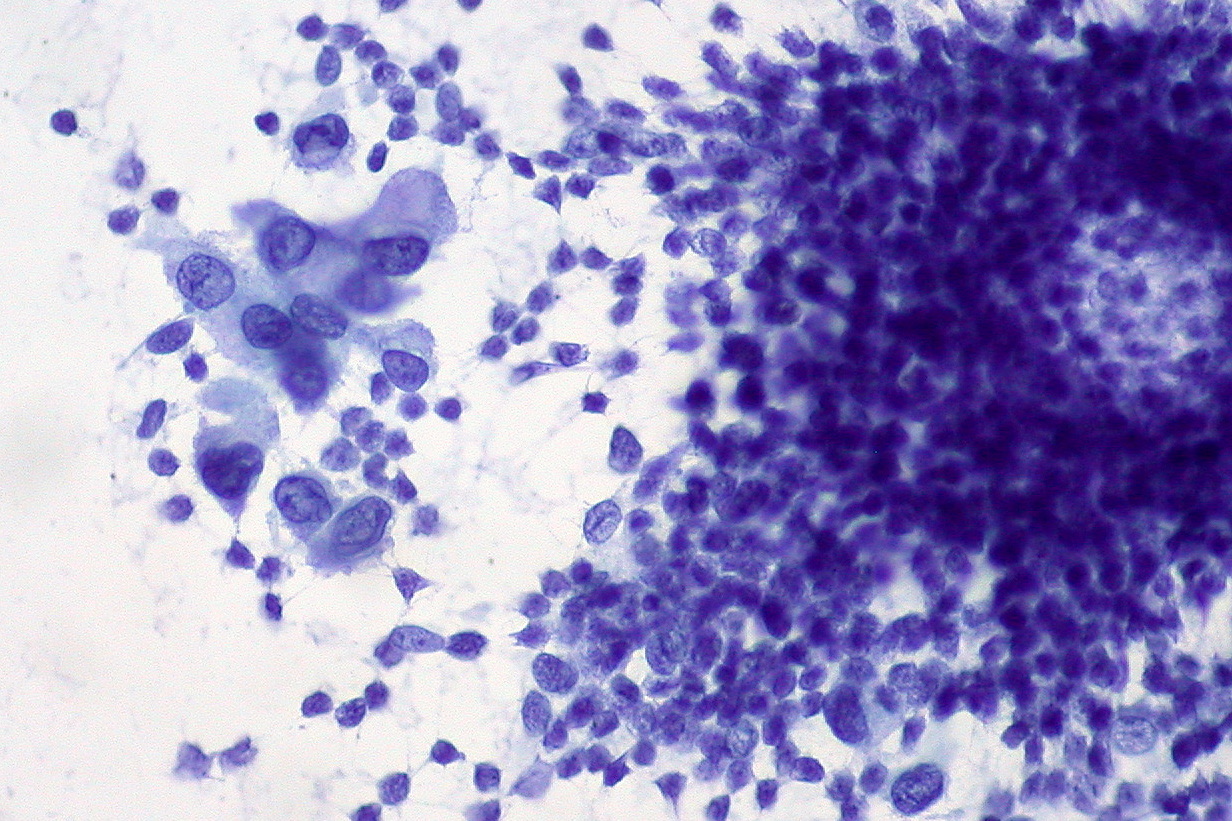
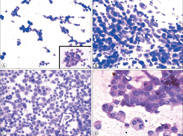
- Carcinoma versus lymphoma: malignant epithelial cells form syncytial sheets with ill-defined cell borders. Large cell lymphoma can form cohesive-appearing aggregates, but individual cell borders remain discernible and lymphoglandular bodies are associated with the lesional cells (Nga 2024).
- Poorly differentiated carcinoma: dispersed cells of poorly differentiated or undifferentiated carcinoma can mimic lymphoma (Nga 2024).
- Small cell carcinoma: evenly granular chromatin. Large cell lymphoma shows coarser chromatin or prominent nucleoli (Nga 2024).
- Undifferentiated nasopharyngeal carcinoma: syncytial sheets of cells with oval to elongated nuclei, vesicular chromatin and central prominent nucleoli. The sheets can resemble epithelioid granulomas, and true granulomas can also occur. Some aspirates appear discohesive and mimic lymphoma (Nga 2024).
- Cystic SCC: a hypocellular cystic aspirate from a large neck mass is a red flag. A cell block with immunocytochemistry can identify sparse squamous cells (Nga 2024).
Ancillary testing in metastatic carcinoma in lymph nodes on cytology
- First panel: CAM5.2 or pancytokeratin for carcinoma and CD3 and CD20 for lymphoma. Synaptophysin and chromogranin identify small cell carcinoma, and EBER in situ hybridization identifies undifferentiated nasopharyngeal carcinoma (Nga 2024).
- Pleomorphic large cells: a broad panel of cytokeratins, Melan-A/S100, CD45, CD3, CD20 and vimentin, with SALL4 for germ cell tumors (Nga 2024).
- HPV testing of nodal SCC on FNA (CAP 2025 guideline; Lewis 2025):
- HR-HPV testing is performed on FNAs of nodal SCC from patients with clinical findings of an oropharyngeal or sinonasal primary, or with SCC of unknown primary (Faquin 2025).
- HPV-specific testing is the recommended method. p16 immunohistochemistry is acceptable only in selected circumstances. A negative FNA result is followed by testing of tissue when tissue becomes available (Faquin 2025).
- p16 immunohistochemistry on cell blocks is neither an effective screening tool nor an effective stand-alone method (Faquin 2025).
- HPV RNA in situ hybridization, DNA PCR and liquid-based DNA amplification can each be used as a stand-alone test on FNA. The assay covers HPV 16 and 33 and other high-risk types, including 18, 26, 35 and 58. DNA in situ hybridization is no longer recommended (Faquin 2025).
- Surrogate markers on cell blocks: in 40 metastatic SCC sampled by FNA, p16 had a specificity of 78.7% for high-risk HPV (Jannapureddy 2010).
- Thyroid metastases: thyroglobulin measurement in the needle washout is covered in thyroid lymph node thyroglobulin.
Immunohistochemistry by likely primary
| Primary | Supporting markers | Notes and pitfalls |
|---|---|---|
| Oropharynx (HPV-associated SCC) | High-risk HPV by RNA ISH, PCR or liquid-based testing | p16 alone is unreliable on cytology (Faquin 2025) |
| Nasopharynx | EBER in situ hybridization | Syncytial sheets resemble granulomas (Nga 2024) |
| Colorectum | CDX2, SATB2, CK20; CK7 negative | In 275 colorectal lung metastases: CDX2 99%, SATB2 98%, CK20 83%, CK7 2% (Malmros 2024); SATB2 sensitivity 93% (Dragomir 2014) |
| Breast | GATA3, TRPS1 | TRPS1 in 86% and GATA3 in 21% of metaplastic triple-negative carcinomas; TRPS1 in 98% and GATA3 in 95% of ER-positive carcinomas (Ai 2021) |
| Thyroid, kidney, female genital tract | PAX8 | PAX8 in 91% of thyroid tumors, 90% of renal cell carcinomas, 98% of endometrial adenocarcinomas and 99% of high-grade serous ovarian carcinomas; negative in all lung adenocarcinomas; positive in a subset of thymic tumors (Laury 2011) |
| Prostate | NKX3.1, PSA | NKX3.1 sensitivity 98.6% for metastatic prostatic adenocarcinoma; PSA can be weak, focal or absent in poorly differentiated tumors (Gurel 2010) |
| Urothelium | GATA3; NKX3.1 negative | GATA3 is highly expressed in urothelial carcinoma (Ai 2021); urinary bladder carcinomas were NKX3.1 negative (Gurel 2010) |
| Lung adenocarcinoma | TTF-1, napsin A, CK7 | CK7 in 99% of primary lung adenocarcinomas (Malmros 2024); TTF-1 and napsin A (Yatabe 2019) |
Sources as cited in each row.
Sources of diagnostic error in metastatic carcinoma in lymph nodes on cytology
- NKX3.1 in airway cells: NKX3.1 nuclear staining was present in 87.5% of transbronchial lymph node FNAs from patients without prostate cancer. The staining was in benign bronchial epithelial cells and seromucinous glands (Gillam 2026).
- NKX3.1 specificity: in 349 nonprostatic tumors, including urinary bladder carcinomas, only 1 invasive lobular breast carcinoma expressed NKX3.1, a specificity of 99.7% (Gurel 2010). GATA3 positivity and NKX3.1 negativity support urothelial carcinoma (Ai 2021; Gurel 2010).
- Lymphoma mimicking carcinoma: DLBCL can form cohesive-appearing aggregates. Undifferentiated nasopharyngeal carcinoma can appear discohesive (Nga 2024).
- Hodgkin lymphoma: classic Hodgkin lymphoma has been misdiagnosed as metastatic lymphoepithelial carcinoma, and metastatic nasopharyngeal carcinoma has been misdiagnosed as Hodgkin lymphoma (Nga 2024).
Reporting of metastatic carcinoma in lymph nodes on cytology
- Malignant category: published ROM 98–100% (Ehinger 2026).
- Suspicious for malignancy: applies to features suggestive of metastatic disease when cellularity, sample quality or ancillary studies are insufficient (Ehinger 2026). The WHO system estimates its ROM at 88–100% (typical 93%) (Yuil Valdes 2026). Published series report 82–100% (Ehinger 2026).
- Sydney system: in 1458 aspirates, 551 of which had histologic control, the ROM was 99.8% for malignant and 100% for suspicious. These figures cover all malignancies (Caputo 2022).
Metastatic Melanoma in Lymph Nodes on FNA
Definition. Metastatic melanoma in a lymph node is a malignant melanocytic neoplasm that has spread to the node from a primary melanoma elsewhere. Unequivocal metastatic melanoma is reported in the Malignant category of the WHO Reporting System for Lymph Node, Spleen and Thymus Cytopathology, with a published ROM of 98–100% (Ehinger 2026). Related topics: Metastatic Carcinoma in Lymph Nodes (Cytology), Metastatic Tumors to the Lung (Cytology) and dermatopathic lymphadenopathy.
Clinical features of metastatic melanoma in lymph nodes on cytology
- Accuracy of FNA: in a meta-analysis of 10 studies (5518 cases), FNA for suspected metastatic melanoma had a pooled sensitivity of 0.97. The positive likelihood ratio was 58 and the negative likelihood ratio 0.03. The authors concluded that FNA is the first-line option for a suspicious mass in a patient with a history of melanoma (Hall 2013).
- Unknown primary: FNA of metastatic melanoma can establish the diagnosis before the primary lesion is identified in up to 20% of cases (Lindsey 2016).
- BRAF testing: BRAF and MEK inhibitors are approved for BRAF V600E-mutated melanoma. In a meta-analysis of 21 studies (1687 cases), VE1 immunohistochemistry had a pooled sensitivity of 0.96 and a specificity of 1.00 for BRAF V600E in melanoma tissue (Anwar 2016).
Cytomorphology of metastatic melanoma in lymph nodes on cytology
- Smear pattern: poorly cohesive cells (Lindsey 2016).
- Cell shape: frankly malignant epithelioid and spindled cells are the most common (Lindsey 2016).
- Melanin: diagnostic when present, and present in about 50% of cases (Lindsey 2016).
- Accessory features: eccentric nuclei, nucleoli and intranuclear pseudoinclusions. Cytoplasmic vacuoles can usually be found on Romanowsky-stained smears (Lindsey 2016).
- Nodal melanocytic inclusions: rare small fragments of bland nonlymphoid cells from a capsular nodal nevus can mimic metastasis on FNA. In one reported case without cell block material, the aspirate was placed in the atypical category (Cozzolino 2026).
Ancillary testing in metastatic melanoma in lymph nodes on cytology
- Sensitivity of melanocytic markers in cytology cell blocks (Saad 2024, 93 melanomas):
- SOX10 100%
- PRAME 86%
- HMB45 84%
- Melan-A 82%
- S100 71%
- PRAME specificity: PRAME was expressed in 23% of carcinomas, 33% of sarcomas and 11% of hematologic malignancies. Its specificity was 82% (Saad 2024).
- Spindle cell melanoma: 4 of 10 spindle cell melanomas were PRAME negative (Saad 2024).
- SOX10 in other tissues: SOX10 stained 12% of breast carcinomas and normal breast lobules. Benign nevi also express SOX10, so SOX10 does not separate benign from malignant melanocytic lesions (Mohamed 2013).
- Nodal nevus versus metastasis: in 45 nodal melanocytic deposits, all 30 nodal nevi were PRAME negative and all 15 melanoma metastases were diffusely PRAME positive. PRAME showed higher sensitivity than reported for HMB45 (Lezcano 2020). PRAME positivity has been reported in a nodal nevus (Cozzolino 2026), so PRAME is interpreted with morphology and location.
Differential diagnosis of metastatic melanoma in lymph nodes on cytology
| Entity | Distinguishing features |
|---|---|
| Nodal nevus | Bland melanocytes, usually capsular; PRAME negative (Lezcano 2020) |
| Dermatopathic lymphadenopathy | Melanin-containing histiocytes and Langerhans cells with a dermatologic history (Nga 2024) |
| Langerhans cell histiocytosis | Grooved nuclei; S100 and CD1a positive; negative for other melanocytic markers (Nga 2024) |
| Poorly differentiated carcinoma | Keratin positive; SOX10 positive in some breast carcinomas (Nga 2024; Mohamed 2013) |
| Large cell lymphoma and syncytial classic Hodgkin lymphoma | CD45, CD20 or CD30 positive; lymphoglandular bodies (Nga 2024) |
| Follicular dendritic cell sarcoma | CD21, CD23, CD35 and CXCL13 positive (Nga 2024) |
Reporting of metastatic melanoma in lymph nodes on cytology
- Malignant category: published ROM 98–100% (Ehinger 2026).
- Atypical category: applies to uncharacterized nonlymphoid fragments when immunocytochemistry is not possible (Cozzolino 2026).
Pattern Approach to Lymph Node Cytology
Definition. The pattern approach sorts a lymph node aspirate by its dominant cell population before ancillary testing. A stepwise approach combines the clinical findings, the low-power pattern (reactive, metastatic or lymphoma), the high-power population pattern and disease-specific findings (Chong 2023). The WHO Reporting System for Lymph Node, Spleen and Thymus Cytopathology defines four patterns for hematolymphoid lesions: mixed lymphoid cell; predominantly small/intermediate cell; predominantly intermediate/large/pleomorphic/blastic cell; and single, very large, atypical cell. The same analysis applies to splenic and thymic smears. Granulomatous inflammation, histiocytic and dendritic cell neoplasms and metastases are treated as separate patterns (Ehinger 2026). Lymphoma entities follow WHO-HAEM5 and ICC 2022 (Alaggio 2022; Campo 2022). Related topics: Sydney System and WHO Reporting System for Lymph Node Cytopathology, Normal and Reactive Lymph Node Cytology, Lymph Node FNA Triage for Flow Cytometry, Cultures and Molecular Testing and Spleen and Thymus FNA and Pediatric Lymph Node Cytology.
Clinical use of the pattern approach in lymph node cytology
- Clinical context first: cytomorphologic diagnoses, with or without ancillary tests, are made within the clinical context. Careful analysis of the clinical setting precedes the final category (Ehinger 2026).
- Common patterns: the mixed lymphoid cell pattern and the predominantly small/intermediate cell pattern are the most common in routine practice. Without flow cytometry, the distinction of lymphoma from benign lymphadenopathy in these patterns can be difficult (Ehinger 2026).
- Partial involvement: limited involvement of a node can produce a mixed, small/intermediate or single very large cell pattern in a high-grade lymphoma (Ehinger 2026).
- Unbiased review: one approach is to review the slides first without the clinical context or flow cytometry results, and then to correlate them (Ehinger 2026).
- Performance with flow cytometry: in 446 cases, fine-needle aspiration cytology with flow cytometry had a sensitivity of 94.9% and a specificity of 99.4% for non-Hodgkin lymphoma. A subtype was confirmed in 67 of 70 cases with histology (Zeppa 2010). In low-grade lymphomas, cytology with flow cytometry had a sensitivity of 98% and a specificity of 100% (Schmid 2011).
- Risk of malignancy by category: in 349 aspirates for suspected lymphoma, the ROM was 58.3% for inadequate, 6.4% for benign, 69.2% for atypical, 96.7% for suspicious and 99.3% for malignant (Makarenko 2022).
Low-power patterns in lymph node cytology
- Reactive pattern: an evenly distributed smear of normal cellularity with some lymphoid clusters, inflammatory cells, and necrosis or granulomas in specific infections (Chong 2023). Details are in Normal and Reactive Lymph Node Cytology.
- Metastatic pattern: an irregularly distributed hypercellular smear with prominent epithelial clusters, often with necrosis (Chong 2023).
- Lymphoma pattern: an evenly distributed hypercellular smear with monotonous hyperchromatic lymphoid clusters and few reactive inflammatory cells (Chong 2023).
High-power population patterns in lymph node cytology
| Pattern | Definition | Benign examples | Neoplastic examples |
|---|---|---|---|
| Two-cell population | Two distinct cell populations | Reactive hyperplasia with small and large lymphocytes | Follicular lymphoma, Hodgkin lymphoma, anaplastic large cell lymphoma, metastatic carcinoma; rarely T-cell/histiocyte-rich large B-cell lymphoma |
| Continuous range | A dominant population of atypical lymphocytes of variable size with eosinophils and other inflammatory cells | Viral lymphadenitis | Peripheral T-cell lymphoma, angioimmunoblastic type |
| Monotonous small | Small atypical cells with few reactive cells | Reactive small lymphocytes | Mantle cell lymphoma, small lymphocytic lymphoma, small cell carcinoma, small round cell sarcoma |
| Monotonous large | Mostly large atypical cells | Immunoblastic reactions | Diffuse large B-cell lymphoma, Burkitt lymphoma (medium-sized cells), poorly differentiated squamous cell carcinoma, undifferentiated nasopharyngeal carcinoma |
Source: Chong 2023.
- Small round cell tumors have slightly larger nuclei than low-grade B-cell lymphomas (Chong 2023).
- Continuous range pattern: this pattern arises mainly from T-cell lymphomas, and T-cell receptor clonality testing is required for a conclusive diagnosis (Chong 2023).
WHO system patterns and the diagnostic algorithm for hematolymphoid lesions
| WHO pattern | Benign lesions with this pattern | Lymphomas | Critical ancillary tests | Subtype on cytology |
|---|---|---|---|---|
| Mixed lymphoid cell | Reactive follicular hyperplasia; post-vaccination and viral lymphadenitis | Classic follicular lymphoma; marginal zone lymphoma; nodal T-follicular helper cell lymphoma, angioimmunoblastic type | Flow cytometry; cyclin D1; MYD88 PCR; T-cell clonality | Yes for follicular and marginal zone lymphoma; no for angioimmunoblastic type |
| Predominantly small/intermediate cell | Reactive small lymphocytes | Small lymphocytic lymphoma; mantle cell lymphoma; lymphoplasmacytic lymphoma; plasma cell neoplasms; mastocytosis | Flow cytometry; cyclin D1, SOX11 or t(11;14); MYD88; serum electrophoresis | Yes |
| Predominantly intermediate/large/pleomorphic/blastic | Kikuchi lymphadenitis, lupus lymphadenitis, infectious mononucleosis | Lymphoblastic lymphoma; large B-cell lymphomas; Burkitt lymphoma; anaplastic large cell lymphoma; peripheral T-cell lymphoma; myeloid sarcoma | Flow cytometry; EBER; FISH; CD30; TRBC1 or T-cell receptor PCR | Yes for most; rarely for PTCL, NOS |
| Single, very large, atypical cell | Warthin-Finkeldey polykaryocytes; Rosai-Dorfman disease | Classic Hodgkin lymphoma; nodular lymphocyte-predominant Hodgkin lymphoma; T-cell/histiocyte-rich large B-cell lymphoma; lymphomatoid granulomatosis | Immunocytochemistry on cell block; histology | Classic Hodgkin lymphoma only |
Source: Ehinger 2026.
The stepwise diagnostic algorithm combines both pattern systems with the clinical findings and ancillary tests.
| Step | Finding | Action or category |
|---|---|---|
| 1. Adequacy | Low cellularity, drying artifact or obscuring blood | Inadequate; repeat FNA or core biopsy |
| 2. Clinical review | Age, site, size, B symptoms, history of cancer, ultrasound | Sets the pretest probability of lymphoma |
| 3. Low power | Reactive, metastatic or lymphoma pattern | Directs triage of material |
| 4. High power | Population pattern | Defines the differential diagnosis |
| 5. Disease-specific findings | Granulomas, Reed-Sternberg cells, Russell bodies, lymphoglandular bodies, necrosis | Narrows the differential diagnosis |
| 6. Ancillary tests | Flow cytometry, cell block immunocytochemistry, clonality and FISH | Benign, atypical, suspicious or malignant category with subtype |
Sources: Chong 2023; Ehinger 2026.
Mixed lymphoid cell pattern in lymph node cytology
- Composition: small and intermediate lymphoid cells with some centroblasts or immunoblasts, with or without tingible-body macrophages. Many eosinophils, plasma cells and histiocytes also produce a mixed pattern (Ehinger 2026).
- Flow cytometry discordance: lambda-monotypic B cells are more likely to be malignant than kappa-monotypic B cells. Classic follicular lymphoma shows dimmer CD19, CD20 and CD38 than reactive hyperplasia (Ehinger 2026).
- Infectious mononucleosis: aspirates contain more large immunoblasts than a usual reactive node, and two of ten cases in one series were cytologically suspicious for lymphoma. Serologic confirmation and resolution of adenopathy are required; otherwise excision follows (Stanley 1990). The immunoblasts are mostly MUM1-positive, CD10-negative, BCL6-negative B cells with polytypic light chains, and Reed-Sternberg-like cells are CD30 positive and CD15 negative (Louissaint 2012).
- Progressive transformation of germinal centers: present in 10–15% of nodes with reactive follicular hyperplasia, with a male predominance of 3:1 and a cervical location in almost half. It can resemble nodular lymphocyte-predominant Hodgkin lymphoma and is not premalignant (Hicks 2002). Immunoarchitectural patterns separate the two on histology (Hartmann 2015).
- COVID-19 post-vaccination lymphadenopathy: of 26 aspirates in a pooled review, 21 were benign and 3 atypical; all were confirmed by follow-up or excision (Caputo 2023). Aspirates show follicular hyperplasia with germinal center elements and an increased proportion of large lymphocytes (Tan 2021). CD8-predominant T cells and striking mitoses were reported (García-Molina 2021). Nodes appeared 3–33 days after vaccination, usually ipsilateral to the injection site (Hagen 2021).
- Kimura disease: a polymorphous lymphoid population with many eosinophils, collagenous fragments, endothelial cells and occasional polykaryocytes (Deshpande 2002).
- Kikuchi lymphadenitis: in 173 aspirates, all cytologic samples contained intracellular apoptotic debris within crescentic and phagocytic macrophages. Cell blocks showed clusters of CD123-positive plasmacytoid monocytes. Flow cytometry and cell blocks showed nonspecific lymphoid hyperplasia in nine cases suspicious for lymphoma (Wang 2020). Small and large atypical lymphocytes with phagocytic histiocytes and extracellular debris, without neutrophils, are diagnostic when the clinical features are typical (Hsueh 1993).
- Immune deficiency: a mixed pattern with monotypic B cells by flow cytometry in a clinical setting of a polymorphic lymphoproliferative disorder of immune deficiency or dysregulation is best categorized as atypical (Ehinger 2026).
- Angioimmunoblastic type: absence of tingible-body macrophages, atypical lymphoid cells and singly scattered epithelioid cells were the strongest predictors against reactive hyperplasia (Gupta 2023). Histology is required to assess dendritic meshworks and vascular proliferation (Ehinger 2026).
Predominantly small/intermediate cell pattern in lymph node cytology
- Entities: small lymphocytic lymphoma, mantle cell lymphoma, lymphoplasmacytic lymphoma, plasma cell neoplasms and mastocytosis. Each can be diagnosed on cytology with flow cytometry and the critical ancillary tests (Ehinger 2026).
- Resting lymph node: a node that persists after lymphadenitis has resolved yields mainly small lymphocytes without centroblasts or tingible-body macrophages. It presents as a single persistent node that neither progresses nor regresses (Ehinger 2026). When too few cells remain for flow cytometry, such an aspirate is categorized as atypical.
- Small lymphocytic lymphoma: small cells with prolymphocytes and paraimmunoblasts and a CD19-positive, CD20-dim, CD5-positive, CD23-positive, CD200-positive phenotype with dim monotypic light chains. Without cyclin D1, SOX11 or t(11;14) testing, a sign-out as low-grade B-cell lymphoma, likely SLL/CLL, is appropriate (Ehinger 2026).
- Mantle cell lymphoma: a predominance of centrocyte-like cells. Nuclear cyclin D1 establishes the diagnosis, although proliferation-center cells of SLL can also show cyclin D1, and cyclin D1-negative cases need SOX11 or FISH (Ehinger 2026).
- Nonlymphoid small cells: small cell carcinoma and small round cell sarcomas fall in this pattern. Epithelial evidence supports carcinoma; otherwise lymphoma and mesenchymal tumors are excluded with ancillary studies (Chong 2023).
- Lymphoglandular bodies: in 132 aspirates of malignant tumors, 6 of 104 nonlymphoid malignancies had easily identifiable lymphoglandular bodies, including small cell carcinoma, melanoma and seminoma (Flanders 1993). More than 100 lymphoglandular bodies per 10 high-power fields occurred in 100% of non-Hodgkin lymphomas and in 4.8% of nonlymphoid round cell tumors on Romanowsky stains (Francis 1994).
- Lymphoplasmacytic lymphoma: small lymphoid cells with lymphoplasmacytoid features, nuclear pseudoinclusions of immunoglobulin and round dense cytoplasmic globules. The cells are CD5 and CD10 negative, marginal zone lymphoma is the main differential diagnosis, and MYD88 L265P testing with IgM paraprotein analysis supports the diagnosis (Ehinger 2026).
- Plasma cell neoplasms: rare in nodes. An aberrant phenotype with CD56, CD117 or light chain restriction separates them from benign plasma cell lesions (Ehinger 2026).
- Mastocytosis: mast cells with pale eccentric cytoplasm and metachromatic granules, or hypogranulated and spindle forms. Aberrant CD25, CD2 or CD30 expression by flow cytometry or immunocytochemistry is diagnostic (Ehinger 2026).
- Discordant results: monotypic B or T cells in an aspirate that appears benign are categorized as atypical or suspicious, depending on the size of the monotypic population (Ehinger 2026).
Disease-specific findings in lymph node cytology
| Finding | Significance | Pitfall |
|---|---|---|
| Reed-Sternberg cells | Hodgkin lymphoma | Reed-Sternberg-like cells in infectious mononucleosis and other lymphomas |
| Russell bodies and Mott cells | Mature B-cell lymphoma with plasmacytic differentiation, such as lymphoplasmacytic lymphoma | Plasma cell neoplasms show the same plasmacytic features |
| Lymphoglandular bodies | Lymphoid tissue | Present in some small cell carcinomas, melanomas, seminomas and myeloid sarcomas |
| Ill-defined granulomas | Tuberculosis and other infections | Small granulomas occur in classic Hodgkin lymphoma, angioimmunoblastic type, nasopharyngeal and squamous cell carcinoma, and seminoma |
| Crescentic histiocytes with karyorrhexis | Kikuchi lymphadenitis | Necrosis in high-grade lymphoma |
| Emperipolesis | Rosai-Dorfman disease | Large histiocytes read as Reed-Sternberg cells |
| Eosinophils | Kimura disease, Hodgkin lymphoma, T-cell lymphoma, Langerhans cell histiocytosis | Eosinophils alone do not establish a diagnosis |
Sources: Chong 2023; Nga 2024; Flanders 1993; Singhal 2015; Deshpande 1998; Wang 2020.
Predominantly intermediate/large/pleomorphic/blastic cell pattern in lymph node cytology
- Category: most cases are categorized as malignant, with or without flow cytometry. High-grade lymphoma cells are fragile, so the malignant category can be assigned without monotypic cells by flow cytometry (Ehinger 2026).
- Benign mimics: Kikuchi disease, lupus lymphadenitis and infectious mononucleosis produce this pattern. A granulomatous reaction within a high-grade lymphoma can obscure the malignant cells (Ehinger 2026).
- Myeloid sarcoma: in 16 cytology cases, 14 were secondary to acute myeloid leukemia. Blasts had round or cleaved nuclei with indistinct nucleoli, and flow cytometry was supportive in 11. Lymphoglandular bodies, which suggest lymphoma, were present in almost half (Singhal 2015).
- Discordant flow cytometry: large monotypic cells by flow cytometry in a case first read as infectious mononucleosis support malignancy, particularly in the large-cell pattern (Ehinger 2026).
Single, very large, atypical cell pattern in lymph node cytology
- Definition: very large atypical cells that do not resemble any lymphoid cell, in an inflammatory background of lymphocytes, histiocytes, plasma cells or eosinophils (Ehinger 2026).
- Classic Hodgkin lymphoma on cytology: classic Hodgkin lymphoma is among the most frequently missed initial diagnoses because Hodgkin and Reed-Sternberg cells can be sparse. A provisional diagnosis is often possible and is confirmed by immunocytochemistry for CD30, CD15 and weak PAX5. Subtyping requires histology. Flow cytometry for Hodgkin and Reed-Sternberg cells on small biopsy and cytology specimens had a sensitivity of 95.4% and a specificity of 98.2% (Chan 2022; Ehinger 2026).
- Nodular lymphocyte-predominant Hodgkin lymphoma on cytology: scattered very large multilobated cells in a background of small lymphocytes and histiocytes, usually with few eosinophils and plasma cells. The resemblance to T-cell/histiocyte-rich large B-cell lymphoma prevents a definitive cytologic diagnosis (Ehinger 2026). FNA with core or cell block was definitive in 7 of 13 cases at initial diagnosis and excision in 13 of 13; the disease accounted for less than 0.5% of FNAs for suspected lymphoma, and flow cytometry showed a light chain-restricted B-cell population in 1 of 11 involved specimens (Gupta 2020).
- Benign mimics: Warthin-Finkeldey polykaryocytes and Rosai-Dorfman disease (Ehinger 2026). Rosai-Dorfman disease shows large histiocytes with emperipolesis, and one aspirate was misread as Hodgkin lymphoma (Deshpande 1998).
- T-cell/histiocyte-rich large B-cell lymphoma: a background of numerous small T cells and histiocytes with fewer than 10% scattered large B cells that mimic Reed-Sternberg cells. In 5 aspirates, small mature lymphocytes made up more than 90% of cells, and cytospin immunocytochemistry showed light chain restriction in all 5 cases (Tani 1998).
Nonlymphoid and uncommon patterns in lymph node cytology
- Metastatic sarcoma: among 1772 sarcoma patients, 2.6% had nodal metastasis, most often angiosarcoma, embryonal rhabdomyosarcoma and epithelioid sarcoma (Fong 1993). In 33 aspirated nodes with metastatic sarcoma, the most common types were Kaposi sarcoma, angiosarcoma and rhabdomyosarcoma (Saoud 2023).
- Germ cell tumor: SALL4 was strongly expressed in seminoma, embryonal carcinoma and yolk sac tumor, and OCT4 in seminoma and embryonal carcinoma (Cao 2009).
- Metastasis within lymphoid patterns: poorly differentiated carcinoma with discohesive cells can produce a two-cell pattern. Adenoid or squamous differentiation at high power supports carcinoma (Chong 2023). Basaloid squamous cell carcinoma and undifferentiated nasopharyngeal carcinoma are included in the differential diagnosis of the monotonous large-cell pattern (Chong 2023). Details are in Metastatic Carcinoma in Lymph Nodes (Cytology).
- Uncommon lymphomas: classic Hodgkin lymphoma, mediastinal large B-cell lymphoma and T-cell/histiocyte-rich large B-cell lymphoma were often difficult to diagnose on FNA, even with flow cytometry and cell block immunohistochemistry. The FNA diagnosis of peripheral T-cell lymphoma depended on cell block immunohistochemistry (Mayall 2003).
Reporting categories and patterns in lymph node cytology
- Category and pattern: the report gives the WHO system category and, when a subtype is not reached, the pattern and the favored diagnosis. Examples are "low-grade B-cell lymphoma, likely MZL" and "FL, likely classic FL" when not all differentials are excluded (Ehinger 2026).
- Benign category: unequivocal benign features, with a specific benign process named when possible. The published ROM of the benign category ranges from 2% to 16% (Ehinger 2026).
- Atypical category: predominantly benign features with minimal features that raise the possibility of malignancy, or material limited by poor smearing, blood or ultrasound gel (Ehinger 2026).
- Specific wording for indeterminate cases: cases that favor a lineage are described as "AUS, favor poorly differentiated carcinoma", "ALUS, favor atypical lymphoproliferative disease" or "ALUS, cannot exclude low-grade lymphoma" (Chong 2023).
- Management by category: repeat FNA with material for ancillary studies, core needle biopsy or excision follows an atypical or suspicious result (Sydney System and WHO Reporting System for Lymph Node Cytopathology).
Limits of the pattern approach in lymph node cytology
- Concordance with histology: in a prospective comparison of 103 patients, FNA with immunophenotyping and histology were concordant in 76. Major discordance occurred in 10, including Hodgkin lymphoma versus anaplastic large cell lymphoma in 3 (Landgren 2004).
- Clinician perspective: in one lymphoma clinic, only 29% of initial FNAs gave a specific classified diagnosis (Hehn 2004). The authors judged FNA unhelpful for primary lymphoma diagnosis in that setting.
- Excision: classic Hodgkin lymphoma subtyping, nodular lymphocyte-predominant Hodgkin lymphoma, T-cell/histiocyte-rich large B-cell lymphoma and the angioimmunoblastic type require tissue architecture (Ehinger 2026).
Spleen and Thymus FNA and Pediatric Lymph Node Cytology
Definition. The WHO Reporting System for Lymph Node, Spleen and Thymus Cytopathology applies its five categories (inadequate/insufficient/nondiagnostic, benign, atypical, suspicious for malignancy and malignant) to fine-needle aspiration biopsy (FNAB) of the spleen and thymus as well as lymph nodes (Cozzolino 2026). Splenic and thymic FNAB is performed under ultrasound or computed tomography guidance. Published data on its accuracy are limited, and its use is not yet standard practice. Its diagnostic value increases with rapid on-site evaluation (ROSE), flow cytometry, immunocytochemistry and molecular testing (Cozzolino 2026). The pattern analysis used for lymph nodes applies to splenic and thymic smears (Ehinger 2026). Children and adolescents have a different spectrum of lymphadenopathy, and FNA performs well in this group (Ronchi 2021). Related topics: Sydney System and WHO Reporting System for Lymph Node Cytopathology, Pattern Approach to Lymph Node Cytology and Lymph Node FNA Triage for Flow Cytometry, Cultures and Molecular Testing.
Clinical use of splenic FNA
- Indications: splenomegaly, focal splenic lesions, infections, lymphomas and other neoplasms, including staging and monitoring of disease (Cozzolino 2026).
- Role: splenic and thymic FNAB can avoid unnecessary surgery and guide therapy (Cozzolino 2026).
- Concerns: sample adequacy, diagnostic accuracy and complications, especially hemorrhage, limit wider use (Cozzolino 2026).
- Meta-analysis: image-guided percutaneous biopsy of the spleen had a pooled sensitivity of 87.0% and specificity of 96.4%. The pooled major complication rate was 2.2%, and 1.3% with needles of 18 gauge or smaller. Hemorrhage was the most common complication, followed by pain (McInnes 2011).
- Multicenter experience: in 398 ultrasound-guided splenic biopsies, overall accuracy was 90.9%: 84.9% for cytology, 88.3% for microhistology and 90.3% for both. For suspected splenic lymphoma, core biopsy was more accurate than cytology (90.9% versus 68.5%). There were no deaths, major complications were below 1% and all complications 5.2% (Civardi 2001).
- Operator experience: a higher number of procedures performed by the operator was significantly associated with better overall accuracy, and the complication rate did not depend on it (Civardi 2001).
- Evidence base: the diagnostic accuracy arm of the meta-analysis included 4 studies (639 patients), and the complication arm 9 studies (741 patients). Needle biopsy of the spleen was considered a favorable alternative to splenectomy (McInnes 2011).
- Flow cytometry: in 18 splenic aspirates with a 22-gauge needle, one or two passes were sufficient for smears and flow cytometry. Flow cytometry showed light chain restriction in all 10 non-Hodgkin lymphomas, quantified myeloid cells in myeloid metaplasia and showed polytypic B cells in reactive hyperplasia. No complications occurred (Zeppa 2003).
- Subtyping: CD19 with CD5 or CD10 coexpression on splenic aspirates identified the lymphoma subtype in two cases, and cytospin immunocytochemistry was concordant in four of six cases (Zeppa 2003).
- Endoscopic ultrasound route: transgastric EUS-guided aspiration with a 19-gauge needle obtained diagnostic material in 5 of 5 splenic tumors: lymphoma (2), sarcoidosis (2) and inflammatory pseudotumor (1) (Iwashita 2009).
Splenic lesions on FNA
- Lymphomas: WHO-HAEM5 groups the splenic B-cell lymphomas and leukemias as hairy cell leukemia, splenic marginal zone lymphoma, splenic diffuse red pulp small B-cell lymphoma and splenic B-cell lymphoma/leukemia with prominent nucleoli. The last entity is new and includes the former hairy cell leukemia variant and some cases of B-cell prolymphocytic leukemia (Alaggio 2022).
- Splenic marginal zone lymphoma: the predominant differential diagnosis of a small B-cell lymphoma in splenic FNA. The cytomorphology resembles classic follicular lymphoma, with centrocyte-like cells, small lymphocytes, monocytoid cells and more frequent tingible-body macrophages; flow cytometry is typically CD5 and CD10 negative (Ehinger 2026). Diagnostic, staging and therapeutic criteria for splenic marginal zone lymphoma were proposed by an international group (Matutes 2008).
- Large cell lymphomas: diffuse large B-cell lymphoma and DLBCL/high-grade B-cell lymphoma with MYC and BCL2 rearrangements occur in the spleen and are subtyped with flow cytometry, EBER and FISH (Ehinger 2026).
- Vascular and stromal tumors: WHO-HAEM5 recognizes splenic vascular-stromal tumors: littoral cell angioma, splenic hamartoma and sclerosing angiomatoid nodular transformation (Alaggio 2022). Littoral cell angioma arises from the sinusoidal lining (littoral) cells of the red pulp and is unique to the spleen. It has been diagnosed by EUS-guided FNA with immunohistochemistry on the cell block (Nagarajan 2011).
- Granulomatous and inflammatory lesions: sarcoidosis and inflammatory pseudotumor are diagnosable on splenic aspirates (Iwashita 2009). Granulomatous lymphadenitis is covered in Granulomatous Lymphadenitis (Cytology).
- Myeloid proliferations: myeloid metaplasia (extramedullary hematopoiesis) is identified by smear morphology and quantified by flow cytometry (Zeppa 2003).
| Splenic FNA finding | Main diagnosis | Key ancillary test |
|---|---|---|
| Small and centrocyte-like B cells with monocytoid cells | Splenic marginal zone lymphoma | Flow cytometry: CD5 and CD10 negative, light chain restricted |
| Monotonous small B cells | Other small B-cell lymphomas | Flow cytometry with CD5, CD10 and CD23; cyclin D1 |
| Large atypical lymphoid cells | Large B-cell lymphoma | Flow cytometry, EBER, FISH |
| Myeloid precursors and megakaryocytes | Myeloid metaplasia (extramedullary hematopoiesis) | Flow cytometric quantification of myeloid cells |
| Epithelioid histiocyte clusters | Sarcoidosis or infection | Special stains and cultures |
| Vascular lining cells | Littoral cell angioma or other vascular-stromal tumor | Immunohistochemistry on the cell block |
| Polymorphous lymphocytes and plasma cells | Reactive hyperplasia | Polytypic B cells |
Sources: Alaggio 2022; Ehinger 2026; Iwashita 2009; Nagarajan 2011; Zeppa 2003.
Clinical use of thymic and mediastinal FNA
- Indications: thymic FNAB is used mainly to diagnose thymic hyperplasia, thymoma, thymic carcinoma and lymphoma. Separating these entities and their subtypes is difficult (Cozzolino 2026).
- Mediastinal FNAB series: in 189 aspirates from four university centers, 71% of diagnoses were neoplastic. Lymphoma and thymoma were the most frequent primary malignancies. Metastatic tumors accounted for 60% of neoplasms, two-thirds from the lung and mostly small cell carcinoma. Sensitivity and specificity for neoplasm were 87–88%, and the positive predictive value was 97%. The primary tumor of 16% of metastases was not identified, and ancillary tests were performed in 27% of cases (Powers 1996).
- Thymic epithelial tumors: in 22 thymic tumors aspirated before resection, the accuracy of a cytologic diagnosis of thymoma and of thymic carcinoma was 100%. Vague terminology and incomplete information caused difficulties, and immunostains identified the epithelial and lymphocytic components. FNA is used less for primary thymic tumors than for mediastinal metastases because of concern about sampling error, the rarity of thymic tumors, the multiplicity of mediastinal lesions and limited experience (Zakowski 2010).
- Classification: the 2021 WHO classification of thymic epithelial tumors covers thymomas, thymic carcinomas and thymic neuroendocrine tumors, with mediastinal germ cell and mesenchymal tumors. Thymomas and most thymic carcinomas lack targetable mutations, have a low tumor mutational burden and typically have high PD-L1 expression. Genetic translocations, including YAP1::MAML2, were recognized in metaplastic thymomas and rare B2 and B3 thymomas (Marx 2022).
- Thymic carcinoma: usually invasive and often advanced at presentation. Thymic carcinoma lacks unique morphologic and immunophenotypic features, which complicates separation from metastatic carcinoma. The AJCC 8th edition TNM staging included thymic carcinomas in 2017 (Roden 2022).
- Mediastinal sarcomas: published FNA experience consists mostly of small series of vascular sarcomas, liposarcomas, malignant peripheral nerve sheath tumor, synovial sarcoma and Ewing sarcoma (Lott-Limbach 2020).
T-lymphoblastic lymphoma as a mediastinal mass in children and adolescents
- Presentation: T-lymphoblastic lymphoma presents in children and adolescents with a rapidly enlarging mediastinal mass, dyspnea and cervical lymphadenopathy that require a quick diagnosis (Bhaker 2015).
- Diagnosis on cytology: combined cytomorphology and flow cytometry diagnosed T-lymphoblastic lymphoma in aspirates and pleural or pericardial fluids and avoided a biopsy (Bhaker 2015).
- Speed: the rapid turnaround of flow cytometry allows patients with large anterior mediastinal masses or superior vena cava syndrome to begin treatment promptly (Gorczyca 2004).
- Outcome: event-free and overall survival for pediatric lymphoblastic lymphoma both exceed 80% with current therapy. Treatment of T-LBL with large mediastinal tumors is complex and toxic, and outcomes of relapsed or refractory disease remain poor (Temple 2023).
- T-ALL versus T-LBL: the two are distinguished clinically by the extent of bone marrow involvement, with ≥25% marrow blasts defining T-cell acute lymphoblastic leukemia by convention in treatment protocols; WHO-HAEM5 gives no strict threshold. NOTCH1 fusions were found only in pediatric T-LBL and were associated with a higher risk of relapse (Te Vrugt 2025).
Flow cytometry of mediastinal and thymic aspirates
- Yield: in 100 mediastinal lesions, flow cytometry was nondiagnostic in 5%. Excluding Hodgkin lymphoma, its sensitivity for mediastinal tumors was about 92%. The diagnosis was reached by flow cytometry alone in 31 of 32 B-cell lymphomas, 7 of 8 T-lymphoblastic leukemias/lymphomas and 11 of 11 thymomas or thymic hyperplasias (Gorczyca 2004).
- Thymocytes versus T-lymphoblastic lymphoma: the pattern of surface CD3 expression against forward scatter reliably separated immature thymocytes of thymoma or thymic hyperplasia from T-lymphoblastic leukemia/lymphoma (Gorczyca 2004).
- Noninformative results: flow cytometry gave no false-positive results. It was noninformative in 3 diffuse large B-cell lymphomas, 1 peripheral T-cell lymphoma and 3 carcinomas (Gorczyca 2004).
- Thymoma pitfall: thymoma and indolent T-lymphoblastic proliferation are part of the differential diagnosis of T-lymphoblastic leukemia/lymphoma (Wang 2026). Keratin-positive epithelial cells on the cell block identify thymoma.
- Mediastinal large B-cell lymphomas: primary mediastinal large B-cell lymphoma is the large B-cell lymphoma expected in the thymus (Ehinger 2026). Classic Hodgkin lymphoma and mediastinal large B-cell lymphoma are sclerotic and often yield scanty neoplastic cells (Mayall 2003).
| Thymic or mediastinal lesion | Cytologic features | Ancillary findings |
|---|---|---|
| Thymic hyperplasia | Small lymphocytes; few epithelial cells | Immature and mature thymocytes in a continuous maturation pattern by flow cytometry |
| Lymphocyte-rich thymoma | Small lymphocytes with cohesive epithelial cells | Keratin-positive epithelium; immature thymocytes by flow cytometry |
| Thymic carcinoma | Malignant epithelial cells | Carcinoma markers; separation from metastasis requires clinical and imaging correlation |
| T-lymphoblastic lymphoma | Monotonous blasts with scant cytoplasm | TdT, cytoplasmic CD3, CD1a; abnormal surface CD3 and scatter pattern |
| Primary mediastinal large B-cell lymphoma | Large cells, often scanty because of sclerosis | B-cell markers; subtyping by immunocytochemistry |
| Classic Hodgkin lymphoma | Rare Reed-Sternberg cells in an inflammatory background | CD30 and CD15 on the cell block |
| Metastatic small cell carcinoma | Small cells with molding | INSM1, keratin |
Sources: Gorczyca 2004; Marx 2022; Mayall 2003; Powers 1996; Roden 2022; Zakowski 2010.
Pediatric lymph node FNA
- Accuracy: in 76 patients aged up to 19 years, 57 aspirates were non-neoplastic, 18 malignant and 1 suspicious. Sensitivity was 93%, specificity 100%, positive predictive value 100%, negative predictive value 98% and accuracy 98.6%. ROSE, a cell block, immunocytochemistry and flow cytometry supported this performance. FNA allowed correct triage of reactive, inflammatory and neoplastic lymphadenopathies with minimal complications (Ronchi 2021).
- Head and neck masses in children: in 257 patients with 338 FNABs, the final diagnoses were reactive lymphadenopathy (38.5%), benign thyroid colloid nodule (12.1%), malignancy (8.2%) and atypical mycobacterial infection (5.8%). Sensitivity was 94.6% and specificity 97.7%. The complication rate was 2.1%, and 73% of procedures used general anesthesia or sedation. Negative FNAB results avoided 95.3% of unnecessary operations (Huyett 2016).
- Mediastinal nodes: in children, endobronchial and esophageal ultrasound-guided needle aspiration had a pooled sampling adequacy of 98%, a diagnostic yield of 61%, a major complication rate of 0.6% and a minor complication rate of 3.5% These techniques are first-line modalities for sampling mediastinal lymphadenopathy in children (Madan 2021).
Benign pediatric lymphadenopathy on FNA
- Reactive hyperplasia is the most common diagnosis (Huyett 2016). The cytology is covered in Normal and Reactive Lymph Node Cytology.
- Progressive transformation of germinal centers: 20% of cases occur in children, with a mean age of 11 years, and recurrence is more common in children (50% versus 23%). Progressive transformation of germinal centers is associated with Hodgkin lymphoma in 30–35% of patients and can precede, follow or accompany it. The authors judged the association nebulous and PTGC not premalignant (Hicks 2002).
- Kikuchi lymphadenitis: in 173 patients, 72.8% were aged 21–40 years and 75.7% were female; almost all had painful cervical nodes. Clusters of CD123-positive plasmacytoid monocytes on the cell block support the diagnosis (Wang 2020). The cytology is described under Pattern Approach to Lymph Node Cytology.
- Mycobacterial infection: atypical mycobacterial infection accounted for 5.8% of pediatric head and neck FNABs (Huyett 2016; Granulomatous Lymphadenitis (Cytology)).
- Langerhans cell histiocytosis: in 47 cytology cases, most patients were aged 0–5 years and the head and neck, including cervical nodes, was the most common site. Giant cells were present in 78%, mild eosinophilia in 61% and necrosis in 38%. Mitoses numbered 1–2 per 10 high-power fields in 25.5%, and no abnormal mitoses were seen (Phulware 2019). In 37 aspirated cases, the sites were bone (28), head and neck soft tissue (6) and lymph nodes (3), and 84% were diagnosed as Langerhans cell histiocytosis on FNA. On cell block, CD1a was positive in 24 of 24 cases and S100 in 22 of 23 (Hang 2017).
Pediatric lymphomas on FNA
- Lymphoblastic lymphoma: the second most common non-Hodgkin lymphoma in children, adolescents and young adults, accounting for 25–35% of cases. T-lymphoblastic lymphoma makes up 70–80% and precursor B-lymphoblastic lymphoma 20–25% (Temple 2023; Lymphoblastic Lymphoma in Cytology).
- Pediatric-type follicular lymphoma: all 21 follicular lymphomas in patients younger than 40 years that lacked a BCL2 abnormality had a Ki-67 fraction >30%, were stage I and did not progress or recur. In 58 adults, the 13 cases with the same profile were stage I and none progressed or relapsed (Louissaint 2012).
- Pediatric nodal marginal zone lymphoma: a distinct entity in WHO-HAEM5 and a provisional entity listed under nodal marginal zone lymphoma in ICC 2022 (Alaggio 2022; Campo 2022; Laurent 2023; Marginal Zone Lymphoma in Cytology).
- Other lymphomas in children: Burkitt lymphoma, anaplastic large cell lymphoma and classic Hodgkin lymphoma are covered in Burkitt Lymphoma (Cytology), Peripheral T-Cell Lymphomas and Anaplastic Large Cell Lymphoma in Cytology and Classic Hodgkin Lymphoma (Cytology).
| Age group | Common benign causes | Common malignant causes |
|---|---|---|
| Children | Reactive hyperplasia; atypical mycobacterial infection; Langerhans cell histiocytosis (mostly 0–5 years) | Lymphoblastic lymphoma; Burkitt lymphoma; anaplastic large cell lymphoma; neuroblastoma and other small round cell tumors |
| Adolescents | Reactive hyperplasia; infectious mononucleosis | Classic Hodgkin lymphoma; T-lymphoblastic lymphoma; pediatric-type follicular lymphoma |
| Young adults | Reactive hyperplasia; Kikuchi lymphadenitis (mostly 21–40 years) | Classic Hodgkin lymphoma |
Sources: Huyett 2016; Phulware 2019; Temple 2023; Wang 2020; Dharmalingam 2023; Louissaint 2012; Molyneux 2012; Wang 2019.
Differential diagnosis of small round cells in pediatric aspirates
- Lymphoma versus small round cell tumors: small round cell tumors have slightly larger nuclei than low-grade B-cell lymphomas (Chong 2023). Lymphoglandular bodies were abundant in all non-Hodgkin lymphomas and rare in nonlymphoid round cell tumors (Francis 1994).
- Neuroblastoma: the most common extracranial malignant neoplasm of early childhood; on cytology it is a small round cell tumor positive for neuroendocrine markers (Dharmalingam 2023).
- Panel: CD45, TdT, CD99, desmin or myogenin, neuroendocrine markers and keratin on the cell block, with flow cytometry for lymphoid neoplasms.
Reporting of splenic, thymic and pediatric FNA
- Categories: the five WHO system categories apply to the spleen and thymus, with key diagnostic criteria, risk of malignancy and standardized reporting (Cozzolino 2026).
- Thymic reports state the cell types identified and the immunostain results clearly, because vague terminology caused management difficulties (Zakowski 2010).
- Further sampling and testing: core biopsy is preferred when splenic lymphoma is suspected and cytology is limited (Civardi 2001). Pediatric-type follicular lymphoma is defined by the absence of a BCL2 abnormality and a high proliferation index, so BCL2 testing and Ki-67 are part of the workup (Louissaint 2012).
References
- Ai D, Yao J, Yang F, et al. TRPS1: a highly sensitive and specific marker for breast carcinoma, especially for triple-negative breast cancer. Mod Pathol. 2021;34(4):710–719. doi:10.1038/s41379-020-00692-8. PMID 33011748
- Al-Abbadi MA, Barroca H, Bode-Lesniewska B, et al. A proposal for the performance, classification, and reporting of lymph node fine-needle aspiration cytopathology: the Sydney system. Acta Cytol. 2020;64(4):306–322. doi:10.1159/000506497. PMID 32454496
- Alaggio R, Amador C, Anagnostopoulos I, et al. The 5th edition of the World Health Organization Classification of Haematolymphoid Tumours: lymphoid neoplasms. Leukemia. 2022;36(7):1720–1748. PMID 35732829. doi:10.1038/s41375-022-01620-2
- Anwar MA, Murad F, Dawson E, Abd Elmageed ZY, Tsumagari K, Kandil E. Immunohistochemistry as a reliable method for detection of BRAF-V600E mutation in melanoma: a systematic review and meta-analysis of current published literature. J Surg Res. 2016;203(2):407–415. doi:10.1016/j.jss.2016.04.029. PMID 27363650
- Bandargal S, Florianova L, Dmitrienko S, et al. Fine needle aspirate flow cytometry's ancillary utility in diagnosing non-Hodgkin lymphoma in the head and neck. J Otolaryngol Head Neck Surg. 2024;53:19160216241296127. doi:10.1177/19160216241296127. PMID 39511788
- Bhaker P, Das A, Rajwanshi A, et al. Precursor T-lymphoblastic lymphoma: speedy diagnosis in FNA and effusion cytology by morphology, immunochemistry, and flow cytometry. Cancer Cytopathol. 2015;123(9):557–565. PMID 26243719. doi:10.1002/cncy.21584
- Campo E, Jaffe ES, Cook JR, et al. The International Consensus Classification of Mature Lymphoid Neoplasms: a report from the Clinical Advisory Committee. Blood. 2022;140(11):1229–1253. PMID 35653592. doi:10.1182/blood.2022015851
- Cao D, Li J, Guo CC, et al. SALL4 is a novel diagnostic marker for testicular germ cell tumors. Am J Surg Pathol. 2009;33(7):1065–1077. PMID 19390421. doi:10.1097/PAS.0b013e3181a13eef
- Caputo A, Caleo A, Cozzolino I, et al. COVID-19 post-vaccination lymphadenopathy: A review of the use of fine needle aspiration cytology. Cytopathology. 2023;34(5):423–432. PMID 36807950. doi:10.1111/cyt.13221
- Caputo A, Ciliberti V, D'Antonio A, et al. Real-world experience with the Sydney System on 1458 cases of lymph node fine needle aspiration cytology. Cytopathology. 2022;33(2):166–175. doi:10.1111/cyt.13079. PMID 34817104
- Caputo A, Fraggetta F, Cretella P, et al. Digital examination of LYmph node CYtopathology Using the Sydney system (DELYCYUS): an international, multi-institutional study. Cancer Cytopathol. 2023;131(11):679–692. doi:10.1002/cncy.22741. PMID 37418195
- Chan A, Scarpa Carniello JV, Gao Q, et al. Role of flow cytometric immunophenotyping for classic Hodgkin lymphoma in small biopsy and cytology specimens. Arch Pathol Lab Med. 2022;146(4):462–468. PMID 34293084. doi:10.5858/arpa.2020-0795-OA
- Chhieng DC, Cangiarella JF, Symmans WF, Cohen JM. Fine-needle aspiration cytology of Hodgkin disease: a study of 89 cases with emphasis on false-negative cases. Cancer. 2001;93(1):52–59. doi:10.1002/1097-0142(20010225)93:1<52::aid-cncr9007>3.0.co;2-3. PMID 11241266
- Chong Y, Park G, Cha HJ, et al. A stepwise approach to fine needle aspiration cytology of lymph nodes. J Pathol Transl Med. 2023;57(4):196–207. doi:10.4132/jptm.2023.06.12. PMID 37460394
- Civardi G, Vallisa D, Bertè R, et al. Ultrasound-guided fine needle biopsy of the spleen: high clinical efficacy and low risk in a multicenter Italian study. Am J Hematol. 2001;67(2):93–99. PMID 11343380. doi:10.1002/ajh.1085
- Cozzolino I, Barroca H, Bode-Lesniewska B, et al. Integrating clinical, imaging, and cytopathological data in lymph node fine needle aspiration biopsy: from the Sydney system to the World Health Organization reporting system for lymph node, spleen, and thymus cytopathology. J Am Soc Cytopathol. 2026;15(4):223–237. doi:10.1016/j.jasc.2026.04.003. PMID 42140836 (Cozzolino 2026c)
- Cozzolino I, Calaminici M, Ronchi A, et al. The World Health Organization Reporting System for Lymph Node, Spleen, and Thymus Cytopathology: part 2 – spleen and thymus. Acta Cytol. 2026;70(2):201–210. PMID 41086121. doi:10.1159/000548853
- Cozzolino I, Ehinger M, Calaminici M, et al. The World Health Organization Reporting System for Lymph Node, Spleen, and Thymus Cytopathology: part 1 – lymph node. Acta Cytol. 2026;70(2):185–200. doi:10.1159/000548199. PMID 40875665 (Cozzolino 2026a)
- Cozzolino I, Ehinger M, Lin O, et al. The atypical and suspicious for malignancy categories of the WHO Reporting System for Lymph Node, Spleen, and Thymus Cytopathology: review of their diagnostic utility, limitations, and clinical impact. Diagn Cytopathol. 2026;54(6):449–460. doi:10.1002/dc.70118. PMID 41963772 (Cozzolino 2026b)
- Deshpande AH, Nayak S, Munshi MM, et al. Kimura's disease. Diagnosis by aspiration cytology. Acta Cytol. 2002;46(2):357–363. PMID 11917585. doi:10.1159/000326734
- Deshpande V, Verma K. Fine needle aspiration (FNA) cytology of Rosai Dorfman disease. Cytopathology. 1998;9(5):329–335. PMID 9800131. doi:10.1046/j.1365-2303.1998.00101.x
- Dharmalingam P, Chenniappan AD, Alashetty S, et al. Neuroblastoma in uncommon age group - A case series diagnosed on cytology. Diagn Cytopathol. 2023;51(10):629–635. PMID 37326472. doi:10.1002/dc.25189
- Dragomir A, de Wit M, Johansson C, Uhlen M, Pontén F. The role of SATB2 as a diagnostic marker for tumors of colorectal origin: results of a pathology-based clinical prospective study. Am J Clin Pathol. 2014;141(5):630–638. doi:10.1309/AJCPWW2URZ9JKQJU. PMID 24713733
- Ehinger M, Calaminici M, Cozzolino I, Zeppa P, Field AS. Practical implications of the World Health Organization Reporting System for Lymph Node, Spleen, and Thymus Cytopathology: categories and ancillary testing for subtyping of hematolymphoid tumors on FNA biopsy cytopathology using a pattern-based approach. Cancer Cytopathol. 2026;134(1):e70066. doi:10.1002/cncy.70066. PMID 41432088
- Faquin WC, Lewis JS Jr, Beadle B, et al. High-risk HPV testing in head and neck carcinoma: key updates from the 2025 College of American Pathologists guideline. Head Neck Pathol. 2025;19(1):115. doi:10.1007/s12105-025-01847-2. PMID 41021082
- Ferry JA, Hill B, Hsi ED. Mature B, T and NK-cell, plasma cell and histiocytic/dendritic cell neoplasms: classification according to the World Health Organization and International Consensus Classification. J Hematol Oncol. 2024;17(1):51. PMID 38978094. doi:10.1186/s13045-024-01570-5
- Flanders E, Kornstein MJ, Wakely PE, et al. Lymphoglandular bodies in fine-needle aspiration cytology smears. Am J Clin Pathol. 1993;99(5):566–569. PMID 8388161. doi:10.1093/ajcp/99.5.566
- Fong Y, Coit DG, Woodruff JM, et al. Lymph node metastasis from soft tissue sarcoma in adults. Analysis of data from a prospective database of 1772 sarcoma patients. Ann Surg. 1993;217(1):72–77. PMID 8424704. doi:10.1097/00000658-199301000-00012
- Francis IM, Das DK, al-Rubah NA, et al. Lymphoglandular bodies in lymphoid lesions and non-lymphoid round cell tumours: a quantitative assessment. Diagn Cytopathol. 1994;11(1):23–27. PMID 7956655. doi:10.1002/dc.2840110107
- García-Molina F, Cegarra-Navarro MF, Andrade-Gonzales RJ, et al. Cytologic and histologic features of COVID-19 post-vaccination lymphadenopathy. Cytojournal. 2021;18:34. PMID 35126612. doi:10.25259/Cytojournal_21_2021
- Gillam J, McMullen P, Barkan GA. NKX3.1 in the airways: a diagnostic trap in transbronchial fine needle aspirations. J Am Soc Cytopathol. 2026;15(3):171–180. doi:10.1016/j.jasc.2025.12.002. PMID 41582076
- Gorczyca W, Tugulea S, Liu Z, et al. Flow cytometry in the diagnosis of mediastinal tumors with emphasis on differentiating thymocytes from precursor T-lymphoblastic lymphoma/leukemia. Leuk Lymphoma. 2004;45(3):529–538. PMID 15160915. doi:10.1080/10428190310001598008
- Gupta P, Gupta N, Bal A, et al. Cytomorphological characterisation of angioimmunoblastic T cell lymphoma: a case-control study. J Clin Pathol. 2023;76(5):320–326. PMID 34697030. doi:10.1136/jclinpath-2021-207887
- Gupta P, Gupta N, Kumar P, et al. Assessment of risk of malignancy by application of the proposed Sydney system for classification and reporting lymph node cytopathology. Cancer Cytopathol. 2021;129(9):701–718. doi:10.1002/cncy.22432. PMID 33830657
- Gupta S, Long SR, Natkunam Y, et al. Role of FNA with core biopsy or cell block in patients with nodular lymphocyte-predominant Hodgkin lymphoma. Cancer Cytopathol. 2020;128(8):570–579. PMID 32343479. doi:10.1002/cncy.22286
- Gurel B, Ali TZ, Montgomery EA, et al. NKX3.1 as a marker of prostatic origin in metastatic tumors. Am J Surg Pathol. 2010;34(8):1097–1105. doi:10.1097/PAS.0b013e3181e6cbf3. PMID 20588175
- Hagen C, Nowack M, Messerli M, et al. Fine needle aspiration in COVID-19 vaccine-associated lymphadenopathy. Swiss Med Wkly. 2021;151:w20557. PMID 34286849. doi:10.4414/smw.2021.20557
- Hall BJ, Schmidt RL, Sharma RR, Layfield LJ. Fine-needle aspiration cytology for the diagnosis of metastatic melanoma: systematic review and meta-analysis. Am J Clin Pathol. 2013;140(5):635–642. doi:10.1309/AJCPWSDDHLLW40WI. PMID 24124141
- Hang JF, Siddiqui MT, Ali SZ. Fine needle aspiration of Langerhans cell histiocytosis: a cytopathologic study of 37 cases. Acta Cytol. 2017;61(2):96–102. PMID 28407623. doi:10.1159/000468505
- Hartmann S, Winkelmann R, Metcalf RA, et al. Immunoarchitectural patterns of progressive transformation of germinal centers with and without nodular lymphocyte-predominant Hodgkin lymphoma. Hum Pathol. 2015;46(11):1655–1661. PMID 26410017. doi:10.1016/j.humpath.2015.07.006
- Hehn ST, Grogan TM, Miller TP. Utility of fine-needle aspiration as a diagnostic technique in lymphoma. J Clin Oncol. 2004;22(15):3046–3052. PMID 15284254. doi:10.1200/JCO.2004.02.104
- Hicks J, Flaitz C. Progressive transformation of germinal centers: review of histopathologic and clinical features. Int J Pediatr Otorhinolaryngol. 2002;65(3):195–202. PMID 12242134. doi:10.1016/s0165-5876(02)00176-3
- Hsueh EJ, Ko WS, Hwang WS, et al. Fine-needle aspiration of histiocytic necrotizing lymphadenitis (Kikuchi's disease). Diagn Cytopathol. 1993;9(4):448–452. PMID 8261853. doi:10.1002/dc.2840090415
- Huyett P, Monaco SE, Choi SS, et al. Utility of fine-needle aspiration biopsy in the evaluation of pediatric head and neck masses. Otolaryngol Head Neck Surg. 2016;154(5):928–935. PMID 26956199. doi:10.1177/0194599816631728
- IAC-IARC-WHO Joint Editorial Board. WHO Reporting System for Lymph Node, Spleen, and Thymus Cytopathology. IAC-IARC-WHO Cytopathology Reporting Systems, 1st ed., vol. 3. Lyon: International Agency for Research on Cancer; 2024. ISBN 978-92-832-4522-3.
- Iwashita T, Yasuda I, Tsurumi H, et al. Endoscopic ultrasound-guided fine needle aspiration biopsy for splenic tumor: a case series. Endoscopy. 2009;41(2):179–182. PMID 19214901. doi:10.1055/s-0028-1119474
- Jannapureddy S, Cohen C, Lau S, Beitler JJ, Siddiqui MT. Assessing for primary oropharyngeal or nasopharyngeal squamous cell carcinoma from fine needle aspiration of cervical lymph node metastases. Diagn Cytopathol. 2010;38(11):795–800. doi:10.1002/dc.21293. PMID 20014308
- Landgren O, Porwit MacDonald A, Tani E, et al. A prospective comparison of fine-needle aspiration cytology and histopathology in the diagnosis and classification of lymphomas. Hematol J. 2004;5(1):69–76. PMID 14745433. doi:10.1038/sj.thj.6200316
- Laurent C, Cook JR, Yoshino T, et al. Follicular lymphoma and marginal zone lymphoma: how many diseases? Virchows Arch. 2023;482(1):149–162. PMID 36394631. doi:10.1007/s00428-022-03432-2
- Laury AR, Perets R, Piao H, et al. A comprehensive analysis of PAX8 expression in human epithelial tumors. Am J Surg Pathol. 2011;35(6):816–826. doi:10.1097/PAS.0b013e318216c112. PMID 21552115
- Lewis JS Jr, Beadle B, Bishop JA, et al. Human papillomavirus testing in head and neck carcinomas: guideline update. Arch Pathol Lab Med. 2025;149(6):e115–e150. doi:10.5858/arpa.2024-0388-CP. PMID 40126379
- Lezcano C, Pulitzer M, Moy AP, Hollmann TJ, Jungbluth AA, Busam KJ. Immunohistochemistry for PRAME in the distinction of nodal nevi from metastatic melanoma. Am J Surg Pathol. 2020;44(4):503–508. doi:10.1097/PAS.0000000000001393. PMID 31633488
- Lindsey KG, Ingram C, Bergeron J, Yang J. Cytological diagnosis of metastatic malignant melanoma by fine-needle aspiration biopsy. Semin Diagn Pathol. 2016;33(4):198–203. doi:10.1053/j.semdp.2016.04.004. PMID 27199077
- Lott-Limbach AA, Wakely PE. Mediastinal sarcomas: experience using fine needle aspiration cytopathology. Mediastinum. 2020;4:14. PMID 35118282. doi:10.21037/med-20-30
- Louissaint A, Ackerman AM, Dias-Santagata D, et al. Pediatric-type nodal follicular lymphoma: an indolent clonal proliferation in children and adults with high proliferation index and no BCL2 rearrangement. Blood. 2012;120(12):2395–2404. PMID 22855608. doi:10.1182/blood-2012-05-429514
- Louissaint A, Ferry JA, Soupir CP, et al. Infectious mononucleosis mimicking lymphoma: distinguishing morphological and immunophenotypic features. Mod Pathol. 2012;25(8):1149–1159. PMID 22627742. doi:10.1038/modpathol.2012.70
- Madan K, Iyer H, Madan NK, et al. Efficacy and safety of EBUS-TBNA and EUS-B-FNA in children: A systematic review and meta-analysis. Pediatr Pulmonol. 2021;56(1):23–33. PMID 33073498. doi:10.1002/ppul.25124
- Makarenko VV, DeLelys ME, Hasserjian RP, et al. Lymph node FNA cytology: diagnostic performance and clinical implications of proposed diagnostic categories. Cancer Cytopathol. 2022;130(2):144–153. PMID 34661975. doi:10.1002/cncy.22523
- Malmros K, Lindholm A, Vidarsdottir H, et al. Diagnostic gastrointestinal markers in primary lung cancer and pulmonary metastases. Virchows Arch. 2024;485(2):347–357. doi:10.1007/s00428-023-03583-w. PMID 37349623
- Marx A, Chan JKC, Chalabreysse L, et al. The 2021 WHO Classification of Tumors of the Thymus and Mediastinum: What Is New in Thymic Epithelial, Germ Cell, and Mesenchymal Tumors? J Thorac Oncol. 2022;17(2):200–213. PMID 34695605. doi:10.1016/j.jtho.2021.10.010
- Matutes E, Oscier D, Montalban C, et al. Splenic marginal zone lymphoma proposals for a revision of diagnostic, staging and therapeutic criteria. Leukemia. 2008;22(3):487–495. PMID 18094718. doi:10.1038/sj.leu.2405068
- Mayall F, Darlington A, Harrison B. Fine needle aspiration cytology in the diagnosis of uncommon types of lymphoma. J Clin Pathol. 2003;56(11):821–825. PMID 14600125. doi:10.1136/jcp.56.11.821
- McInnes MD, Kielar AZ, Macdonald DB. Percutaneous image-guided biopsy of the spleen: systematic review and meta-analysis of the complication rate and diagnostic accuracy. Radiology. 2011;260(3):699–708. PMID 21693659. doi:10.1148/radiol.11110333
- Mitra SK, Misra RK, Rai P. Cytomorphological patterns of tubercular lymphadenitis and its comparison with Ziehl-Neelsen staining and culture in eastern UP (Gorakhpur region): cytological study of 400 cases. J Cytol. 2017;34(3):139–143. doi:10.4103/JOC.JOC_207_15. PMID 28701826
- Mohamed A, Gonzalez RS, Lawson D, Wang J, Cohen C. SOX10 expression in malignant melanoma, carcinoma, and normal tissues. Appl Immunohistochem Mol Morphol. 2013;21(6):506–510. doi:10.1097/PAI.0b013e318279bc0a. PMID 23197006
- Molyneux EM, Rochford R, Griffin B, et al. Burkitt's lymphoma. Lancet. 2012;379(9822):1234–1244. PMID 22333947. doi:10.1016/S0140-6736(11)61177-X
- Nagarajan P, Cai G, Padda MS, et al. Littoral cell angioma of the spleen diagnosed by endoscopic ultrasound-guided fine-needle aspiration biopsy. Diagn Cytopathol. 2011;39(5):318–322. PMID 21488173. doi:10.1002/dc.21384
- Nga ME. Pitfalls in lymph node fine needle aspiration cytology. Acta Cytol. 2024;68(3):260–280. doi:10.1159/000535906. PMID 38118434
- Phulware RH, Guleria P, Iyer VK, et al. Cytological diagnosis of Langerhans cell histiocytosis: a series of 47 cases. Cytopathology. 2019;30(4):413–418. PMID 31017324. doi:10.1111/cyt.12709
- Powers CN, Silverman JF, Geisinger KR, et al. Fine-needle aspiration biopsy of the mediastinum. A multi-institutional analysis. Am J Clin Pathol. 1996;105(2):168–173. PMID 8607440. doi:10.1093/ajcp/105.2.168
- Reagans R, Li Z. Approach to mediastinal cytopathology: a diagnostic overview. Mediastinum. 2026;10:21. doi:10.21037/med-2025-1-58. PMID 42427473
- Roden AC, Ahmad U, Cardillo G, et al. Thymic carcinomas: a concise multidisciplinary update on recent developments from the Thymic Carcinoma Working Group of the International Thymic Malignancy Interest Group. J Thorac Oncol. 2022;17(5):637–650. PMID 35227908. doi:10.1016/j.jtho.2022.01.021
- Ronchi A, Caputo A, Pagliuca F, et al. Lymph node fine needle aspiration cytology (FNAC) in paediatric patients: why not? Diagnostic accuracy of FNAC in a series of heterogeneous paediatric lymphadenopathies. Pathol Res Pract. 2021;217:153294. PMID 33290901. doi:10.1016/j.prp.2020.153294
- Saad M, Cantley R, Hao W, et al. Performance of preferentially expressed antigen in melanoma (PRAME) immunohistochemistry for metastatic melanoma in cytology specimens. Diagn Cytopathol. 2024;52(7):362–368. doi:10.1002/dc.25313. PMID 38558495
- Saoud C, Lam H, Ali SZ. Fine-needle aspiration of sarcomas metastatic to lymph nodes: a cytomorphologic study over a 10-year period. Acta Cytol. 2023;67(5):493–506. PMID 37497908. doi:10.1159/000531958
- Schmid S, Tinguely M, Cione P, et al. Flow cytometry as an accurate tool to complement fine needle aspiration cytology in the diagnosis of low grade malignant lymphomas. Cytopathology. 2011;22(6):397–406. PMID 20735454. doi:10.1111/j.1365-2303.2010.00801.x
- Sewell WA, Al-Abbadi MA, Geddie WR, et al. The power of flow cytometry in the cytopathologic diagnosis of lymphoid lesions. Acta Cytol. Published online January 8, 2026:1–10. doi:10.1159/000550290. PMID 41505355
- Singhal RL, Monaco SE, Pantanowitz L. Cytopathology of myeloid sarcoma: a study of 16 cases. J Am Soc Cytopathol. 2015;4(2):98–103. PMID 31051716. doi:10.1016/j.jasc.2014.10.001
- Stanley MW, Steeper TA, Horwitz CA, et al. Fine-needle aspiration of lymph nodes in patients with acute infectious mononucleosis. Diagn Cytopathol. 1990;6(5):323–329. PMID 1963402. doi:10.1002/dc.2840060507
- Tan NJH, Tay KXJ, Wong SBJ, et al. COVID-19 post-vaccination lymphadenopathy: report of cytological findings from fine needle aspiration biopsy. Diagn Cytopathol. 2021;49(12):E467-E470. PMID 34432391. doi:10.1002/dc.24863
- Tani E, Johansson B, Skoog L. T-cell-rich B-cell lymphoma: fine-needle aspiration cytology and immunocytochemistry. Diagn Cytopathol. 1998;18(1):1–4. PMID 9451551. doi:10.1002/(sici)1097-0339(199801)18:1<1::aid-dc1>3.0.co;2-k
- Te Vrugt M, Newman H, Teachey DT, et al. T-cell lymphoblastic lymphoma in children, adolescents and young adults. Br J Haematol. 2025;207(4):1213–1221. PMID 40808247. doi:10.1111/bjh.70053
- Temple WC, Mueller S, Hermiston ML, et al. Diagnosis and management of lymphoblastic lymphoma in children, adolescents and young adults. Best Pract Res Clin Haematol. 2023;36(1):101449. PMID 36907639. doi:10.1016/j.beha.2023.101449
- Wang HW, Balakrishna JP, Pittaluga S, Jaffe ES. Diagnosis of Hodgkin lymphoma in the modern era. Br J Haematol. 2019;184(1):45–59. doi:10.1111/bjh.15614. PMID 30407610
- Wang M, Yu X, Yang Y. Diagnosing Kikuchi's disease on fine needle aspiration biopsy. Cytopathology. 2020;31(1):22–25. PMID 31302946. doi:10.1111/cyt.12754
- Wang WJ, Khoury JD, Fang H, et al. Practical approaches to the diagnosis and differential diagnosis of T-lymphoblastic leukemia/lymphoma. Hum Pathol. 2026:106198. PMID 42248336. doi:10.1016/j.humpath.2026.106198
- WHO Classification of Tumours Editorial Board. Haematolymphoid Tumours. WHO Classification of Tumours, 5th ed., vol. 11. Lyon: International Agency for Research on Cancer; 2024. ISBN 978-92-832-4520-9.
- Yatabe Y, Dacic S, Borczuk AC, et al. Best practices recommendations for diagnostic immunohistochemistry in lung cancer. J Thorac Oncol. 2019;14(3):377–407. doi:10.1016/j.jtho.2018.12.005. PMID 30572031
- Yuil Valdes AG, Hafeez S, Lynn TJ, Khoury JD. Real-world application of the WHO Reporting System for Lymph Node Cytopathology in the context of the 5th edition of the WHO Classification of Hematolymphoid Neoplasms. Acta Cytol. Published online April 16, 2026:1–25. doi:10.1159/000551968. PMID 41989978
- Zakowski MF, Huang J, Bramlage MP. The role of fine needle aspiration cytology in the diagnosis and management of thymic neoplasia. J Thorac Oncol. 2010;5(10 Suppl 4):S281-5. PMID 20859120. doi:10.1097/JTO.0b013e3181f23e19
- Zeppa P, Picardi M, Marino G, et al. Fine-needle aspiration biopsy and flow cytometry immunophenotyping of lymphoid and myeloproliferative disorders of the spleen. Cancer. 2003;99(2):118–127. PMID 12704692. doi:10.1002/cncr.11011
- Zeppa P, Vigliar E, Cozzolino I, et al. Fine needle aspiration cytology and flow cytometry immunophenotyping of non-Hodgkin lymphoma: can we do better? Cytopathology. 2010;21(5):300–310. PMID 20132132. doi:10.1111/j.1365-2303.2009.00725.x
Image credits
- Metastatic adenocarcinoma in a transbronchial FNA of a hilar lymph node. Image: Ed Uthman, Wikimedia Commons, CC BY 2.0.
- Supraclavicular lymph node FNA. A, metastatic lung adenocarcinoma with papillary fragments; inset, an acinus (10× objective). B, metastatic squamous cell carcinoma from the tongue (20×). C, non-Hodgkin lymphoma (20×). D, metastatic high-grade urothelial carcinoma (40×). H&E. Image: Qayoom Sumaira et al., PMC open access (Turk Patoloji Derg. 2025 May 31;41(2):59–64. doi: 10.5146/tjpath.2025.13817) Figure 1, CC BY.
