Library › Salivary gland fine-needle aspiration and the Milan System
Salivary gland fine-needle aspiration and the Milan System
Last reviewed 2026-11-04 by a board-certified pathologist. 106 references.
The Milan System for Reporting Salivary Gland Cytopathology (MSRSGC)
Definition. The Milan System for Reporting Salivary Gland Cytopathology (MSRSGC) is the standard system for reporting salivary gland fine-needle aspiration (FNA). It places each aspirate in one of six diagnostic categories. Each category has a defined risk of malignancy (ROM) and a management recommendation. The current version is the 2nd edition (2023). The first edition was published in 2018. Tumor names follow the WHO Classification of Head and Neck Tumours, 5th edition (WHO 2024; Skálová 2022).
Clinical use of the Milan System for Reporting Salivary Gland Cytopathology
- Role of FNA: salivary FNA answers two management questions. The first is whether a lesion is non-neoplastic or neoplastic. The second, for a neoplasm, is whether it is benign or malignant. The answers guide the choice between observation and surgery and the extent of surgery (Straccia et al. 2026).
- Case mix: most salivary gland nodules arise in the parotid gland and are benign. Pleomorphic adenoma and Warthin tumor are the most common entities (Straccia et al. 2026).
- Limits of FNA: tumor heterogeneity, cystic change and overlapping cytomorphology limit subtyping and grading on smears alone (Straccia et al. 2026). Cytology cannot assess invasive or perineural growth (Wan et al. 2023).
- Changes in the 2nd edition (Rossi et al. 2024):
- ROM values refined from systematic reviews and meta-analyses
- a new chapter on salivary gland imaging
- updated ancillary testing
- nomenclature aligned with the WHO classification of head and neck tumours, 5th edition
- The category names are unchanged from the first edition (Rossi and Griffith 2025).
Specimen adequacy in the Milan System for Reporting Salivary Gland Cytopathology
- No fixed cell count. The first edition suggested a minimum of 60 lesional cells. The 2nd edition removed this threshold because the evidence did not support it (Onyszczuk and Drozdzowska 2025).
- Adequacy is judged in context. A specimen is adequate when it supports an informative interpretation in the clinical and radiologic setting (Straccia et al. 2026).
- Non-mucinous cyst fluid without epithelial cells (watery proteinaceous fluid with histiocytes, lymphocytes and debris) is non-diagnostic.
- Mucin without epithelial cells is AUS. A low-grade mucoepidermoid carcinoma cannot be excluded (Rossi and Griffith 2025).
Diagnostic categories of the Milan System, 2nd edition
| Category | Definition and examples | ROM (2nd edition) | Management |
|---|---|---|---|
| I. Non-diagnostic | Insufficient or non-representative material; non-mucinous cyst fluid without epithelial cells | 15% | Clinical and radiologic correlation; repeat FNA with imaging guidance; core needle biopsy if inadequacy persists |
| II. Non-neoplastic | Sialadenitis, sialadenosis, reactive lymph node, lymphoepithelial cyst, granulomas | 11% | Clinical follow-up and radiologic correlation |
| III. Atypia of undetermined significance (AUS) | Atypia insufficient for a neoplasm or for malignancy; mucin without epithelium; scant oncocytic or oncocytoid cells; metaplastic changes | 30% | Clinical and radiologic correlation; an explanatory note states the limits of the specimen |
| IVa. Neoplasm: benign | A specific benign neoplasm: pleomorphic adenoma, Warthin tumor and other specific benign neoplasms | <3% | Surgery or clinical follow-up |
| IVb. Neoplasm: salivary gland neoplasm of uncertain malignant potential (SUMP) | A neoplasm is present, but the differential includes benign and malignant tumors; no high-grade features | 35% | Ancillary testing when possible, to reach a more specific category |
| V. Suspicious for malignancy | Features highly suggestive of malignancy that are insufficient for a definitive diagnosis | 83% | Surgery |
| VI. Malignant | Diagnostic features of malignancy; tumor type and grade (low or high) given when possible | >98% | Surgery; extent depends on tumor type and grade |
- ROM values are from the 2nd edition (Rossi et al. 2024; Straccia et al. 2026, Table 1).
- Pooled data agree: a meta-analysis of 35 studies (10,706 FNAs, 7168 with histologic follow-up) found ROMs of 11.4%, 10.9%, 30.5%, 2.8%, 37.7%, 83.8% and 97.7% for categories I, II, III, IVa, IVb, V and VI (Wang et al. 2022).
- The first edition ROMs differed: for example, about 20% for AUS and 60% for suspicious for malignancy (Onyszczuk and Drozdzowska 2025; Arashloo et al. 2025).
- AUS should be under 10% of all salivary gland aspirates (Onyszczuk and Drozdzowska 2025).
- Reporting the ROM is optional in the 2nd edition; each category is reported by its name (Faquin and Rossi 2023).
- Grade guides the extent of surgery for malignant aspirates. A high-grade carcinoma is usually treated by radical excision, with possible facial nerve sacrifice and neck dissection. A low-grade carcinoma is usually treated by limited conservative excision (Faquin and Rossi 2023).
- ROM depends on the denominator. ROM calculated from resected cases is higher than ROM calculated from the whole category. The non-diagnostic category shows this most clearly (Straccia et al. 2026).
Typical findings and entities by Milan category
| Category | Typical findings | Examples of entities | Related unit |
|---|---|---|---|
| I. Non-diagnostic | Blood, normal salivary elements from a mass, non-mucinous cyst fluid without epithelium | Cystic Warthin tumor, cystic carcinoma, missed lesion | Salivary Gland Fine-Needle Aspiration Technique, Diagnostic Accuracy and Ancillary Testing |
| II. Non-neoplastic | Inflammatory cells, granulomas, enlarged acini, crystals, reactive lymphoid tissue | Sialadenitis, sialadenosis, lymphoepithelial cyst, reactive intraparotid lymph node | Normal Salivary Gland and Non-Neoplastic Salivary Lesions in Cytology |
| III. AUS | Mucin without epithelium, scant oncocytic or basaloid cells, atypical lymphoid cells, reactive or metaplastic atypia | Mucocele, low-grade mucoepidermoid carcinoma, Warthin tumor with metaplasia | Normal Salivary Gland and Non-Neoplastic Salivary Lesions in Cytology |
| IVa. Benign neoplasm | Classic features of a specific benign tumor | Pleomorphic adenoma, Warthin tumor, oncocytoma, lipoma | Pleomorphic Adenoma, Warthin Tumor |
| IVb. SUMP | Cellular neoplasm without a specific diagnosis and without high-grade features | Cellular pleomorphic adenoma, basal cell adenoma, oncocytoma, low-grade carcinomas | Oncocytic and Basaloid Salivary Gland Lesions in Cytology |
| V. Suspicious for malignancy | Features of a carcinoma or lymphoma that are quantitatively or qualitatively insufficient | Scant high-grade carcinoma, adenoid cystic carcinoma without confirmation | Adenoid Cystic Carcinoma |
| VI. Malignant | Diagnostic malignant features, with tumor type and grade | Mucoepidermoid, adenoid cystic, acinic cell, secretory and salivary duct carcinoma; lymphoma; metastasis | Acinic Cell, Secretory, Salivary Duct and Other Salivary Gland Carcinomas in Cytology, Mucoepidermoid Carcinoma |
Sources: Faquin and Rossi 2023; Rossi 2024.
Subtypes of salivary gland neoplasm of uncertain malignant potential (SUMP)
| SUMP subtype | Cytologic pattern | Main differential |
|---|---|---|
| Cellular basaloid neoplasm | Monotonous basaloid cells with fibrillary, hyaline or no matrix | Cellular pleomorphic adenoma, basal cell adenoma, adenoid cystic carcinoma, basal cell adenocarcinoma, epithelial-myoepithelial carcinoma |
| Cellular oncocytic or oncocytoid neoplasm | Oncocytic or granular cells without classic Warthin or oncocytoma features | Oncocytoma, Warthin tumor, oncocytic mucoepidermoid carcinoma, acinic cell carcinoma, secretory carcinoma |
| Cellular neoplasm with clear cell features | Clear or vacuolated cytoplasm | Epithelial-myoepithelial carcinoma, hyalinizing clear cell carcinoma, clear cell myoepithelial tumors, metastatic renal cell carcinoma |
| Cellular neoplasm with mixed or unspecified features | Two or more patterns, or no dominant pattern | Pleomorphic adenoma variants, myoepithelial tumors |
The report gives a differential diagnosis and the predicted tumor grade (Rossi and Griffith 2025). Studies differ on the ROM of each subtype. In the largest multi-institutional series (742 SUMP aspirates among 9,938 FNAs), the ROM of oncocytic and oncocytoid SUMP was 52.5% and that of basaloid SUMP 36.5% (Hang 2022). Clear cell SUMP had the highest ROM in a three-institution series (Xia 2024). Subtype data are tabulated under oncocytic and basaloid salivary gland lesions in cytology (Oncocytic and Basaloid Salivary Gland Lesions in Cytology).
Relation of the Milan System to the WHO Reporting System for Head and Neck Cytopathology
The International Academy of Cytology, the International Agency for Research on Cancer and the WHO (IAC-IARC-WHO) have developed a reporting system for head and neck cytopathology. It has six categories: insufficient/inadequate/non-diagnostic, benign, atypical, neoplasm of uncertain malignant potential, suspicious for malignancy and malignant (Kowalewski 2025).
| Milan category | WHO head and neck cytopathology category | ROM in the Institut Curie comparison |
|---|---|---|
| Non-diagnostic | Insufficient/inadequate/non-diagnostic | 50% (2 cases) in both systems |
| Non-neoplastic | Benign | Milan 16.8%; WHO benign 7.1% |
| Benign neoplasm | Benign | Milan 4.3%; WHO benign 7.1% |
| AUS | Atypical | No cases |
| SUMP | Neoplasm of uncertain malignant potential | 50% (2 cases) in both systems |
| Suspicious for malignancy | Suspicious for malignancy | 56.1% in both systems |
| Malignant | Malignant | 98.2% in both systems |
Source: Kowalewski 2025 (2,218 aspirates; 1,356 with histology).
- Main difference: the WHO system merges non-neoplastic lesions and benign neoplasms into one benign category. This merger simplifies reporting. The benign category does not distinguish a non-neoplastic lesion from a benign neoplasm that needs excision (Kowalewski 2025).
- Performance: excluding non-diagnostic and indeterminate neoplasm categories, both systems had a sensitivity of 93.3% and a specificity of 93.9% (Kowalewski 2025).
CAP protocol for salivary gland carcinoma resections
The College of American Pathologists (CAP) protocol for carcinomas of the major and minor salivary glands, version 1.0.0.0 (posted April 2026; required for accreditation from January 2027), replaced the Major Salivary Glands protocol. It applies to resection specimens of the parotid, submandibular, sublingual and minor salivary glands and uses AJCC Version 9 staging (CAP 2026). Cytology specimens are reported with the Milan System.
Ancillary testing within the Milan System for Reporting Salivary Gland Cytopathology
- Cell blocks allow immunohistochemistry, fluorescence in situ hybridization (FISH) and next-generation sequencing. A non-contributory cell block suggests a sampling problem (Straccia et al. 2026).
- Markers and alterations that establish specific diagnoses (Straccia et al. 2026):
- PLAG1 and HMGA2: pleomorphic adenoma and carcinoma ex pleomorphic adenoma
- MYB immunostain and MYB::NFIB or MYBL1::NFIB fusion: adenoid cystic carcinoma
- CRTC1::MAML2: mucoepidermoid carcinoma
- ETV6::NTRK3: secretory carcinoma
- EWSR1::ATF1: hyalinizing clear cell carcinoma
- DOG1 and NR4A3: acinic cell carcinoma
- androgen receptor and HER2: salivary duct carcinoma
- Ancillary results can reclassify a case into a more specific category. For example, an aspirate suspicious for malignancy can be reclassified as malignant when ancillary testing confirms a specific carcinoma (Onyszczuk and Drozdzowska 2025).
- Core needle biopsy is a selective next step after repeated non-diagnostic results, persistent AUS or SUMP, or when architecture would change surgical planning (Straccia et al. 2026).
Common errors in assigning Milan categories
| Situation | Correct category | Key point |
|---|---|---|
| Mucin only, no epithelium | AUS | Low-grade mucoepidermoid carcinoma is in the differential |
| Watery cyst fluid with histiocytes, no epithelium | Non-diagnostic | Non-mucinous cyst contents |
| Classic pleomorphic adenoma or Warthin tumor | Benign neoplasm | A specific benign diagnosis is required |
| Cellular basaloid neoplasm with scant matrix | SUMP | Differential includes basal cell adenoma, cellular pleomorphic adenoma and adenoid cystic carcinoma |
| Oncocytic neoplasm with significant mucin | SUMP | Mucoepidermoid carcinoma is in the differential (Rossi and Griffith 2025) |
Milan System for Reporting Salivary Gland Cytopathology, 2nd edition category lookup
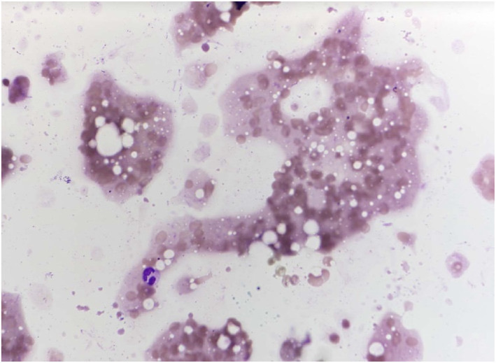
Normal Salivary Gland and Non-Neoplastic Salivary Lesions in Cytology
Definition. Non-neoplastic salivary lesions are inflammatory, degenerative, cystic and lymphoid processes that enlarge a salivary gland without a neoplasm. In the Milan System for Reporting Salivary Gland Cytopathology (MSRSGC), 2nd edition, they are reported in the non-neoplastic category (category II), which has a risk of malignancy (ROM) of 11% (Faquin and Rossi 2023; Rossi 2024). Cysts with mucin and no epithelium are reported as atypia of undetermined significance (AUS). Metastases and lymphomas that involve the gland are reported as suspicious for malignancy or malignant. The categories themselves are described under the Milan System for Reporting Salivary Gland Cytopathology (The Milan System for Reporting Salivary Gland Cytopathology).
Normal salivary gland components in fine-needle aspirates
Normal salivary tissue is a common finding in aspirates of small nodules and of glands that are only diffusely enlarged. Its presence alone does not explain a discrete mass. In a series of 201 non-neoplastic aspirates, 12 contained normal salivary gland only and 5 contained fatty infiltration (Malhotra 2005).
| Component | Cytomorphology | Significance |
|---|---|---|
| Serous acinar cells | Grape-like acinar clusters attached to small ducts; abundant finely granular or vacuolated cytoplasm; small round basal nuclei | Normal parotid parenchyma; individual acinar cells resemble acinic cell carcinoma |
| Mucous acinar cells | Clusters of cells with pale, foamy, mucin-filled cytoplasm | Normal submandibular and sublingual parenchyma |
| Ductal cells | Flat cohesive honeycomb sheets and tubules of cuboidal cells with dense cytoplasm and uniform nuclei | Normal ducts; prominent in sialadenitis with acinar atrophy |
| Oncocytes | Cells with abundant granular eosinophilic cytoplasm | Oncocytic metaplasia increases with age; the differential includes oncocytoma and Warthin tumor |
| Adipose tissue | Mature fat in fragments or single adipocytes | Normal stroma; abundant in older patients and in lipomatosis |
| Lymphoid cells | Polymorphous small and large lymphocytes, tingible-body macrophages | Intraparotid lymph node or lymphoid stroma of a lesion |
| Skeletal muscle | Striated fibers | Contaminant from the masseter or sternocleidomastoid |
Sialadenitis in salivary gland cytology
Malhotra 2005 classified 146 of 201 non-neoplastic aspirates as inflammatory: 42 acute, 89 chronic and 15 granulomatous. The acute group included abscesses, acute obstructive sialadenitis and acute infective sialadenitis. The granulomatous group included 10 cases of tuberculosis, 3 of sarcoidosis and 2 foreign-body granulomas (Malhotra 2005).
| Type | Cytomorphology | Main mimic |
|---|---|---|
| Acute sialadenitis and abscess | Neutrophils, necrotic debris, bacteria; few degenerated acinar or ductal cells | Necrotic carcinoma; infarcted Warthin tumor |
| Chronic sialadenitis | Low cellularity; cohesive ductal groups; scant or absent acini; lymphocytes and plasma cells; fibrous stromal fragments; sometimes squamous or mucous metaplasia | Low-grade mucoepidermoid carcinoma; basaloid neoplasm |
| Chronic sclerosing sialadenitis (Küttner tumor) | Tubular ductal structures surrounded by collagen or lymphoplasmacytic cells, stromal fragments, lymphoid background, few or no acini | Carcinoma, especially in patients with a history of head and neck cancer |
| Granulomatous sialadenitis | Epithelioid histiocytes in aggregates, multinucleated giant cells; caseous necrosis in tuberculosis | Necrotizing carcinoma; metastatic squamous cell carcinoma with keratin granulomas |
| Lymphoepithelial sialadenitis | Polymorphous lymphoid cells with clusters of ductal cells infiltrated by lymphocytes | Extranodal marginal zone (MALT) lymphoma |
- Chronic sclerosing sialadenitis presents as a hard submandibular mass that clinically mimics a neoplasm. Cheuk 2002 described 7 cases: 6 men and 1 woman, aged 47–72 years, 3 with bilateral disease. The aspirates were paucicellular to moderately cellular and lacked acini (Cheuk 2002).
- History of malignancy is a source of error. Of 4 patients with chronic sclerosing sialadenitis, 3 had a prior squamous cell carcinoma. Their aspirates were interpreted as squamous cell carcinoma, basaloid neoplasm or salivary gland neoplasm. Resection showed sialadenitis in all 4 (Leon 2016).
- Küttner tumor and IgG4-related disease: in 13 cases of chronic sclerosing sialadenitis, 11 of 12 evaluable cases had increased IgG4-positive plasma cells (mean 229 per high-power field; IgG4/IgG ratio 0.86), and 3 patients had systemic IgG4-related disease (Geyer 2010). Chronic sclerosing sialadenitis is therefore regarded as a manifestation of IgG4-related disease in most cases. The consensus histologic thresholds for the salivary glands are more than 100 IgG4-positive plasma cells per high-power field and an IgG4/IgG ratio above 40%, with serum IgG4 as supporting evidence (Deshpande 2012).
- Sialolith-associated chronic sialadenitis is a separate process. Of 129 resected submandibular glands with chronic sialadenitis, 3 exceeded 50 IgG4-positive plasma cells per high-power field. In those 3, the IgG4 cells were part of a non-specific infiltrate around ducts that had contained sialoliths (Harrison 2013).
- Interlaboratory performance: in the College of American Pathologists nongynecologic program, granulomatous sialadenitis was among the benign entities with a high false-positive rate (Hughes 2005).
Sialadenosis in salivary gland cytology
Sialadenosis is a non-inflammatory, non-neoplastic, usually bilateral and recurrent enlargement of the parotid glands, associated with hormonal, nutritional and neurogenic disorders. Smears are cellular, with enlarged acini in clusters, numerous naked nuclei from disrupted acinar cells and few or no inflammatory cells. The main mimic is acinic cell carcinoma: sialadenosis retains the lobular acinar arrangement with attached ducts, and acinic cell carcinoma forms sheets and loose clusters of acinar-type cells without ducts. Sialadenosis is reported as non-neoplastic and does not require surgery (Ascoli 1993).
Crystals in salivary gland aspirates
Crystalloids were found in 37 of 664 screened salivary aspirates (5.6%). The full study set of 91 aspirates with crystalloids comprised these 37 screened cases and 54 archival cases. Amylase crystalloids formed 75% of these, tyrosine crystalloids 11% and collagenous crystalloids 3% (Sun 2021).
| Crystal | Morphology and stains | Associated lesions |
|---|---|---|
| Amylase crystalloids | Geometric, rectangular to rhomboid, non-birefringent; visible with Romanowsky and Papanicolaou stains; amylase activity confirms the nature of the material | Sialadenitis, obstructed ducts, cysts, oncocytic cysts and Warthin tumor |
| Tyrosine-rich crystals of oncocytic lesions | Needle-shaped or tabular, pink on H&E; not birefringent; red-brown with Millon reagent; PAS, trichrome, Congo red and alcian blue negative | Warthin tumor, oncocytic cystadenoma and oncocytoma |
| Tyrosine-rich and collagenous crystalloids of pleomorphic adenoma | Radiating crystalloids within or near the tumor | Pleomorphic adenoma |
Sources: Boutonnat 2000; Gilcrease 1998; Sun 2021.
- Diagnostic categories in the crystal series: of 91 aspirates with crystalloids, 30 (33%) were non-diagnostic, 42 (46%) non-neoplastic or benign, 10 (11%) benign neoplasm and 9 (10%) AUS (Sun 2021).
- Histologic follow-up: oncocytic cyst or cystadenoma, pleomorphic adenoma, developmental cyst, lymphoepithelial cyst and Warthin tumor were found at resection (Sun 2021).
- Significance: crystalloids favor a benign process. Their presence does not establish the category on its own. The category is assigned from the cellular findings (Sun 2021).
Cystic salivary lesions and the cystic neck mass
Cyst fluid is frequently hypocellular, and many neoplasms show cystic change. Basal cell adenoma, canalicular adenoma, oncocytoma, sebaceous adenoma, intraductal papilloma, epithelial-myoepithelial carcinoma, intraductal carcinoma and secretory carcinoma can present as cystic lesions (Vegni 2023). Mucoepidermoid carcinoma and Warthin tumor are the most frequent cystic neoplasms. They are described under mucoepidermoid carcinoma (Mucoepidermoid Carcinoma) and Warthin tumor (Warthin Tumor).
| Lesion | Cyst contents and cells | Milan category |
|---|---|---|
| Mucus retention cyst and mucocele | Thin or thick mucin, muciphages, few ductal cells | AUS for mucin without epithelium from a major gland; mucin from a minor gland favors a mucocele |
| Lymphoepithelial cyst | Anucleate and nucleated squames, keratinous debris, polymorphous lymphocytes | Non-neoplastic |
| Branchial cleft cyst | Mature and degenerated squamous cells, keratin debris, inflammatory cells, cholesterol crystals | Not a salivary lesion; correlation with HPV status in adults |
| Cystic metastatic HPV-associated squamous cell carcinoma | Basaloid nonkeratinizing tumor cells, often few, in cyst debris | Malignant or suspicious for malignancy |
| Non-mucinous cyst fluid without epithelium | Watery proteinaceous fluid, histiocytes, lymphocytes, debris | Non-diagnostic |
- Lymphoepithelial cysts in HIV infection: of 495 salivary aspirates from HIV-positive patients, 115 (23.2%) were benign lymphoepithelial cysts and 168 (33.9%) were reactive lymphadenopathy. Lymphoma accounted for 13 aspirates and Kaposi sarcoma for 3 (Michelow 2012).
- Lymphoepithelial cysts also occur without HIV infection (Choudhury 2023).
- Minor salivary gland mucinous lesions: mucoceles are common in the minor glands. The high prevalence of mucoceles at this site favors a benign interpretation of mucin in a minor gland aspirate (Velez Torres 2024).
Cystic squamous lesions of the lateral neck
The differential diagnosis of a lateral neck cyst in an adult includes a branchial cleft cyst and a cystic nodal metastasis of HPV-associated oropharyngeal carcinoma. The parotid tail and upper neck overlap, so the aspirate can be labeled as parotid.
- Cystic metastases among excised branchial cleft cysts: of 436 excised lateral branchial cleft cysts, 13 (3%) were cystic metastases. In patients older than 18 years the rate was 3.3% (Hammarstedt Nordenvall 2022).
- HPV DNA testing of the aspirate: all 176 cysts finally diagnosed as branchial cleft cysts were HPV DNA negative. HPV DNA was present in 100 of 128 (78.1%) aspirates of solitary cystic neck metastases (Jörtsö 2025).
- p16 immunostaining: metastatic oropharyngeal nonkeratinizing squamous cell carcinoma showed diffuse, strong p16 staining. Branchial cleft cysts showed only focal staining of the superficial epithelium. Keratinizing carcinomas of the larynx and oral cavity were p16 negative, so a negative result does not exclude carcinoma (Pai 2009).
Lymphoid lesions of the salivary glands in cytology
The parotid contains intraparotid lymph nodes, and lymphoid tissue accompanies Warthin tumor, lymphoepithelial cysts and several carcinomas.
- Intraparotid lymph node: polymorphous lymphocytes with tingible-body macrophages and lymphohistiocytic aggregates. In the College of American Pathologists program, intraparotid lymph node had a 36% false-positive rate (Hughes 2005).
- Lymphoepithelial sialadenitis occurs in Sjögren syndrome and HIV infection. Lymphocytes infiltrate ductal cell clusters. Primary Sjögren syndrome is associated with a markedly increased risk of non-Hodgkin lymphoma (pooled standardized incidence ratio 18.8) (Zintzaras 2005).
- Extranodal marginal zone lymphoma (MALT lymphoma) arises in a background of lymphoepithelial sialadenitis. It shows a monotonous population of small to intermediate lymphoid cells, monocytoid cells and plasma cells. Flow cytometry or immunostains on cell block establish clonality.
- Flow cytometry with FNA: in 1,402 head and neck aspirates with combined cytology and flow cytometry, sensitivity for non-Hodgkin lymphoma was 95.5% and specificity 99.9%. Only 7 of 13 Hodgkin lymphomas were detected. Rapid on-site evaluation (ROSE) lowered the rate of samples with too few cells for flow cytometry from 16.4% to 7.0% (Crous 2019).
- Report: a reactive lymph node is non-neoplastic. A clonal lymphoid population is reported as malignant. Atypical lymphoid cells without clonality studies are reported as AUS.
Metastases to the salivary glands in cytology
Metastases represent about 8% of malignant parotid lesions, and about 80% arise from squamous cell carcinomas of the head and neck region (Calo 2024). The institutional series below reflects a referral population, in which metastases formed a larger share of malignant aspirates. Most reach the parotid through intraparotid lymph nodes.
- Institutional series: of 630 parotid aspirates, 80 (12.4%) were malignant and 53 of these were metastases. Melanoma (24%), squamous cell carcinoma (22.6%) and renal cell carcinoma (19%) were the most frequent. Breast, lung, intestinal and testicular carcinomas, solitary fibrous tumor and Merkel cell carcinoma also occurred (Calo 2024).
- Milan categories: 48 of the 53 metastases were classified as malignant and 5 as suspicious for malignancy. A known primary tumor was present in 40 (75.4%) patients (Calo 2024).
- Immunocytochemistry: clinical history, cytomorphology and immunocytochemistry on cell blocks identified all 53 metastases (Calo 2024).
| Metastasis | Cytologic features | Useful markers |
|---|---|---|
| Cutaneous squamous cell carcinoma | Keratinized cells, keratin debris, necrosis | p40, p63, CK5/6; no MAML2 rearrangement |
| Melanoma | Discohesive epithelioid or spindle cells, macronucleoli, intranuclear pseudoinclusions, pigment | SOX10, S100, Melan-A, HMB-45, PRAME |
| Merkel cell carcinoma | Small round cells, nuclear molding, finely granular chromatin | CK20 (dot-like), synaptophysin, INSM1 |
| Renal cell carcinoma, clear cell type | Clear or vacuolated cytoplasm, vascular fragments | PAX8, CA9, CD10 |
| Breast carcinoma | Ductal cells in clusters and single files | GATA3, ER; GATA3 is also positive in salivary duct carcinoma |
Differential diagnosis of non-neoplastic salivary findings that mimic neoplasms
| Non-neoplastic finding | Neoplasm it mimics | Separating features |
|---|---|---|
| Normal acini | Acinic cell carcinoma | Lobular acini attached to ducts; low cellularity in proportion to the lesion |
| Sialadenosis | Acinic cell carcinoma | Bilateral diffuse enlargement; enlarged acini with ducts; naked nuclei |
| Chronic sialadenitis with squamous metaplasia | Mucoepidermoid carcinoma, squamous cell carcinoma | Inflammation, fibrosis, bland metaplastic cells, absent acini |
| Chronic sclerosing sialadenitis | Basaloid neoplasm, carcinoma | Ducts surrounded by collagen; lymphoplasmacytic background |
| Reactive intraparotid lymph node | Lymphoma, Warthin tumor, acinic cell carcinoma with lymphoid stroma | Polymorphous lymphocytes; tingible-body macrophages; no oncocyte sheets |
| Lymphoepithelial cyst | Warthin tumor, cystic squamous cell carcinoma | Squames and lymphocytes without oncocytes; bland squamous cells |
| Granulomatous sialadenitis | Necrotic carcinoma | Epithelioid histiocytes and giant cells; cultures and special stains |
| Mucus retention cyst | Low-grade mucoepidermoid carcinoma | Mucin with muciphages only; reported as AUS in major glands |
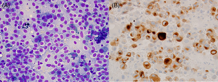
Salivary Gland Fine-Needle Aspiration Technique, Diagnostic Accuracy and Ancillary Testing
Definition. Salivary gland fine-needle aspiration (FNA) is the sampling of a salivary mass with a fine needle for cytologic diagnosis, usually under ultrasound guidance. The material supports smears, liquid-based preparations, cell blocks, flow cytometry and molecular tests. The Milan System for Reporting Salivary Gland Cytopathology (MSRSGC), 2nd edition, reports the result in six categories and recommends ancillary testing to reclassify a case into a more specific category (Faquin and Rossi 2023; Rossi 2024). The categories are described under the Milan System for Reporting Salivary Gland Cytopathology (The Milan System for Reporting Salivary Gland Cytopathology).
Imaging before salivary gland FNA
- Ultrasound is the first imaging test for a superficial salivary mass and guides the needle to the solid or most suspicious part of a lesion.
- MRI defines deep-lobe extension, perineural spread and the relation to the facial nerve (Faquin and Rossi 2023).
- Diffusion-weighted MRI: the mean apparent diffusion coefficient reflects cellularity and histologic type. Combining diffusion-weighted imaging with other MRI sequences improves the separation of benign and malignant parotid tumors (Kato 2023).
- Imaging and cytology are interpreted together. The Milan System judges adequacy in the clinical and radiologic setting and recommends radiologic correlation for non-diagnostic and non-neoplastic results (Faquin and Rossi 2023).
FNA technique and specimen preparation for salivary gland lesions
- Sampling: several passes with a fine needle, by aspiration or capillary technique. Cystic lesions are drained and any residual solid component is resampled under ultrasound.
- Direct smears: air-dried smears stained with a Romanowsky stain show matrix, mucin and cytoplasmic granules. Alcohol-fixed Papanicolaou-stained smears show nuclear detail and keratinization.
- Needle rinse: the rinse is used for a cell block, a liquid-based preparation, flow cytometry or molecular testing.
- Complications: no complications occurred after FNA in a meta-analysis of six studies that compared FNA with core needle biopsy (Cho 2020). Infarction of Warthin tumor after FNA produces necrosis, squamous metaplasia and fibrosis, which can mimic carcinoma at resection (Stárek 2002).
| Preparation | Strengths | Limitations |
|---|---|---|
| Air-dried smear, Romanowsky stain | Shows matrix (fibrillary, hyaline globules), mucin, zymogen granules; used for ROSE | Nuclear detail is less sharp |
| Alcohol-fixed smear, Papanicolaou stain | Nuclear chromatin, nucleoli, keratinization | Matrix is pale; mucin is less visible |
| Liquid-based preparation (ThinPrep, SurePath) | Clean background; residual material for immunocytochemistry | Matrix fragments and mucin are altered and cells appear smaller |
| Cell block | Immunohistochemistry, in situ hybridization, FISH, sequencing | Depends on cellularity; a non-contributory block suggests a sampling problem |
| Flow cytometry | Clonality of lymphoid populations | Needs a dedicated pass in a cell-preserving medium such as RPMI; marginal zone lymphoma is often missed or nondiagnostic (Crapanzano 2003) |
Sources: Faquin and Rossi 2023; Rarick 2014; Tabibi 2022; Straccia 2026.
Rapid on-site evaluation of salivary gland FNA
- Agreement with the final diagnosis: of 386 salivary FNAs with ROSE, agreement between ROSE and the final cytologic diagnosis was good to excellent (simple kappa 0.76; linear weighted kappa 0.81). Major discrepancies that changed management occurred in 10 (2.7%) cases (Wangsiricharoen 2017).
- Effect on non-diagnostic rate: in a two-center series of 549 patients, one center had a non-diagnostic rate of 16.2% and the center that used ROSE had none. The use of ROSE was associated with fewer non-diagnostic results and higher FNA accuracy (Xu 2024).
- Triage for flow cytometry: in head and neck lymphoid lesions, ROSE lowered the rate of samples with too few cells for flow cytometry from 16.4% to 7.0% (Crous 2019).
Liquid-based cytology in salivary gland FNA
- Morphologic differences: ThinPrep preparations of salivary aspirates show specific artifacts in cells, extracellular material and architecture. With knowledge of these differences, ThinPrep is used without loss of accuracy in adequately cellular samples (Rarick 2014).
- Performance with the Milan System: in 129 SurePath cases with histologic follow-up, sensitivity and specificity were 95.1% and 88.8% by one method of calculation and 81% and 77.7% by a second (Sadullahoğlu 2019).
- Ancillary use: liquid-based preparations and cell blocks show mucin and glandular differentiation in oncocytic aspirates (Araneta 2026).
Diagnostic accuracy of salivary gland FNA, core needle biopsy and frozen section
| Method | Evidence | Result |
|---|---|---|
| FNA, malignancy | Meta-analysis, 64 studies, 6,169 cases | Area under the summary ROC curve 0.96; for neoplasia 0.99 (Schmidt 2011a) |
| FNA, parotid, prospective studies | Meta-analysis, 63 studies with data, 5,647 patients | Sensitivity 88.2% (95% CI 50.9–98.2%), specificity 99.5%; non-diagnostic 5.3%, indeterminate 14.7% (Liu 2016) |
| FNA with the Milan System | Meta-analysis, 35 studies, 10,706 FNAs | ROM by category (I, II, III, IVA, IVB, V, VI): 11.4%, 10.9%, 30.5%, 2.8%, 37.7%, 83.8%, 97.7% (Wang 2022) |
| FNA, interlaboratory surveys | 6,249 participant responses | Sensitivity 73%, specificity 91% (Hughes 2005) |
| Ultrasound-guided core needle biopsy | Meta-analysis, 5 studies, 277 cases | Sensitivity 92%, specificity 100%, inadequacy 1.2% (Schmidt 2011b) |
| Core needle biopsy versus FNA | Meta-analysis, 6 studies, 1,924 procedures | Fewer non-diagnostic results and higher sensitivity and specificity with core biopsy; hematoma in 0.3% of core biopsies (Cho 2020) |
| Core needle biopsy, head and neck | Meta-analysis, 16 studies, 1,291 lesions | Accuracy 94% for neoplasia and 96% for malignancy (Novoa 2012) |
| Frozen section, parotid | Meta-analysis, 13 studies, 1,880 cases | Sensitivity 90%, specificity 99% (Schmidt 2011c) |
- FNA identifies a neoplasm more accurately than it identifies malignancy (Schmidt 2011a).
- Heterogeneity is high. Pooled estimates vary across studies and centers, and the I² statistic exceeded 70% in most analyses (Liu 2016).
- Core needle biopsy is a selective second step after a non-diagnostic, AUS or SUMP result (Straccia 2026).
- Frozen section is accurate and consistent across centers. Its result determines the extent of resection during surgery (Schmidt 2011c).
Interobserver reproducibility of the Milan System
- MIRST: 555 qualified participants reviewed 75 images before the 2018 publication. Overall agreement with the reference interpretation was 42%. Agreement was highest for benign neoplasm (58.9%) and non-diagnostic (49.2%), and lowest for SUMP (23.6%) and suspicious for malignancy (22.7%) (Kurtycz 2020).
- Basaloid neoplasms: average agreement among seven cytopathologists was 46% for 45 basaloid aspirates (Layfield 2022).
Immunohistochemistry on salivary gland cell blocks
Cell-block studies improved the diagnosis in 6 of 10 non-neoplastic cases, 6 of 6 benign neoplasms and 59 of 60 malignant cases in a three-institution series (Tabibi 2022).
| Marker | Expected pattern | Main use |
|---|---|---|
| PLAG1 | Nuclear; positive in 96% of pleomorphic adenomas | Pleomorphic adenoma and carcinoma ex pleomorphic adenoma; a negative result makes pleomorphic adenoma unlikely (Katabi 2018) |
| HMGA2 | Nuclear | Pleomorphic adenoma without PLAG1 alteration (Jo and Krane 2018) |
| MYB | Nuclear; 80% of adenoid cystic carcinomas positive on smears, all pleomorphic adenomas negative | Adenoid cystic carcinoma (Pusztaszeri 2014) |
| β-catenin | Nuclear; 82% of basal cell adenomas | Basal cell adenoma (Jo 2016) |
| LEF1 | Nuclear | Favors basal cell adenoma or pleomorphic adenoma against adenoid cystic carcinoma (Schmitt 2017) |
| NR4A3 | Nuclear; specific for acinic cell carcinoma | Acinic cell carcinoma (Haller 2019b; Nguyen 2020) |
| DOG1 | Apical or canalicular membranous | Acinic cell carcinoma; negative in Warthin tumor and oncocytoma (Schmitt 2014) |
| Pan-TRK | Diffuse nuclear in secretory carcinoma; focal nuclear staining (<10% of cells) occurs in some other tumors, and pleomorphic adenoma shows membranous or cytoplasmic staining | Secretory carcinoma (Hung 2019) |
| S100, mammaglobin | Diffuse | Secretory carcinoma (Wiles 2022) |
| Androgen receptor | Nuclear | Salivary duct carcinoma (Moriki 2001) |
| HER2 | Membranous; ERBB2 amplification | High-grade salivary duct carcinoma; trastuzumab eligibility (Stevens 2015; Takahashi 2019) |
| RAS Q61R | Membranous and cytoplasmic in myoepithelial cells | Epithelial-myoepithelial carcinoma (Nakaguro 2021) |
| p63, p40, SOX10 | Nuclear | Myoepithelial and basal cells; SOX10 negative in benign oncocytic tumors (Schmitt 2015) |
Molecular alterations used in salivary gland FNA
| Alteration | Tumor | Detection on FNA |
|---|---|---|
| PLAG1 or HMGA2 rearrangement | Pleomorphic adenoma; carcinoma ex pleomorphic adenoma | FISH; PLAG1 and HMGA2 immunostains (Katabi 2015) |
| CRTC1::MAML2, CRTC3::MAML2 | Mucoepidermoid carcinoma | MAML2 FISH or RNA sequencing (Tonon 2003) |
| MYB::NFIB, MYBL1::NFIB | Adenoid cystic carcinoma | MYB FISH; MYB immunostain (Persson 2009) |
| Enhancer hijacking of NR4A3 by t(4;9)(q13;q31) | Acinic cell carcinoma | NR4A3 immunostain (Haller 2019a) |
| ETV6::NTRK3 | Secretory carcinoma | ETV6 FISH; pan-TRK immunostain (Skálová 2010) |
| EWSR1::ATF1 | Hyalinizing clear cell carcinoma | EWSR1 FISH (Tanguay 2013) |
| PRKD1 E710D; PRKD1, PRKD2 or PRKD3 rearrangement | Polymorphous adenocarcinoma, classic and cribriform subtypes | Sequencing of smears (Andreasen 2018); FISH (Weinreb 2014) |
| HRAS codon 61 mutation | Epithelial-myoepithelial carcinoma | Sequencing; RAS Q61R immunostain (Urano 2019) |
| CTNNB1 mutation | Basal cell adenoma | Sequencing; nuclear β-catenin (Jo 2016) |
| NCOA4::RET | Intercalated duct-type intraductal carcinoma | RNA sequencing (Viswanathan 2021) |
- MAML2 rearrangement and grade: the translocation was present in 75% of low- and intermediate-grade and 46% of high-grade mucoepidermoid carcinomas, and in none of 36 mimics (Seethala 2010). A negative MAML2 result therefore does not exclude high-grade mucoepidermoid carcinoma.
- FISH on cell blocks: of 51 salivary FNAs with FISH for PLAG1, MYB, MAML2 or ETV6, 15 were subclassified after testing. All subclassified cases with excision had concordant FNA, FISH and excision diagnoses (Darras 2019).
- Next-generation sequencing: in an ex vivo study of 32 tumors, a hotspot panel detected somatic mutations with a sensitivity of 71% and specificity of 94% for malignancy. Cytology alone had a sensitivity of 64% and specificity of 100% in the same cases (Yamamoto 2020).
- WHO 5th edition definitions include the recurrent molecular alterations of mucoepidermoid carcinoma, adenoid cystic carcinoma, secretory carcinoma, polymorphous adenocarcinoma, hyalinizing clear cell carcinoma, mucinous adenocarcinoma and microsecretory adenocarcinoma (Skálová 2022).
- Ancillary testing and category: ancillary studies allow a specific diagnosis in many SUMP and suspicious aspirates (Jo and Krane 2018; Dubucs 2019).
Other reporting systems for salivary gland cytology
| System | Categories | Relation to the Milan System |
|---|---|---|
| WHO Reporting System for Head and Neck Cytopathology (IAC-IARC-WHO) | Insufficient/inadequate/non-diagnostic; benign; atypical; neoplasm of uncertain malignant potential; suspicious for malignancy; malignant | Merges the Milan non-neoplastic and benign neoplasm categories into one benign category (Kowalewski 2025a) |
| Osaka Medical College (OMC) classification | 11 categories that include histologic type and malignancy grade | Adds histologic type and grade to the category (Taniuchi 2021) |
| IAC-IARC-WHO Reporting System for Soft Tissue Cytopathology | Soft tissue categories | Applied to mesenchymal tumors of the salivary glands (Kowalewski 2025b) |
- WHO system and Milan System compared: in 2,218 salivary FNAs, 1,356 with histology, the WHO benign category had a ROM of 7.1%. This category combines the Milan non-neoplastic category (ROM 16.8%) and benign neoplasm category (ROM 4.3%). Excluding non-diagnostic and indeterminate neoplasm categories, both systems had a sensitivity of 93.3% and a specificity of 93.9% (Kowalewski 2025a).
- OMC classification: of 1,175 patients classified by the OMC system, 729 (62.0%) were placed in the benign histologic type category, with an accuracy of 93.4%. Malignancy grade was assigned in 87 (7.4%) patients with an accuracy of 88.5% (Taniuchi 2021; Taniuchi 2022).
- Soft tissue and bone tumors accounted for 86 of 2,219 (3.9%) salivary FNAs. FNA correctly identified 42 of 44 malignant tumors (95.5%) (Kowalewski 2025b).
Salivary gland FNA in special sites and age groups
Minor salivary gland FNA
Minor salivary gland aspirates come from the palate, buccal space, floor of mouth, lip, tongue and maxillary sinus. In 43 minor gland aspirates classified by the Milan System, the risk of neoplasm was 87% and the ROM 39%; SUMP (15 cases) was the most frequent category. Mucoceles are frequent in the minor glands, so mucin alone at this site favors a benign process (Velez Torres 2024).
Pediatric salivary gland FNA
Of 477 pediatric aspirates from 22 institutions, 34.6% were non-neoplastic, 27.5% benign neoplasm, 12.4% malignant and 10.3% non-diagnostic. The ROM was 5.9% for non-diagnostic, 9.1% for non-neoplastic, 35.7% for AUS, 3.3% for benign neoplasm, 31.8% for SUMP and 100% for suspicious for malignancy and malignant. Mucoepidermoid carcinoma was the most frequent malignancy and acinic cell carcinoma the second; pleomorphic adenoma was the most frequent benign neoplasm (Maleki 2022).
Oncocytic and Basaloid Salivary Gland Lesions in Cytology
Definition. Oncocytic and basaloid lesions are two cytomorphologic patterns of salivary gland aspirates. Each pattern contains benign and malignant tumors that share cell types. Oncocytic cells contain abundant granular eosinophilic cytoplasm packed with mitochondria. Oncocytoid cells have abundant cytoplasm of other origin, such as zymogen granules or vacuoles. Basaloid cells are small cells with scant cytoplasm and a high nuclear-to-cytoplasmic ratio (Lubin 2019; Cantley 2019). An aspirate that shows a neoplasm of either pattern without the features of a specific entity is reported in the Milan category salivary gland neoplasm of uncertain malignant potential (SUMP) (Faquin and Rossi 2023). The cytology of Warthin tumor, pleomorphic adenoma, adenoid cystic carcinoma and mucoepidermoid carcinoma is described in their own units (Warthin Tumor, Pleomorphic Adenoma, Adenoid Cystic Carcinoma, Mucoepidermoid Carcinoma).
Oncocytic and oncocytoid salivary lesions in cytology
True oncocytic lesions include oncocytosis (nodular oncocytic hyperplasia), oncocytoma and oncocytic carcinoma. Oncocytoid lesions include acinic cell carcinoma, secretory carcinoma, mucoepidermoid carcinoma with oncocytic change and salivary duct carcinoma (Lubin 2019). In the WHO 5th edition, oncocytic carcinoma is placed in the category of salivary carcinoma not otherwise specified and emerging entities (Skálová 2022).
| Entity | Architecture | Cells | Background | Usual Milan category |
|---|---|---|---|---|
| Oncocytosis and oncocytoma | Compact globoid clusters ("bunch of grapes"); fibrovascular cores | Uniform oncocytes with granular cytoplasm and central round nuclei | Clean | Benign neoplasm when classic; SUMP when cellular without context |
| Warthin tumor | Cohesive flat sheets | Oncocytes in honeycomb arrangement | Lymphocytes and granular cystic debris | Benign neoplasm |
| Oncocytic mucoepidermoid carcinoma | Discohesive clusters and single cells | Oncocytoid cells; rare intracytoplasmic mucin; intermediate cells | Mucin, cyst debris | SUMP, suspicious or malignant |
| Warthin-like mucoepidermoid carcinoma | Sheets in a cystic lymphoid background | Squamoid and mucous cells; bilayered oncocytes absent | Lymphocytes, mucin | Often misclassified as benign neoplasm (Warthin tumor); SUMP, suspicious or malignant when recognized |
| Acinic cell carcinoma | Sheets, acinar groups, naked nuclei | Polygonal cells with granular or vacuolated cytoplasm | Clean or lymphoid | SUMP, suspicious or malignant |
| Secretory carcinoma | Papillary groups, clusters, single cells | Vacuolated cytoplasm | Mucin or proteinaceous material | SUMP, suspicious or malignant |
| Oncocytic carcinoma and oncocytic salivary duct carcinoma | Clusters with necrosis | Pleomorphic oncocytoid cells, prominent nucleoli | Necrosis | Suspicious or malignant |
Sources: Araneta 2026; Ishibashi 2015; Lubin 2019; Saoud 2024.
- Cohesion separates benign from low-grade malignant oncocytic tumors. In 49 aspirates with surgical follow-up, all 43 benign neoplasm diagnoses were confirmed as Warthin tumor or oncocytoma. Warthin tumor showed cohesive flat clusters with cystic debris. Oncocytoma showed compact globoid clusters in a clean background. Oncocytic mucoepidermoid carcinoma formed discohesive clusters and single cells, and cell blocks showed focal glands and rare intracytoplasmic mucin (Araneta 2026).
- Warthin-like mucoepidermoid carcinoma: of 15 tumors first diagnosed as metaplastic Warthin tumor, 5 had CRTC1::MAML2. The bilayered oncocytic epithelium of Warthin tumor was always present in fusion-negative tumors and absent in fusion-positive tumors (Ishibashi 2015).
- Acinic cell carcinoma shows large cells with abundant cytoplasm and is a recognized mimic of oncocytoma and Warthin tumor (Saoud 2024).
- Interlaboratory performance: oncocytoma had an 18% false-positive rate for malignancy in College of American Pathologists surveys (Hughes 2005).
Immunohistochemistry of oncocytic salivary lesions on cell block
| Marker | Warthin tumor | Oncocytoma | Acinic cell carcinoma | Mucoepidermoid carcinoma |
|---|---|---|---|---|
| DOG1 | Negative (100%) | Negative | Positive in 70% of cell blocks and 93% of resections | Variable, often negative |
| p63 | Positive in 87.5% of cell blocks | Basal cells positive | Negative (100% of cell blocks) | Positive in squamoid and intermediate cells |
| SOX10 | Negative | Negative | Positive in 61.5% of cell blocks; all resections | Variable, often negative |
| S100 | Negative | Negative | Positive in fewer than 10% | Negative |
| MAML2 rearrangement | Absent | Absent | Absent | Present in most |
Sources: Schmitt 2014; Schmitt 2015; Ishibashi 2015.
- SOX10 and DOG1 identify acinic cell carcinoma. Neither marker was expressed by benign oncocytic tumors on cell block or resection (Schmitt 2015).
- A negative SOX10 and DOG1 result does not establish a benign tumor, because mucoepidermoid carcinoma is often negative for both markers (Schmitt 2015).
- NR4A3 and pan-TRK identify acinic cell carcinoma and secretory carcinoma. These carcinomas are described under other salivary gland carcinomas in cytology (Acinic Cell, Secretory, Salivary Duct and Other Salivary Gland Carcinomas in Cytology).
Basaloid salivary neoplasms in cytology
Basaloid neoplasms show monotonous and usually bland basaloid epithelium. They are separated by a second cell population such as myoepithelial cells, by the type of acellular matrix and by cytologic atypia (Cantley 2019). Pleomorphic adenoma and adenoid cystic carcinoma have characteristic findings that allow a specific diagnosis. Other basaloid tumors are frequently classified only as SUMP (Cantley 2019). Basal cell adenoma and basal cell adenocarcinoma cannot be separated on FNA, because the distinction rests on invasion; both are classified as SUMP, basaloid subtype (Faquin and Rossi 2023).
| Entity | Cells | Matrix | Key separating findings |
|---|---|---|---|
| Cellular pleomorphic adenoma | Basaloid epithelial and plasmacytoid or spindle myoepithelial cells | Fibrillary chondromyxoid stroma with embedded cells | PLAG1 or HMGA2 positive |
| Basal cell adenoma | Monotonous basaloid cells; three-dimensional and sharp-angled clusters; peripheral palisading | Hyaline basement membrane-like material in and around clusters | Nuclear β-catenin; CTNNB1 mutation |
| Basal cell adenocarcinoma | Basaloid cells with more atypia, mitoses or necrosis | Hyaline material | Invasion on histology; cytology overlaps with basal cell adenoma |
| Adenoid cystic carcinoma | Basaloid cells with angular hyperchromatic nuclei | Sharply outlined hyaline globules and cylinders | MYB strong and diffuse; MYB::NFIB |
| Canalicular adenoma | Cuboidal to columnar cells in beaded cords | Myxoid stroma | Upper lip of older women; S100, GFAP and SOX10 positive |
| Epithelial-myoepithelial carcinoma | Ductal cells with clear myoepithelial cells; naked nuclei | Hyaline or basement membrane material | HRAS codon 61 mutation |
| Sebaceous lymphadenoma | Scant basaloid clusters with rare sebaceous cells | None | Predominant lymphoid background |
Sources: Cantley 2019; Jo 2016; Kawahara 2007; Sheldon 2023; Thompson 2015.
- Basal cell adenoma, solid type: three-dimensional clusters were present in 71% of cases, sharp-angled small clusters in 86%, basement membrane-like material in 71% and crushed cells in 86%. In 3 of 5 cystic basal cell adenomas the aspirate lacked basaloid tumor cells (Kawahara 2007).
- Misdiagnosis as basal cell adenoma: 5 aspirates interpreted as basal cell adenoma proved to be 2 pleomorphic adenomas, 2 lymphoepithelial cysts and 1 basal cell adenocarcinoma (Kawahara 2007).
- Canalicular adenoma is a minor salivary gland tumor. Of 67 cases, 46 involved the upper lip and 13% were multifocal. No major gland examples were found (Thompson 2015).
- Sebaceous neoplasms: sebaceous adenoma shows multivacuolated polygonal cells and allows a specific diagnosis. Sebaceous lymphadenoma shows mainly lymphocytes with scant basaloid clusters and received a non-specific basaloid neoplasm diagnosis (single cases; Sheldon 2023).
- Monomorphic adenoma had a 53% false-positive rate for malignancy in College of American Pathologists surveys (Hughes 2005).
- Interobserver agreement for basaloid neoplasms in the Milan System was 46%, and the SUMP category did not correlate with tumor type (Layfield 2022).
Matrix patterns in basaloid salivary aspirates
| Matrix on Romanowsky stain | Appearance | Tumors |
|---|---|---|
| Fibrillary chondromyxoid | Feathery magenta stroma with frayed edges and embedded myoepithelial cells | Pleomorphic adenoma |
| Hyaline globules and cylinders | Sharply outlined spheres and tubes surrounded by basaloid cells | Adenoid cystic carcinoma; also some basal cell adenomas and epithelial-myoepithelial carcinomas |
| Membranous or basement membrane-like | Dense hyaline material at the edges and within cell clusters | Basal cell adenoma, basal cell adenocarcinoma |
| Absent or minimal matrix | Cellular basaloid groups only | Cellular pleomorphic adenoma, basal cell adenoma, solid adenoid cystic carcinoma |
Sources: Cantley 2019; Kawahara 2007.
Immunohistochemistry of basaloid salivary neoplasms
- Nuclear β-catenin: present in 18 of 22 (82%) basal cell adenomas and absent in all adenoid cystic carcinomas and pleomorphic adenomas. Four of 5 basal cell adenomas tested had the CTNNB1 p.I35T mutation (Jo 2016).
- LEF1: on cell block, 60% of basal cell adenomas and 51% of pleomorphic adenomas were positive, and 91% of adenoid cystic carcinomas were negative. LEF1 positivity favored a benign neoplasm with a specificity of 92% on cell block (Schmitt 2017).
- MYB: 80% of adenoid cystic carcinomas were positive on alcohol-fixed smears, and all pleomorphic adenomas were negative (Pusztaszeri 2014). Strong diffuse nuclear MYB labeling had a sensitivity of 83% and a specificity of 100% for adenoid cystic carcinoma on cytology. Weak or focal labeling also occurred in other basaloid lesions (Sun 2021).
- PLAG1: positive in 96% of pleomorphic adenomas (Katabi 2018).
SUMP subtypes and risk of malignancy for oncocytic and basaloid aspirates
The Milan System subclassifies SUMP as basaloid, oncocytic or oncocytoid, clear cell and mixed or not otherwise specified (Faquin and Rossi 2023).
| Study | SUMP cases with follow-up | Basaloid ROM | Oncocytic ROM | Other subtypes |
|---|---|---|---|---|
| Hang 2018 | 59 | 40.0% | 61.1% (oncocytic and squamoid) | Myoepithelial 18.8% |
| Chowsilpa 2019 | 65 | 38.5% | 7.7% | Overall ROM 33.8%; risk of neoplasm 95.4% |
| Manucha 2021 | 36 | 23% | 58.8% | Basaloid without matrix 8.3%; with nonfibrillary matrix 60% |
| Hang 2022 | 329 | 36.5% | 52.5% | SUMP not otherwise specified 31.9% |
| Xia 2024 | 101 | 17.1% | 20.5% | All clear cell SUMP malignant; overall 20.8% |
- The largest series found a higher ROM for oncocytic SUMP. In 9,938 consecutive aspirates from 10 institutions, 742 (7.5%) were SUMP. The ROM of oncocytic and oncocytoid SUMP (52.5%) was significantly higher than that of basaloid SUMP (36.5%). The type of matrix or background did not change the ROM (Hang 2022).
- Clear cell SUMP had the highest ROM. All clear cell SUMP cases with resection were malignant in a three-institution series (Xia 2024).
- The report states the subtype and the differential diagnosis, for example "SUMP, cellular basaloid neoplasm; differential includes basal cell adenoma, cellular pleomorphic adenoma and adenoid cystic carcinoma" (Faquin and Rossi 2023).
Differential diagnosis of oncocytic and basaloid salivary aspirates
| Finding | Favors benign | Favors malignant |
|---|---|---|
| Cohesion | Cohesive flat sheets or globoid clusters | Discohesive clusters and single cells |
| Background | Clean, or lymphoid with granular cystic debris | Mucin, necrosis |
| Second cell population | Myoepithelial cells in chondromyxoid stroma | Clear myoepithelial cells around ductal cells; intermediate cells |
| Matrix | Fibrillary stroma; basement membrane-like material | Sharply outlined hyaline globules |
| Nuclei | Uniform, smooth | Angular, hyperchromatic, or pleomorphic with prominent nucleoli |
| Markers | PLAG1, nuclear β-catenin, LEF1 | MYB strong and diffuse, NR4A3, nuclear pan-TRK, MAML2 rearrangement |
Sources: Araneta 2026; Cantley 2019; Jo 2016; Schmitt 2017; Sun 2021.
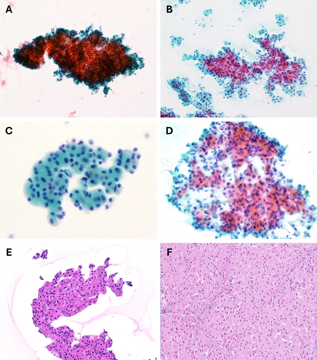
Acinic Cell, Secretory, Salivary Duct and Other Salivary Gland Carcinomas in Cytology
Definition. Salivary carcinomas diagnosed or suspected on fine-needle aspiration (FNA), apart from mucoepidermoid carcinoma and adenoid cystic carcinoma, include acinic cell carcinoma, secretory carcinoma, salivary duct carcinoma, carcinoma ex pleomorphic adenoma, epithelial-myoepithelial carcinoma, myoepithelial carcinoma, polymorphous adenocarcinoma, hyalinizing clear cell carcinoma and intraductal carcinoma. The WHO 5th edition includes the recurrent molecular alterations of several of these tumors in their definitions (Skálová 2022). Low-grade examples are frequently reported as salivary gland neoplasm of uncertain malignant potential (SUMP) or suspicious for malignancy. The Milan System reports a malignant aspirate with the tumor type and grade when possible (Faquin and Rossi 2023). Mucoepidermoid carcinoma and adenoid cystic carcinoma are described in their own units (Mucoepidermoid Carcinoma, Adenoid Cystic Carcinoma).
Grade of salivary carcinomas reported on FNA
| Grade | Carcinomas | Cytologic features |
|---|---|---|
| Usually low grade | Acinic cell carcinoma, secretory carcinoma, epithelial-myoepithelial carcinoma, polymorphous adenocarcinoma, hyalinizing clear cell carcinoma, low-grade intraductal carcinoma | Bland or mildly atypical nuclei, little necrosis; overlap with benign neoplasms |
| Variable | Myoepithelial carcinoma, carcinoma ex pleomorphic adenoma | Depends on the carcinoma type; myoepithelial carcinoma is often bland on cytology |
| Usually high grade | Salivary duct carcinoma, high-grade intraductal carcinoma, carcinomas with high-grade transformation | Pleomorphic nuclei, prominent nucleoli, mitoses, necrosis |
Sources: Faquin and Rossi 2023; Skálová 2022; Argyris 2023; Viswanathan 2021.
Acinic cell carcinoma in cytology
- Molecular basis: a recurrent t(4;9)(q13;q31) places active enhancers upstream of NR4A3 and upregulates this transcription factor (Haller 2019a).
- Cytomorphology: cellular smears of polygonal cells with abundant granular or finely vacuolated cytoplasm and indistinct borders, in sheets, acinar groups and loose clusters. Naked nuclei are frequent. Ducts are absent. The zymogen granules are PAS positive and diastase resistant (Faquin and Rossi 2023). Some tumors have a lymphoid stroma. Acinic cell carcinoma is a known mimic of oncocytoma and Warthin tumor (Saoud 2024).
- NR4A3 immunostaining: nuclear NR4A3 was highly specific and sensitive for acinic cell carcinoma in 64 tumors, including high-grade forms. None of 29 secretory carcinomas or 70 other salivary carcinomas were positive. NR4A3 FISH was less sensitive (84%) (Haller 2019b).
- NR4A3 on FNA cell blocks: all 11 acinic cell carcinomas showed nuclear NR4A3 (NOR-1) staining, and all 59 other tumors were negative (Nguyen 2020).
- DOG1 and SOX10: both are positive in acinic cell carcinoma and negative in benign oncocytic tumors (Schmitt 2015).
- Main mimics: normal acini and sialadenosis (lobular acini with ducts), oncocytoma and Warthin tumor (cohesive oncocytes), and secretory carcinoma (vacuolated cells, S100 and mammaglobin positive).
Secretory carcinoma in cytology
- Definition: a carcinoma with ETV6 rearrangement, most often ETV6::NTRK3, first described as mammary analogue secretory carcinoma. In the original series of 16 tumors, t(12;15)(p13;q25) was present in all analyzable cases but one and absent in all conventional acinic cell carcinomas (Skálová 2010).
- Clinical features: in a multi-institutional series of 40 aspirates, the mean age was 52 years (range 13–80) and 30 tumors arose in the parotid. Nine patients had regional lymph node metastases and 4 had distant metastases (Wiles 2022).
- Cytomorphology: cellular smears of large round cells with intracytoplasmic vacuoles or granules, round to oval nuclei with smooth contours and prominent nucleoli, in clusters, papillary groups and single cells. The background contains inflammatory cells, mucin or proteinaceous material (Wiles 2022).
- Milan categories: of 38 aspirates, 19 (50%) were malignant, 10 (26%) suspicious for malignancy, 7 (18%) SUMP and 2 (6%) AUS (Wiles 2022).
- Immunoprofile: S100 was positive in 23 of 24 tumors and mammaglobin in 18 of 18. ETV6::NTRK3 was detected in 32 of 33 tested tumors (Wiles 2022).
- Pan-TRK: diffuse nuclear staining was present in 9 of 14 (64%) secretory carcinomas. Other tumors showed membranous or cytoplasmic staining, with focal nuclear staining in a few, and all acinic cell carcinomas were negative (Hung 2019). On cytology, all 8 secretory carcinomas showed nuclear pan-TRK and none expressed NR4A3 (Ito 2021).
- Accuracy of cytology alone: in a meta-analysis of 17 studies with 45 molecularly confirmed cases, the sensitivity of FNA for a specific diagnosis of secretory carcinoma was 27.7%. Ancillary studies on cell block increased accuracy (Kala 2024).
- Non-parotid "acinic cell carcinomas": 11 of 14 extraparotid tumors diagnosed as acinic cell carcinoma had an ETV6 translocation and were reclassified as secretory carcinoma (Bishop 2013).
- Therapeutic relevance: among 24 patients with NTRK fusion-positive salivary carcinoma treated with larotrectinib, all with ETV6::NTRK3, the objective response rate was 92% (Le 2022).
Salivary duct carcinoma and carcinoma ex pleomorphic adenoma in cytology
Salivary duct carcinoma
- Cytomorphology: large polygonal cells in groups and singly, with cribriform groups, pseudopapillae, oncocytic change and variable necrosis (Wakely 2022). Nuclei are hyperchromatic with prominent nucleoli. Flat sheets with a cribriform pattern in a necrotic background are characteristic (Moriki 2001).
- Accuracy: of 70 aspirates of salivary duct carcinoma, 86% were classified as malignant or suspicious for malignancy under the Milan System. A specific diagnosis of salivary duct carcinoma was made in 19 (27%). Androgen receptor was positive in all tested cases (Wakely 2022).
- Androgen receptor: all salivary duct carcinomas expressed androgen receptor on smears and sections, and the other salivary tumors were negative (Moriki 2001).
- HER2: high-grade salivary duct carcinomas frequently expressed mammaglobin, and 13 of 15 tested had ERBB2 (HER2) amplification (Stevens 2015). That selected series overstates the general frequency: in a national cohort of 177 salivary duct carcinomas, HER2 was positive in about 30% (44 of 153) and androgen receptor in about 96% (161 of 168) (Boon 2018).
- Targets for systemic therapy: androgen receptor and HER2 expression, tested on the cell block or resection, are the targets for androgen-directed and HER2-directed treatment (Boon 2018; Takahashi 2019).
- Therapeutic relevance: in a phase II trial of trastuzumab and docetaxel in 57 patients with HER2-positive salivary duct carcinoma, the overall response rate was 70.2% (Takahashi 2019).
- Main mimics: metastatic carcinoma (breast, lung, squamous cell carcinoma), high-grade mucoepidermoid carcinoma and oncocytic carcinoma.
Carcinoma ex pleomorphic adenoma
- Cytomorphology: in 10 cases, review showed the carcinoma component in all 10 aspirates and the pleomorphic adenoma component in 6. The carcinoma cells formed variable clusters of polygonal cells with large nuclei in a necrotic background. Salivary duct carcinoma was the carcinoma component in 9 of the 10 (Okano 2020).
- Sampling error: sampling only the benign pleomorphic adenoma component is a recurring cause of a false-negative result in salivary duct carcinoma ex pleomorphic adenoma (Wakely 2022).
- PLAG1 and HMGA2: 86% of carcinomas ex pleomorphic adenoma had PLAG1 or HMGA2 rearrangement or amplification, and 90% of de novo carcinomas lacked them (Katabi 2015).
Epithelial-myoepithelial and myoepithelial carcinomas in cytology
Epithelial-myoepithelial carcinoma
- Histologic basis: biphasic tubules of inner ductal cells and outer clear myoepithelial cells.
- Cytomorphology: two cell populations: cohesive small ductal cells and larger clear myoepithelial cells, often as naked nuclei, with hyaline basement membrane-like material. It is included in the differential diagnosis of basaloid and clear cell SUMP (Faquin and Rossi 2023).
- Stroma-poor epithelial and myoepithelial aspirates: of 20 such aspirates with histologic follow-up, 14 (70%) were benign neoplasms and 6 (30%) were malignant, including 3 epithelial-myoepithelial carcinomas (Chen 2012).
- HRAS mutations: found in 82.7% of 87 epithelial-myoepithelial carcinomas, concentrated in codon 61. No epithelial-myoepithelial carcinoma ex pleomorphic adenoma had an HRAS mutation, and no mimic had one (Urano 2019).
- RAS Q61R immunostain: positive in 65% of epithelial-myoepithelial carcinomas, mainly in myoepithelial cells, and in only 3% of tumors with an epithelial-myoepithelial carcinoma-like component (Nakaguro 2021).
Myoepithelial carcinoma
- Setting: myoepithelial carcinoma commonly develops in a preexisting pleomorphic adenoma (Argyris 2023).
- Cytomorphology: 13 aspirates from 9 patients showed mainly epithelioid or polygonal cells with minimal atypia. Myxoid and chondromyxoid stroma was often the dominant feature in carcinomas ex pleomorphic adenoma (Argyris 2023).
- Accuracy: a specific cytologic diagnosis of myoepithelial carcinoma at the primary site is uncommon. Diagnoses in the series included myoepithelial carcinoma ex pleomorphic adenoma (46%), myoepithelial neoplasm, pleomorphic adenoma, basaloid neoplasm and myxoma (Argyris 2023).
- PLAG1 immunostaining is positive in some de novo myoepithelial carcinomas and does not separate them from carcinoma ex pleomorphic adenoma (Katabi 2018).
Polymorphous adenocarcinoma and hyalinizing clear cell carcinoma in cytology
Polymorphous adenocarcinoma
- Site and differential: polymorphous adenocarcinoma arises mainly in the palatal minor glands. It overlaps cytologically with adenoid cystic carcinoma and pleomorphic adenoma in the same site (Andreasen 2018).
- Cytomorphology: uniform bland cuboidal cells with round to oval nuclei in tubular, papillary and cribriform groups, sometimes with hyaline or myxoid stroma. A specific cytologic diagnosis was made in 38% of 8 aspirates (Wakely 2021).
- PRKD1 E710D: sequencing of smears detected the hotspot mutation in about 50% of polymorphous adenocarcinomas. The mutation was absent in all adenoid cystic carcinomas and pleomorphic adenomas, which gave a specificity of 100% and a sensitivity of 50% (Andreasen 2018).
- Cribriform subtype: PRKD1, PRKD2 or PRKD3 rearrangements were found in 80% of cribriform adenocarcinomas of minor salivary gland and in 6% of classic tumors (Weinreb 2014). In the WHO 5th edition, cribriform adenocarcinoma of salivary gland is a subtype of polymorphous adenocarcinoma (Skálová 2022).
Hyalinizing clear cell carcinoma
Hyalinizing clear cell carcinoma is a low-grade carcinoma of cords and nests of clear cells in a hyalinized stroma, defined by EWSR1::ATF1. Detection of the fusion showed that mucin does not exclude the diagnosis and that tumors with squamous differentiation belong to the same entity; the same fusion occurs in clear cell odontogenic carcinoma. The differential diagnosis includes epithelial-myoepithelial carcinoma, myoepithelial carcinoma, clear cell mucoepidermoid carcinoma and metastatic renal cell carcinoma (Tanguay 2013).
Intraductal carcinoma of salivary gland in cytology
- Low-grade intraductal carcinoma: a cellular, biphasic population of large atypical ductal cells with mildly pleomorphic nuclei and a minor population of small uniform myoepithelial cells in a clean background (Viswanathan 2021).
- High-grade intraductal carcinoma: ductal cells with marked pleomorphism, coarse chromatin and necrosis (Viswanathan 2021).
- Immunoprofile: ductal cells express mammaglobin, androgen receptor and S100. Myoepithelial cells express p63 or calponin (Viswanathan 2021).
- Molecular findings: 4 low-grade tumors had NCOA4::RET fusions, and a high-grade tumor had HRAS and PIK3CA mutations. Definitive classification requires molecular analysis (Viswanathan 2021).
Immunohistochemical and molecular profile of salivary carcinomas on FNA
| Carcinoma | Positive markers | Defining or common alteration |
|---|---|---|
| Acinic cell carcinoma | NR4A3, DOG1, SOX10 | NR4A3 activation by t(4;9)(q13;q31) |
| Secretory carcinoma | S100, mammaglobin, nuclear pan-TRK | ETV6::NTRK3 |
| Salivary duct carcinoma | Androgen receptor (about 96%); HER2 (about 30%); mammaglobin in many high-grade tumors | ERBB2 amplification in a subset |
| Carcinoma ex pleomorphic adenoma | PLAG1 or HMGA2 in many cases | PLAG1 or HMGA2 rearrangement |
| Epithelial-myoepithelial carcinoma | Ductal keratins; p63 and SMA in myoepithelial cells; RAS Q61R | HRAS codon 61 mutation |
| Myoepithelial carcinoma | p63, SOX10, S100, smooth muscle actin, keratins | PLAG1 or HMGA2 when ex pleomorphic adenoma |
| Polymorphous adenocarcinoma | S100, SOX10 | PRKD1 E710D; PRKD1–3 rearrangement in the cribriform subtype |
| Hyalinizing clear cell carcinoma | p63, keratins; S100 negative | EWSR1::ATF1 |
| Intraductal carcinoma | Mammaglobin, androgen receptor, S100 in ductal cells | NCOA4::RET and other RET fusions (intercalated duct type); HRAS and PIK3CA mutations (apocrine type) |
Sources: Boon 2018; Haller 2019b; Hung 2019; Katabi 2015; Moriki 2001; Nakaguro 2021; Stevens 2015; Tanguay 2013; Urano 2019; Viswanathan 2021; Weinreb 2014; Wiles 2022.
Differential diagnosis of salivary carcinomas with vacuolated, granular or clear cytoplasm
| Feature | Acinic cell carcinoma | Secretory carcinoma | Salivary duct carcinoma | Hyalinizing clear cell carcinoma | Epithelial-myoepithelial carcinoma |
|---|---|---|---|---|---|
| Cytoplasm | Granular, zymogen-type, or finely vacuolated | Vacuolated, bubbly; intracytoplasmic secretions | Abundant, granular or oncocytic | Clear to pale | Clear in myoepithelial cells |
| Nuclei | Small, round, bland | Round, smooth, prominent nucleoli | Large, pleomorphic, prominent nucleoli | Bland | Bland ductal and myoepithelial nuclei |
| Background | Naked nuclei, sometimes lymphocytes | Mucin, proteinaceous material | Necrosis | Hyalinized stroma fragments | Hyaline basement membrane material |
| Key marker | NR4A3 | Nuclear pan-TRK, mammaglobin | Androgen receptor | EWSR1 rearrangement | RAS Q61R |
References
- Andreasen S, Melchior LC, Kiss K, et al. The PRKD1 E710D hotspot mutation is highly specific in separating polymorphous adenocarcinoma of the palate from adenoid cystic carcinoma and pleomorphic adenoma on FNA. Cancer Cytopathol. 2018;126(4):275–281. PMID 29266837. doi:10.1002/cncy.21959
- Araneta L, Kuo YJ, Hang JF. Fine-needle aspiration of salivary gland tumors with low-grade oncocytic features: focusing on oncocytoma versus oncocytic mucoepidermoid carcinoma. Diagn Cytopathol. 2026;54(12):901–909. PMID 42639847. doi:10.1002/dc.70202
- Arashloo A, Saffar H, Lotfi M, Ameli F. Effectiveness of the Milan System for Reporting Salivary Gland Cytology: a 5-year retrospective review. Iran J Pathol. 2025;20(1):76–83. doi:10.30699/ijp.2024.2021304.3249. PMID 40060236
- Argyris PP, Wakely PE Jr. Cytopathology of salivary gland myoepithelial carcinoma: a study of 13 cases and review of the literature. J Am Soc Cytopathol. 2023;12(6):461–468. PMID 37270329. doi:10.1016/j.jasc.2023.05.001
- Ascoli V, Albedi FM, De Blasiis R, et al. Sialadenosis of the parotid gland: report of four cases diagnosed by fine-needle aspiration cytology. Diagn Cytopathol. 1993;9(2):151–155. PMID 8513708. doi:10.1002/dc.2840090208
- Bishop JA, Yonescu R, Batista D, Eisele DW, Westra WH. Most nonparotid "acinic cell carcinomas" represent mammary analog secretory carcinomas. Am J Surg Pathol. 2013;37(7):1053–1057. PMID 23681074. doi:10.1097/PAS.0b013e3182841554
- Boon E, Bel M, van Boxtel W, et al. A clinicopathological study and prognostic factor analysis of 177 salivary duct carcinoma patients from The Netherlands. Int J Cancer. 2018;143(4):758–766. PMID 29492965. doi:10.1002/ijc.31353
- Boutonnat J, Ducros V, Pinel C, et al. Identification of amylase crystalloids in cystic lesions of the parotid gland. Acta Cytol. 2000;44(1):51–56. PMID 10667160. doi:10.1159/000326225
- Calo L, Alboni T, Bruno C, et al. Metastases to the parotid glands: an institutional experience. Cytopathology. 2024;35(4):488–496. PMID 38752464. doi:10.1111/cyt.13387
- Cantley RL. Fine-needle aspiration cytology of cellular basaloid neoplasms of the salivary gland. Arch Pathol Lab Med. 2019;143(11):1338–1345. PMID 31509452. doi:10.5858/arpa.2019-0327-RA
- Chen L, Ray N, He H, Hoschar A, Bhuiya T. Cytopathologic analysis of stroma-poor salivary gland epithelial/myoepithelial neoplasms on fine needle aspiration. Acta Cytol. 2012;56(1):25–33. PMID 22236742. doi:10.1159/000333563
- Cheuk W, Chan JK. Kuttner tumor of the submandibular gland: fine-needle aspiration cytologic findings of seven cases. Am J Clin Pathol. 2002;117(1):103–108. PMID 11791589. doi:10.1309/G9T3-22MH-Q7KL-G2DL
- Cho J, Kim J, Lee JS, et al. Comparison of core needle biopsy and fine-needle aspiration in diagnosis of malignant salivary gland neoplasm: systematic review and meta-analysis. Head Neck. 2020;42(10):3041–3050. PMID 32671867. doi:10.1002/hed.26377
- Choudhury S. Benign lymphoepithelial cyst of parotid gland: a pathologist's perspective. J Cytol. 2023;40(4):217–219. PMID 38058667. doi:10.4103/joc.joc_61_23
- Chowsilpa S, An D, Lose H, et al. Risk of malignancy associated with cytomorphology subtypes in the salivary gland neoplasm of uncertain malignant potential (SUMP) category in the Milan System: a bi-institutional study. Cancer Cytopathol. 2019;127(6):377–389. PMID 31116514. doi:10.1002/cncy.22150
- College of American Pathologists. Protocol for the Examination of Specimens From Patients With Carcinomas of the Major and Minor Salivary Glands (Salivary Gland), version 1.0.0.0. Northfield, IL: College of American Pathologists; April 2026. https://www.cap.org/protocols-and-guidelines/cancer-reporting-tools/cancer-protocol-templates
- Crapanzano JP, Lin O. Cytologic findings of marginal zone lymphoma. Cancer. 2003;99(5):301–309. PMID 14579297. doi:10.1002/cncr.11509
- Crous H, Gillam A, Kalokerinos MA, et al. Investigation of lymphoid lesions of the head and neck using combined fine needle aspiration cytology and flow cytometry: accuracy and pitfalls. Cytopathology. 2019;30(4):370–377. PMID 30985042. doi:10.1111/cyt.12706
- Darras N, Mooney KL, Long SR. Diagnostic utility of fluorescence in situ hybridization testing on cytology cell blocks for the definitive classification of salivary gland neoplasms. J Am Soc Cytopathol. 2019;8(3):157–164. PMID 31097292. doi:10.1016/j.jasc.2019.01.006
- Deshpande V, Zen Y, Chan JK, et al. Consensus statement on the pathology of IgG4-related disease. Mod Pathol. 2012;25(9):1181–1192. PMID 22596100. doi:10.1038/modpathol.2012.72
- Dubucs C, Basset C, D'Aure D, et al. A 4-year retrospective analysis of salivary gland cytopathology using the Milan System for Reporting Salivary Gland Cytology and ancillary studies. Cancers (Basel). 2019;11(12):1912. PMID 31805712. doi:10.3390/cancers11121912
- Faquin WC, Rossi ED, Baloch Z, Barkan GA, Foschini MP, Kurtycz DFI, Pusztaszeri MP, Vielh P, eds. The Milan System for Reporting Salivary Gland Cytopathology, 2nd ed. Springer; 2023. doi:10.1007/978-3-031-26662-1
- Geyer JT, Ferry JA, Harris NL, et al. Chronic sclerosing sialadenitis (Küttner tumor) is an IgG4-associated disease. Am J Surg Pathol. 2010;34(2):202–210. PMID 20061932. doi:10.1097/PAS.0b013e3181c811ad
- Gilcrease MZ, Nelson FS, Guzman-Paz M. Tyrosine-rich crystals associated with oncocytic salivary gland neoplasms. Arch Pathol Lab Med. 1998;122(7):644–649. PMID 9674547
- Haller F, Bieg M, Will R, et al. Enhancer hijacking activates oncogenic transcription factor NR4A3 in acinic cell carcinomas of the salivary glands. Nat Commun. 2019;10(1):368. PMID 30664630. doi:10.1038/s41467-018-08069-x
- Haller F, Skálová A, Ihrler S, et al. Nuclear NR4A3 immunostaining is a specific and sensitive novel marker for acinic cell carcinoma of the salivary glands. Am J Surg Pathol. 2019;43(9):1264–1272. PMID 31094928. doi:10.1097/PAS.0000000000001279
- Hammarstedt Nordenvall L, Jörtsö E, von Beckerath M, et al. Prevalence of cystic metastases in a consecutive cohort of surgically removed branchial cleft cysts. Acta Otolaryngol. 2022;142(1):100–105. PMID 34962438. doi:10.1080/00016489.2021.2016951
- Hang JF, Alruwaii F, Zeng BR, Lai CR, Wu HH. Subtyping salivary gland neoplasm of uncertain malignant potential based on cell type demonstrates differential risk of malignancy. Cancer Cytopathol. 2018;126(11):924–933. PMID 30335220. doi:10.1002/cncy.22066
- Hang JF, Lee JJL, Nga ME, et al. Multi-institutional validation of a modified scheme for subcategorizing salivary gland neoplasm of uncertain malignant potential (SUMP). Cancer Cytopathol. 2022;130(7):511–522. PMID 35637575. doi:10.1002/cncy.22593
- Harrison JD, Rodriguez-Justo M. IgG4-related sialadenitis is rare: histopathological investigation of 129 cases of chronic submandibular sialadenitis. Histopathology. 2013;63(1):96–102. PMID 23692045. doi:10.1111/his.12122
- Hughes JH, Volk EE, Wilbur DC; Cytopathology Resource Committee, College of American Pathologists. Pitfalls in salivary gland fine-needle aspiration cytology: lessons from the College of American Pathologists Interlaboratory Comparison Program in Nongynecologic Cytology. Arch Pathol Lab Med. 2005;129(1):26–31. PMID 15628905. doi:10.5858/2005-129-26-PISGFC
- Hung YP, Jo VY, Hornick JL. Immunohistochemistry with a pan-TRK antibody distinguishes secretory carcinoma of the salivary gland from acinic cell carcinoma. Histopathology. 2019;75(1):54–62. PMID 30801752. doi:10.1111/his.13845
- Ishibashi K, Ito Y, Masaki A, et al. Warthin-like mucoepidermoid carcinoma: a combined study of fluorescence in situ hybridization and whole-slide imaging. Am J Surg Pathol. 2015;39(11):1479–1487. PMID 26457352. doi:10.1097/PAS.0000000000000507
- Ito H, Ishida M, Ebisu Y, et al. Utility of an immunocytochemical analysis for pan-Trk in the cytodiagnosis of secretory carcinoma of the salivary gland. Diagn Cytopathol. 2021;49(8):E329–E335. PMID 33885200. doi:10.1002/dc.24750
- Jo VY, Krane JF. Ancillary testing in salivary gland cytology: a practical guide. Cancer Cytopathol. 2018;126(Suppl 8):627–642. PMID 30156767. doi:10.1002/cncy.22010
- Jo VY, Sholl LM, Krane JF. Distinctive patterns of CTNNB1 (β-catenin) alterations in salivary gland basal cell adenoma and basal cell adenocarcinoma. Am J Surg Pathol. 2016;40(8):1143–1150. PMID 27259009. doi:10.1097/PAS.0000000000000669
- Jörtsö E, Marklund L, Harper Hysek M, et al. Fine needle aspiration cytology including the analysis of human papilloma virus (HPV) DNA enhances the diagnostic workup of solitary cystic neck lesions in a population with a high incidence of HPV positive oropharyngeal cancer. Acta Oncol. 2025;64:276–283. PMID 39962709. doi:10.2340/1651-226X.2025.42078
- Kala PS, Gupta M, Thapliyal N. Efficacy of fine-needle aspiration cytology in diagnosing secretory carcinoma of salivary gland: a systematic review and meta-analysis. Acta Cytol. 2024;68(2):83–106. PMID 38228123. doi:10.1159/000536249
- Katabi N, Ghossein R, Ho A, et al. Consistent PLAG1 and HMGA2 abnormalities distinguish carcinoma ex-pleomorphic adenoma from its de novo counterparts. Hum Pathol. 2015;46(1):26–33. PMID 25439740. doi:10.1016/j.humpath.2014.08.017
- Katabi N, Xu B, Jungbluth AA, et al. PLAG1 immunohistochemistry is a sensitive marker for pleomorphic adenoma: a comparative study with PLAG1 genetic abnormalities. Histopathology. 2018;72(2):285–293. PMID 28796899. doi:10.1111/his.13341
- Kato H, Kawaguchi M, Ando T, et al. Current status of diffusion-weighted imaging in differentiating parotid tumors. Auris Nasus Larynx. 2023;50(2):187–195. PMID 35879151. doi:10.1016/j.anl.2022.07.002
- Kawahara A, Harada H, Akiba J, Yokoyama T, Kage M. Fine-needle aspiration cytology of basal cell adenoma of the parotid gland: characteristic cytological features and diagnostic pitfalls. Diagn Cytopathol. 2007;35(2):85–90. PMID 17230571. doi:10.1002/dc.20598
- Kowalewski A, Borowczak J, Brisse HJ, et al. Application of the Milan and IAC-IARC-WHO Systems in diagnosing soft tissue and bone tumors of the salivary glands: the Institut Curie experience. Cancer Cytopathol. 2025;133(9):e70040. PMID 40833868. doi:10.1002/cncy.70040
- Kowalewski A, Borowczak J, Choussy O, Lesnik M, Badois N, Klijanienko J. Comparative analysis of the World Health Organization Reporting System for Head and Neck Cytopathology and the Milan System for Reporting Salivary Gland Cytopathology. Cancer Cytopathol. 2025;133(9):e70041. PMID 40853710. doi:10.1002/cncy.70041
- Kurtycz DFI, Rossi ED, Baloch Z, et al. Milan Interobserver Reproducibility Study (MIRST): Milan System 2018. J Am Soc Cytopathol. 2020;9(3):116–125. PMID 32171680. doi:10.1016/j.jasc.2019.12.002
- Layfield LJ, Esebua M, Pantanowitz L, et al. Salivary gland neoplasms with basaloid features in the era of the Milan system for reporting salivary gland cytology: classification and interobserver agreement. Diagn Cytopathol. 2022;50(7):341–349. PMID 35460214. doi:10.1002/dc.24962
- Le X, Baik C, Bauman J, et al. Larotrectinib treatment for patients with TRK fusion-positive salivary gland cancers. Oncologist. Published online May 10, 2022. PMID 35536733. doi:10.1093/oncolo/oyac080
- Leon ME, Santosh N, Agarwal A, et al. Diagnostic challenges in the fine needle aspiration biopsy of chronic sclerosing sialadenitis (Küttner's tumor) in the context of head and neck malignancy: a series of 4 cases. Head Neck Pathol. 2016;10(3):389–393. PMID 26909791. doi:10.1007/s12105-016-0701-1
- Liu CC, Jethwa AR, Khariwala SS, Johnson J, Shin JJ. Sensitivity, specificity, and posttest probability of parotid fine-needle aspiration: a systematic review and meta-analysis. Otolaryngol Head Neck Surg. 2016;154(1):9–23. PMID 26428476. doi:10.1177/0194599815607841
- Lubin D, Song S, Zafar HM, Baloch Z. The key radiologic and cytomorphologic features of oncocytic and oncocytoid lesions of the salivary gland. Diagn Cytopathol. 2019;47(6):617–636. PMID 30912629. doi:10.1002/dc.24175
- Maleki Z, Saoud C, Viswanathan K, et al. Application of the Milan System for Reporting Salivary Gland Cytopathology in pediatric patients: an international, multi-institutional study. Cancer Cytopathol. 2022;130(5):370–380. PMID 35081269. doi:10.1002/cncy.22556
- Malhotra P, Arora VK, Singh N, et al. Algorithm for cytological diagnosis of nonneoplastic lesions of the salivary glands. Diagn Cytopathol. 2005;33(2):90–94. PMID 16007651. doi:10.1002/dc.20318
- Manucha V, Gonzalez MF, Akhtar I. Analysis of the risk of malignancy associated with the basaloid and oncocytic subtypes of the salivary gland neoplasm of unknown malignant potential (SUMP) category in the Milan system. Cancer Cytopathol. 2021;129(8):603–611. PMID 33788998. doi:10.1002/cncy.22427
- Michelow P, Dezube BJ, Pantanowitz L. Fine needle aspiration of salivary gland masses in HIV-infected patients. Diagn Cytopathol. 2012;40(8):684–690. PMID 22807383. doi:10.1002/dc.21597
- Moriki T, Ueta S, Takahashi T, Mitani M, Ichien M. Salivary duct carcinoma: cytologic characteristics and application of androgen receptor immunostaining for diagnosis. Cancer. 2001;93(5):344–350. PMID 11668470. doi:10.1002/cncr.9050
- Nakaguro M, Tanigawa M, Hirai H, et al. The diagnostic utility of RAS Q61R mutation-specific immunohistochemistry in epithelial-myoepithelial carcinoma. Am J Surg Pathol. 2021;45(7):885–894. PMID 33481388. doi:10.1097/PAS.0000000000001673
- Nguyen L, Chopra S, Laskar DB, et al. NOR-1 distinguishes acinic cell carcinoma from its mimics on fine-needle aspiration biopsy specimens. Hum Pathol. 2020;102:1–6. PMID 32416209. doi:10.1016/j.humpath.2020.05.001
- Novoa E, Gürtler N, Arnoux A, Kraft M. Role of ultrasound-guided core-needle biopsy in the assessment of head and neck lesions: a meta-analysis and systematic review of the literature. Head Neck. 2012;34(10):1497–1503. PMID 22127851. doi:10.1002/hed.21821
- Okano K, Ishida M, Sandoh K, et al. Cytological features of carcinoma ex pleomorphic adenoma of the salivary glands: a diagnostic challenge. Diagn Cytopathol. 2020;48(2):149–153. PMID 31675185. doi:10.1002/dc.24333
- Onyszczuk M, Drozdzowska B. The second edition of the Milan System for Reporting Salivary Gland Cytopathology: system application, outcome, and cytohistological correlation. J Cytol. 2025;42(1):1–10. doi:10.4103/joc.joc_24_24. PMID 40078348
- Pai RK, Erickson J, Pourmand N, Kong CS. p16(INK4A) immunohistochemical staining may be helpful in distinguishing branchial cleft cysts from cystic squamous cell carcinomas originating in the oropharynx. Cancer. 2009;117(2):108–119. PMID 19365840. doi:10.1002/cncy.20001
- Persson M, Andrén Y, Mark J, et al. Recurrent fusion of MYB and NFIB transcription factor genes in carcinomas of the breast and head and neck. Proc Natl Acad Sci U S A. 2009;106(44):18740–18744. PMID 19841262. doi:10.1073/pnas.0909114106
- Pusztaszeri MP, Sadow PM, Ushiku A, Bordignon P, McKee TA, Faquin WC. MYB immunostaining is a useful ancillary test for distinguishing adenoid cystic carcinoma from pleomorphic adenoma in fine-needle aspiration biopsy specimens. Cancer Cytopathol. 2014;122(4):257–265. PMID 24302647. doi:10.1002/cncy.21381
- Rarick JM, Wasman J, Michael CW. The utility of liquid-based cytology in salivary gland fine-needle aspirates: experience of an academic institution. Acta Cytol. 2014;58(6):552–562. PMID 25171320. doi:10.1159/000364854
- Rossi ED, Baloch Z, Barkan G, et al. Second edition of the Milan System for Reporting Salivary Gland Cytopathology: refining the role of salivary gland FNA. Cytopathology. 2024;35(2):188–198. doi:10.1111/cyt.13331. PMID 37971186
- Rossi ED, Griffith CC. Low-grade oncocytoid neoplasm in the Milan system: tips for the diagnosis of Warthin tumor and other differential diagnostic considerations. Cancer Cytopathol. 2025;133(10):e70037. doi:10.1002/cncy.70037. PMID 40931696
- Sadullahoğlu C, Yıldırım HT, Nergiz D, et al. The risk of malignancy according to Milan reporting system of salivary gland fine-needle aspiration with Becton Dickinson SurePath liquid-based processing. Diagn Cytopathol. 2019;47(9):863–868. PMID 31095901. doi:10.1002/dc.24214
- Saoud C, Bailey GE, Graham A, Bonilla LM, Sanchez SI, Maleki Z. Pitfalls in salivary gland cytology. Acta Cytol. 2024;68(3):194–205. PMID 38417405. doi:10.1159/000538069
- Schmidt RL, Hall BJ, Layfield LJ. A systematic review and meta-analysis of the diagnostic accuracy of ultrasound-guided core needle biopsy for salivary gland lesions. Am J Clin Pathol. 2011;136(4):516–526. PMID 21917673. doi:10.1309/AJCP5LTQ4RVOQAIT
- Schmidt RL, Hall BJ, Wilson AR, Layfield LJ. A systematic review and meta-analysis of the diagnostic accuracy of fine-needle aspiration cytology for parotid gland lesions. Am J Clin Pathol. 2011;136(1):45–59. PMID 21685031. doi:10.1309/AJCPOIE0CZNAT6SQ
- Schmidt RL, Hunt JP, Hall BJ, Wilson AR, Layfield LJ. A systematic review and meta-analysis of the diagnostic accuracy of frozen section for parotid gland lesions. Am J Clin Pathol. 2011;136(5):729–738. PMID 22031311. doi:10.1309/AJCP2SD8RFQEUZJW
- Schmitt AC, Cohen C, Siddiqui MT. Expression of SOX10 in salivary gland oncocytic neoplasms: a review and a comparative analysis with other immunohistochemical markers. Acta Cytol. 2015;59(5):384–390. PMID 26619208. doi:10.1159/000441890
- Schmitt AC, Griffith CC, Cohen C, Siddiqui MT. LEF-1: diagnostic utility in distinguishing basaloid neoplasms of the salivary gland. Diagn Cytopathol. 2017;45(12):1078–1083. PMID 28972308. doi:10.1002/dc.23820
- Schmitt AC, McCormick R, Cohen C, Siddiqui MT. DOG1, p63, and S100 protein: a novel immunohistochemical panel in the differential diagnosis of oncocytic salivary gland neoplasms in fine-needle aspiration cell blocks. J Am Soc Cytopathol. 2014;3(6):303–308. PMID 31051720. doi:10.1016/j.jasc.2014.06.001
- Seethala RR, Dacic S, Cieply K, et al. A reappraisal of the MECT1/MAML2 translocation in salivary mucoepidermoid carcinomas. Am J Surg Pathol. 2010;34(8):1106–1121. PMID 20588178. doi:10.1097/PAS.0b013e3181de3021
- Sheldon JD, Wakely PE Jr. Cytopathology of benign sebaceous salivary gland neoplasia: comparison of two analogous yet dissimilar entities. Cytopathology. 2023;34(6):573–580. PMID 37403446. doi:10.1111/cyt.13268
- Skálová A, Hyrcza MD, Leivo I. Update from the 5th edition of the World Health Organization classification of head and neck tumors: salivary glands. Head Neck Pathol. 2022;16(1):40–53. PMID 35312980. doi:10.1007/s12105-022-01420-1
- Skálová A, Vanecek T, Sima R, et al. Mammary analogue secretory carcinoma of salivary glands, containing the ETV6-NTRK3 fusion gene: a hitherto undescribed salivary gland tumor entity. Am J Surg Pathol. 2010;34(5):599–608. PMID 20410810. doi:10.1097/PAS.0b013e3181d9efcc
- Stárek I, Skálová A, Tichý T. Histopathologic changes in parotid gland parenchyma after fine needle aspiration biopsy of a Warthin's tumour. A case report. Pathol Res Pract. 2002;198(12):829–832. PMID 12608661. doi:10.1078/0344-0338-00343
- Stevens TM, Kovalovsky AO, Velosa C, et al. Mammary analog secretory carcinoma, low-grade salivary duct carcinoma, and mimickers: a comparative study. Mod Pathol. 2015;28(8):1084–1100. PMID 26089091. doi:10.1038/modpathol.2015.64
- Straccia P, Fiorentino V, Piermattei A, et al. Optimizing preoperative assessment of salivary gland lesions: updates and practical implications in the Milan System 2nd edition. Virchows Arch. 2026;488(6):1203–1216. doi:10.1007/s00428-026-04508-z. PMID 41949783
- Sun T, Akalin A, Dresser K, Zuo T. The utility of MYB immunohistochemistry (IHC) in fine needle aspiration (FNA) diagnosis of adenoid cystic carcinoma (AdCC). Head Neck Pathol. 2021;15(2):389–394. PMID 32661670. doi:10.1007/s12105-020-01202-7
- Sun T, Faquin WC, Torous VF. Crystalloids in FNA specimens of salivary gland lesions: a retrospective study in a single large institute. Cancer Cytopathol. 2021;129(6):432–438. PMID 33296146. doi:10.1002/cncy.22395
- Tabibi S, Gabrielson M, Saoud C, et al. Ancillary studies on cell blocks from fine needle aspiration specimens of salivary gland lesions: a multi-institutional study. Diagn Cytopathol. 2022;50(5):235–252. PMID 35092649. doi:10.1002/dc.24939
- Takahashi H, Tada Y, Saotome T, et al. Phase II trial of trastuzumab and docetaxel in patients with human epidermal growth factor receptor 2-positive salivary duct carcinoma. J Clin Oncol. 2019;37(2):125–134. PMID 30452336. doi:10.1200/JCO.18.00545
- Tanguay J, Weinreb I. What the EWSR1-ATF1 fusion has taught us about hyalinizing clear cell carcinoma. Head Neck Pathol. 2013;7(1):28–34. PMID 23459838. doi:10.1007/s12105-013-0427-2
- Taniuchi M, Kawata R, Omura S, et al. A novel clinically-oriented classification of fine-needle aspiration cytology for salivary gland tumors: a 20-year retrospective analysis of 1175 patients. Int J Clin Oncol. 2021;26(2):326–334. PMID 33219459. doi:10.1007/s10147-020-01816-5
- Taniuchi M, Terada T, Kawata R. Fine-needle aspiration cytology for parotid tumors. Life (Basel). 2022;12(11):1897. PMID 36431032. doi:10.3390/life12111897
- Thompson LD, Bauer JL, Chiosea S, et al. Canalicular adenoma: a clinicopathologic and immunohistochemical analysis of 67 cases with a review of the literature. Head Neck Pathol. 2015;9(2):181–195. PMID 25141970. doi:10.1007/s12105-014-0560-6
- Tonon G, Modi S, Wu L, et al. t(11;19)(q21;p13) translocation in mucoepidermoid carcinoma creates a novel fusion product that disrupts a Notch signaling pathway. Nat Genet. 2003;33(2):208–213. PMID 12539049. doi:10.1038/ng1083
- Urano M, Nakaguro M, Yamamoto Y, et al. Diagnostic significance of HRAS mutations in epithelial-myoepithelial carcinomas exhibiting a broad histopathologic spectrum. Am J Surg Pathol. 2019;43(7):984–994. PMID 30994537. doi:10.1097/PAS.0000000000001258
- Vegni F, Feraco A, Policardo F, et al. Cystic lesions in the salivary gland. Pitfalls to be avoided on cytology. Cytopathology. 2023;34(6):542–550. PMID 37377125. doi:10.1111/cyt.13263
- Velez Torres JM, Tjendra Y, Curnow P, et al. Utility of the Milan System for Reporting Salivary Gland Cytopathology in fine needle aspiration cytology of minor salivary gland lesions. J Am Soc Cytopathol. 2024;13(5):350–358. PMID 38862316. doi:10.1016/j.jasc.2024.05.001
- Viswanathan K, Sadow PM, Maleki Z, et al. Cytomorphologic features of intraductal salivary gland carcinoma: a multi-institutional study of 13 FNA cases with histologic, molecular, and clinical correlations. Cancer Cytopathol. 2021;129(12):928–946. PMID 34597458. doi:10.1002/cncy.22504
- Wakely PE Jr, Lott-Limbach AA. Classic polymorphous adenocarcinoma: fine needle aspiration cytopathology of eight cases. Cytopathology. 2021;32(6):789–794. PMID 34346533. doi:10.1111/cyt.13046
- Wakely PE. Salivary duct carcinoma: a report of 70 FNA cases and review of the literature. Cancer Cytopathol. 2022;130(8):595–608. PMID 35255198. doi:10.1002/cncy.22568
- Wan Y, Long C, Liu Y, et al. Value of cytopathology in the diagnosis of adenoid cystic carcinoma and an analysis of misdiagnoses. BMC Surg. 2023;23(1):52. doi:10.1186/s12893-023-01945-4. PMID 36894958
- Wang Z, Zhao H, Guo H, An C. Application of the Milan System for Reporting Salivary Gland Cytopathology: a systematic review and meta-analysis. Cancer Cytopathol. 2022;130(11):849–859. doi:10.1002/cncy.22604. PMID 35637572
- Wangsiricharoen S, Lekawanvijit S, Rangdaeng S. Agreement between rapid on-site evaluation and the final cytological diagnosis of salivary gland specimens. Cytopathology. 2017;28(4):321–328. PMID 28419576. doi:10.1111/cyt.12428
- Weinreb I, Zhang L, Tirunagari LM, et al. Novel PRKD gene rearrangements and variant fusions in cribriform adenocarcinoma of salivary gland origin. Genes Chromosomes Cancer. 2014;53(10):845–856. PMID 24942367. doi:10.1002/gcc.22195
- WHO Classification of Tumours Editorial Board. Head and Neck Tumours. WHO Classification of Tumours, 5th ed., vol. 9. IARC; 2024 (print; online edition 2022). ISBN 978-92-832-4514-8.
- Wiles AB, Gabrielson M, Baloch ZW, et al. Secretory carcinoma of the salivary gland, a rare entity: an international multi-institutional study. Cancer Cytopathol. 2022;130(9):684–694. PMID 35385604. doi:10.1002/cncy.22574
- Xia R, Hindi I, Savant D, et al. Salivary gland neoplasm of uncertain malignant potential (SUMP) (Milan IVB) and its subgroups: a multi-institutional analysis of risk of neoplasm and malignancy. Diagn Cytopathol. 2024;52(12):756–762. PMID 39162245. doi:10.1002/dc.25394
- Xu M, Deng L, Peng K, et al. Rapid on-site evaluation improves diagnostic performance of fine-needle aspiration cytology for salivary lesions: comparison of data from two cancer centers in southern China. Diagn Cytopathol. 2024;52(5):243–253. PMID 38263730. doi:10.1002/dc.25277
- Yamamoto S, Sakamoto Y, Nakano S, et al. Next-generation sequencing assay in salivary gland cytology: a pilot study. J Oral Pathol Med. 2020;49(10):1037–1043. PMID 32941702. doi:10.1111/jop.13109
- Zintzaras E, Voulgarelis M, Moutsopoulos HM. The risk of lymphoma development in autoimmune diseases: a meta-analysis. Arch Intern Med. 2005;165(20):2337–2344. PMID 16287762. doi:10.1001/archinte.165.20.2337
Image credits
- Non-diagnostic parotid aspirate, Milan category I. Image: Mayer Marcel et al., PMC open access (Cancer Cytopathol. 2025 Dec 19;134(1):e70070. doi: 10.1002/cncy.70070) FIGURE 1, CC BY.
- Metastatic melanoma in a salivary gland aspirate. A, numerous single cells with round to oval nuclei and moderate cytoplasm, a few containing melanin pigment (Diff-Quik stain, 200×). B, Melan-A positivity on the cell block (200×). Image: Tabibi Seena et al., PMC open access (Diagn Cytopathol. 2022 Jan 29;50(5):235–52. doi: 10.1002/dc.24939) FIGURE 3, CC BY.
- Oncocytoma and nodular oncocytic hyperplasia. A, compact clusters in an organoid, grape-like arrangement (SurePath, Papanicolaou stain, 200×). B, branching and tight clusters (direct smear, Papanicolaou stain, 200×). C, round, uniform, central nuclei with dense granular cytoplasm (SurePath, 400×). D, vessels surrounding the acinar clusters (direct smear, 400×). E, cell block (H&E, 200×). F, resected oncocytoma (H&E, 200×). Image: Araneta Lois et al., PMC open access (Diagn Cytopathol. 2026 Aug 25;54(12):901–9. doi: 10.1002/dc.70202) FIGURE 2, CC BY.