Library › Anal and lower anogenital cytology
Anal and lower anogenital cytology
Last reviewed 2026-11-04 by a board-certified pathologist. 66 references.
Anal Cancer Screening with Anal Cytology
Definition. Anal cancer screening is the testing of asymptomatic people at increased risk of anal squamous cell carcinoma to detect its precursor, anal high-grade squamous intraepithelial lesion (HSIL). Almost all anal squamous cell carcinomas are caused by human papillomavirus (HPV) and are preceded by HSIL (Stier et al. 2024). The screening tests are anal cytology, high-risk HPV (hrHPV) testing with or without HPV16 genotyping, and cytology-hrHPV co-testing. An abnormal screening result leads to high-resolution anoscopy (HRA) with biopsy of visible lesions. The International Anal Neoplasia Society (IANS) published consensus guidelines for anal cancer screening in 2024 (Stier et al. 2024). No WHO reporting system applies to anal cytology. Anal cytology is reported with the Bethesda System terminology used for cervical and vaginal cytology (Bean and Chhieng 2010; Hopp et al. 2023).
Related page: Cytomorphology of Anal Cytology Samples (normal elements, organisms and squamous intraepithelial lesions in anal cytology) Related page: Anal Squamous Cell Carcinoma and Other Anal Malignancies on Cytology (anal squamous cell carcinoma and other malignancies) Related page: Lower Anogenital Cytology Outside the Cervix and Anus (vulvar, vaginal and male genital sampling) Related page: The Bethesda System for Reporting Cervical Cytology (Bethesda System categories shared with anal cytology)
Rationale for anal cancer screening
- General population incidence. The IANS guideline takes the general US population incidence as 1.7 per 100,000 person-years (Stier et al. 2024).
- Evidence that treatment of HSIL prevents cancer. The Anal Cancer/HSIL Outcomes Research (ANCHOR) trial randomized 4,459 people with HIV aged 35 years or older with biopsy-proven anal HSIL to treatment or active monitoring. Anal cancer developed in 9 participants in the treatment arm and 21 in the monitoring arm. The incidence was 173 versus 402 per 100,000 person-years, a 57% reduction (95% CI 6–80%; P = 0.03) (Palefsky et al. 2022).
- Observational data on cytology-based programs. In a cohort of 3,111 people with HIV, the incidence of invasive anal squamous cell carcinoma was 21.9 per 100,000 person-years among participants in a structured anal cytology screening program and 107.0 per 100,000 person-years among those who declined. The propensity-adjusted hazard ratio was 0.17 (95% CI 0.03–0.86) (Revollo et al. 2020).
Anal cancer incidence by risk group
The IANS risk thresholds were based on a meta-analysis of anal cancer incidence in high-risk groups (Clifford et al. 2021) and on US data by sexual orientation and HIV status (Stier et al. 2024).
| Risk group | Summary incidence per 100,000 person-years (95% CI) |
|---|---|
| Men who have sex with men (MSM) with HIV | 85 (82–89) |
| Men with HIV who are not MSM | 32 (30–35) |
| Women with HIV | 22 (19–24) |
| MSM without HIV | 19 (10–36) |
| After vulvar cancer | 48 (38–61) |
| After vaginal cancer | 10 (3–30) |
| After cervical cancer | 9 (8–12) |
| Solid organ transplant recipients, all | 13 (12–15) |
| Solid organ transplant recipients more than 10 years after transplant | 24.5 in males; 49.6 in females |
| Systemic lupus erythematosus | 10 (5–19) |
| Ulcerative colitis | 6 (3–11) |
| Crohn disease | 3 (2–4) |
Source: Clifford et al. 2021.
- Age effect. Incidence in MSM with HIV rose from 16.8 per 100,000 person-years below age 30 years to 107.5 per 100,000 at age 60 years or older (Clifford et al. 2021).
IANS 2024 risk categories and age to begin screening
IANS defined Risk Category A as groups with an incidence of at least 17 per 100,000 person-years, 10-fold the general population incidence. Risk Category B comprises groups with an increased incidence below that threshold, or an unknown incidence. Screening of Category B is offered through shared decision-making, provided that HRA capacity is adequate (Stier et al. 2024).
| Population | Risk category | Start of screening | Incidence cited by IANS (per 100,000 person-years) |
|---|---|---|---|
| MSM and transgender women with HIV | A | Age 35 years | >70 at age 30–44; >100 at age 45 or older |
| Women with HIV | A | Age 45 years | >25 at age 45 or older |
| Men who have sex with women, with HIV | A | Age 45 years | >40 at age 45 or older |
| MSM and transgender women without HIV | A | Age 45 years | >18 at age 45–59; >34 at age 60 or older |
| History of vulvar HSIL or vulvar cancer | A | Within 1 year of diagnosis | >40 |
| Solid organ transplant recipients | A | 10 years after transplant | >25 |
| History of cervical or vaginal cancer | B | Shared decision, age 45 years | 9 |
| History of cervical or vaginal HSIL | B | Shared decision, age 45 years | 8 |
| Perianal warts (male or female) | B | Shared decision, age 45 years | Unknown |
| Persistent cervical HPV16 for more than 1 year | B | Shared decision, age 45 years | Unknown |
| Other immunosuppression (for example, rheumatoid arthritis, lupus, Crohn disease, ulcerative colitis, systemic steroid therapy) | B | Shared decision, age 45 years | 6 |
Source: Stier et al. 2024, Table 1.
- Age 35 years for MSM and transgender women with HIV. IANS cited strong evidence for treatment of HSIL at age 35 years or older and initial data suggesting that treatment before age 35 years may cause more harm than benefit (Stier et al. 2024). In 1,255 MSM with HIV aged 18–34 years, no prevalent or incident anal cancer was detected (Liu et al. 2024).
- Transgender women. Data for transgender women were lacking. The task force applied the recommendations for MSM by analogy (Stier et al. 2024).
- Vulvar warts. The task force voted not to place women with a history of vulvar warts in Category B (Stier et al. 2024).
- Age to stop screening. The guideline does not set an age at which screening ends. The authors list the age to start and stop screening and the screening intervals as knowledge gaps (Stier et al. 2024).
Screening tests in the IANS 2024 guidelines
IANS graded recommendation strength from A to E and evidence quality from I to III, using the system of the US cervical cancer management guidelines (Stier et al. 2024).
| Primary test | Triage test | Grade | Main IANS comments |
|---|---|---|---|
| Cytology | None | BII | The most widely used and evaluated anal screening test. Referral thresholds may vary with HRA capacity. |
| Cytology | hrHPV testing | CII | hrHPV triage of ASC-US or LSIL reduces HRA referral. This strategy is not widely evaluated. |
| hrHPV, with or without genotyping | None | BII | Efficiency is limited where HPV prevalence is high, as in MSM with HIV. HPV16 genotyping may help identify patients at high risk of HSIL or cancer. Adding HPV18 does not appear to improve performance. |
| hrHPV | Cytology | CII | Cytology triage of hrHPV-positive results improves specificity. Observational data are lacking. |
| Cytology-hrHPV co-test | None | BII | No clear benefit over primary hrHPV testing for HSIL. Its negative predictive value is the main advantage. |
| Digital anal rectal examination (DARE) | None | BII | Performed at every screening visit. It is the only screening recommended where HRA is unavailable. |
Source: Stier et al. 2024, Table 2.
- No preferred strategy. Comparative-effectiveness data were insufficient for IANS to prefer one strategy (Stier et al. 2024).
- Longitudinal data. Most anal screening studies were cross-sectional. Prospective studies followed patients for only 2–3 years, so intervals beyond 1–3 years could not be recommended (Stier et al. 2024).
- Order of collection. DARE follows collection of the swab for cytology or hrHPV testing (Stier et al. 2024). In randomized data cited by Wiley et al., water-soluble lubricant produced artifacts and lower cell counts per field in PreservCyt samples (Wiley et al. 2013).
Management of anal screening results in the IANS 2024 guidelines
IANS defined sufficient HRA capacity as HRA available within 6 months of an abnormal screening test. A longer wait defines limited capacity (Stier et al. 2024).
| Strategy and result | Management with sufficient HRA capacity | Modification with limited HRA capacity |
|---|---|---|
| Cytology alone | ||
| NILM | Repeat screening in 12 months | Repeat in 12–24 months |
| ASC-US or LSIL | HRA | Repeat in 12 months |
| ASC-H or HSIL | HRA | HRA |
| Unsatisfactory | Repeat cytology | Repeat cytology |
| Cytology with hrHPV triage of ASC-US or worse | ||
| ASC-US, hrHPV negative | Repeat screening in 12 months | Repeat in 24 months |
| LSIL, hrHPV negative | HRA or repeat screening in 12 months (provider discretion) | Repeat in 12 months |
| ASC-US or LSIL, hrHPV positive | HRA | HRA for HPV16; repeat in 12 months for non-16 hrHPV |
| ASC-H or HSIL, any HPV result | HRA | HRA |
| Primary hrHPV, with or without HPV16 genotyping | ||
| hrHPV negative | Repeat screening in 12–24 months | Repeat in 24 months |
| hrHPV positive | HRA | HRA for HPV16; repeat in 12 months for non-16 hrHPV |
| Primary hrHPV with cytology triage | ||
| hrHPV positive (non-16), NILM | HRA or repeat screening in 12 months (provider discretion) | Repeat in 12 months |
| hrHPV positive, ASC-US or worse | HRA | HRA for ASC-H, HSIL or HPV16; repeat in 12 months for ASC-US or LSIL with non-16 hrHPV |
| HPV16 positive, any cytology | HRA | HRA |
| Cytology-hrHPV co-testing | ||
| NILM, hrHPV negative | Repeat screening in 12–24 months | Repeat in 24 months |
| ASC-US, hrHPV negative | Repeat screening in 12 months | Repeat in 24 months |
| NILM, hrHPV positive (non-16) | HRA or repeat screening in 12 months (provider discretion) | Repeat in 12 months |
| LSIL, hrHPV negative | HRA or repeat screening in 12 months (provider discretion) | Repeat in 12–24 months |
| ASC-US or LSIL, hrHPV positive | HRA | HRA for HPV16; repeat in 12 months for non-16 hrHPV |
| ASC-H or HSIL, any HPV result; HPV16 positive, any cytology | HRA | HRA |
Source: Stier et al. 2024, Table 3 and section 3.2.
Performance of anal screening tests
A meta-analysis of cytology and HPV-related biomarkers formed the evidence base for the IANS test recommendations. The reference standard was histologic anal intraepithelial neoplasia grade 2 or worse (AIN2+) (Clarke et al. 2022).
| Test | Studies | Pooled sensitivity (95% CI) | Pooled specificity (95% CI) |
|---|---|---|---|
| Cytology, ASC-US or worse | 33 | 81.0% (72–87%) | 62.4% (54–70%) |
| Cytology, HSIL threshold | 26 | 21.1% (16–27%) | 96.4% (95–98%) |
| hrHPV testing | 21 | 91.9% (87–95%) | 41.8% (35–49%) |
| Cytology-hrHPV co-testing | 12 | 93.0% (86–97%) | 33.4% (25–43%) |
| HPV16 genotyping | 10 | 45.5% (34–57%) | 83.4% (79–87%) |
| HPV16/18 genotyping | 8 | 44.1% (33–56%) | 77.4% (70–83%) |
| HPV E6/E7 mRNA | 6 | 74.2% (69–79%) | 64.3% (58–70%) |
| p16/Ki-67 dual stain | 4 | 65.8% (39–85%) | 70.3% (55–82%) |
Source: Clarke et al. 2022.
- Effect of the risk group. In MSM with HIV, cytology at the ASC-US threshold had a sensitivity of 85.2% and a specificity of 52.8%. In women, the values were 65.7% and 82.2%. In MSM without HIV, they were 56.6% and 66.5%, from only 4 studies (Clarke et al. 2022).
- hrHPV in MSM with HIV. hrHPV testing had a sensitivity of 96.1% and a specificity of 29.9%, which reflects the high HPV prevalence in this group (Clarke et al. 2022).
- Cytology with concurrent HRA in every participant. In the Study of the Prevention of Anal Cancer (SPANC), 617 homosexual men aged 35 years or older had cytology and HRA at the same visit. Any cytologic abnormality had a sensitivity of 83.2%, a specificity of 52.6%, a positive predictive value of 45.8% and a negative predictive value of 86.7% for histologic HSIL. Specificity rose with age (Jin et al. 2016).
- Cytology as triage of hrHPV. In SPANC, algorithms that referred HPV16-positive men immediately and triaged non-16 hrHPV by cytology kept the sensitivity of hrHPV testing for persistent HSIL. Specificity was about 20% higher and HRA referral about 20% lower (Jin et al. 2025).
- Grading discrepancy. In 200 liquid-based anal samples, cytology had 98% sensitivity for any anal squamous intraepithelial lesion and 50% specificity for the severity of the subsequent biopsy. Patients with ASC-US or LSIL cytology had a 46–56% risk of HSIL at biopsy (Arain et al. 2005).
Reproducibility of anal cytology
- HIV-positive MSM. Two cytopathologists agreed on 66% of 363 liquid-based samples (κ 0.54; linear-weighted κ 0.69). With results dichotomized at ASC-US or worse, agreement was 86% (κ 0.69) (Darragh et al. 2013, interrater).
- HIV-negative MSM. Two readers agreed on 93.3% of 713 slides at the ASC-US threshold (κ 0.82). Agreement was highest for NILM (96.8%) and lowest for ASC-US (76.9%) (Benevolo et al. 2024).
- External proficiency testing. In the College of American Pathologists (CAP) nongynecologic interlaboratory comparison program from 2006 to 2011, concordance was 78.8% for NILM, 85% for LSIL and 80.2% for herpes simplex virus. It was 57.1% for HSIL, 56.2% for squamous cell carcinoma and 41.5% for ameba (Darragh et al. 2013, CAP).
Collection and processing of anal cytology specimens
- Standard technique. A Dacron swab is inserted blindly about 5 cm beyond the anal verge, pressed against the anal wall and rotated while withdrawn over 10–20 seconds, so that the canal is sampled circumferentially. The swab is then placed in liquid preservative. In the protocol of Wiley et al., the swab was first lightly moistened with water (Wiley et al. 2013).
- Anatomic target. The dentate line lies about 5 cm proximal to the anal verge. The anal transformation zone adjoins it and extends about 0.5–1 cm cephalad (Wiley et al. 2013).
- Blind sampling. The anal canal is not visualized during cytology sampling, unlike the cervix. Transformation zone cells are therefore used as an indicator of sample quality (Roberts et al. 2016).
- Preparation. Conventional and liquid-based specimens are both acceptable. Liquid-based specimens are preferred (Bean and Chhieng 2010).
- Self-collection. In a meta-analysis of 10 studies, self-collected and clinician-collected swabs had equivalent adequacy for HPV testing (adequacy ratio 1.01). Self-collected swabs had about 10% lower adequacy for cytology (adequacy ratio 0.91; 95% CI 0.88–0.95). The prevalence of HPV and of cytologic abnormality did not differ significantly (Dyer et al. 2025).
- Patient factors. In SPANC, initial unsatisfactory samples were more common with fewer receptive anal sexual partners, HIV-negative status, never having douched, douching with soapy water and nervousness during the examination. They were less common in men with histologic HSIL (Templeton et al. 2017).
Adequacy criteria for anal cytology
| Element | Criterion | Source |
|---|---|---|
| Squamous cellularity, any preparation | A minimum of 2,000–3,000 nucleated squamous cells | Bean and Chhieng 2010 |
| ThinPrep (PreservCyt) | Fewer than 1–2 nucleated squamous cells per high-power field is unsatisfactory | Criteria of Darragh and Winkler 2011, as applied by Wiley et al. 2013 |
| SurePath | Fewer than 3–6 nucleated squamous cells per high-power field is unsatisfactory | Criteria of Darragh and Winkler 2011, as applied by Wiley et al. 2013 |
| Abnormal cells | A sample with abnormal cells is not unsatisfactory, whatever its cellularity | Templeton et al. 2017 |
| Transformation zone component | At least 10 rectal columnar or squamous metaplastic cells; reported as a quality indicator | Roberts et al. 2016 |
| Obscuring material | Samples composed mostly of anucleate squames, obscured by fecal material or sparsely cellular are unsatisfactory | Zaccarini and Khurana 2017 |
- Counting method. Cellularity per high-power field is estimated from the average count in 10 fields at ×40 objective magnification (Arain et al. 2005; Okayama et al. 2017).
- Transformation zone component and false-negative cytology. In SPANC, an adequate transformation zone component was absent in 32.3% of false-negative cytology samples. It was absent in 11.0% of true-positive and 13.1% of true-negative samples (Roberts et al. 2016).
- Metaplastic cells and sensitivity. Sensitivity for HSIL was 87.5% when 10 or more metaplastic cells were present and 70.2% when they were not (Jin et al. 2016).
- Transformation zone cells are not required for adequacy. A glandular or transitional component was not required for adequacy in the series of Arain et al. (2005). Its absence did not prevent abnormal diagnoses in that series.
Rates and follow-up of unsatisfactory anal cytology
| Series | Population | Unsatisfactory rate | Follow-up findings |
|---|---|---|---|
| Templeton et al. 2017 | 617 gay and bisexual men, Sydney | 9.9% initially; 4.7% on repeat | Fewer unsatisfactory samples in men with histologic HSIL |
| Khattab et al. 2018 | 1,276 ThinPrep samples, 77% HIV positive | 10% | 34% of co-tested patients hrHPV positive; HSIL or worse in 6% within 2 years |
| Morency et al. 2019 | 1,417 anal tests | 17.4% | SIL on biopsy in 68.4% of biopsied unsatisfactory cases |
| Zaccarini and Khurana 2017 | 1,205 tests in high-risk males | 17.8% | 40% of adequate repeats showed an epithelial cell abnormality |
- Follow-up of unsatisfactory samples. In the Cleveland Clinic series, one-fifth of patients with an unsatisfactory sample had a later squamous abnormality, and more than 5% had HSIL or worse within 2 years. The authors recommend prompt repeat cytology or hrHPV testing (Khattab et al. 2018).
Bethesda System terminology in anal cytology
Anal cytology uses the Bethesda System categories: NILM, ASC-US, ASC-H, LSIL, HSIL and squamous cell carcinoma (Bean and Chhieng 2010). Site-specific adequacy criteria apply (Hopp et al. 2023). The morphology of each category in anal samples and the histologic terminology (LAST and the WHO Digestive System Tumours, 6th edition) are described in Cytomorphology of Anal Cytology Samples.
HPV and biomarker testing on anal specimens
- Validation status. A clinical validation study of the BD Onclarity HPV assay on anal specimens, first submitted to ClinicalTrials.gov in March 2024, stated that to that date no HPV test had been clinically validated for anal specimens (ClinicalTrials.gov NCT06327568).
- HPV-negative HSIL. In one laboratory series, 6 patients with high-grade lesions tested hrHPV negative on the anal swab. HPV16 was detected in the biopsy tissue of 4 of them (Russell et al. 2026).
- Biomarker trends. In HIV-positive MSM, the proportion of samples positive for p16/Ki-67 and HPV oncogene mRNA rose with the severity of the cytologic interpretation (P for trend <0.0001). The authors proposed these biomarkers as quality control for anal cytology (Darragh et al. 2013, interrater).
- Dual stain. IANS did not include the p16/Ki-67 dual stain among recommended screening tests (Stier et al. 2024).
Differences between anal and cervical screening
| Feature | Anal screening | Cervical screening |
|---|---|---|
| Target population | Risk-defined groups only (IANS Category A; Category B by shared decision) | General population |
| Sampling | Blind swab of the anal canal | Direct visualization of the cervix |
| Diagnostic procedure after an abnormal test | HRA with biopsy | Colposcopy with biopsy |
| Referral threshold with cytology alone | ASC-US or worse (sufficient HRA capacity) | Risk-based management |
| Longitudinal outcome data | 2–3 years | 10 years or more |
Sources: Stier et al. 2024; Roberts et al. 2016.
IANS 2024 anal cancer screening start lookup
Cytomorphology of Anal Cytology Samples
Definition. Anal cytology samples the squamous epithelium of the anal canal, the anal transformation zone and the distal rectal mucosa. It is interpreted with the Bethesda System categories used for cervical cytology: negative for intraepithelial lesion or malignancy (NILM), atypical squamous cells of undetermined significance (ASC-US), atypical squamous cells, cannot exclude HSIL (ASC-H), low-grade squamous intraepithelial lesion (LSIL), high-grade squamous intraepithelial lesion (HSIL) and squamous cell carcinoma (Bean and Chhieng 2010). Site-specific adequacy criteria and confounding factors distinguish anal from cervical samples. Anal cytology tends to underestimate the grade of the underlying lesion (Hopp et al. 2023).
Related page: Anal Cancer Screening with Anal Cytology (screening populations, IANS management, collection and adequacy)
Related page: Anal Squamous Cell Carcinoma and Other Anal Malignancies on Cytology (invasive squamous cell carcinoma and other anal malignancies)
Related page: Organisms in Cervical Cytology (morphology of organisms shared with cervical cytology)
Related page: lsil (LSIL criteria in cervical cytology)
Related page: hsil (HSIL criteria in cervical cytology)
Normal cellular elements of anal cytology
The anal transformation zone adjoins the dentate line, about 5 cm proximal to the anal verge, and is the site most susceptible to HSIL (Wiley et al. 2013; Roberts et al. 2016). Collection technique is described in Anal Cancer Screening with Anal Cytology.
| Element | Origin | Significance | Source |
|---|---|---|---|
| Nucleated squamous cells | Squamous mucosa of the anal canal | Counted for adequacy: 2,000–3,000 cells overall | Bean and Chhieng 2010 |
| Anucleate squames | Keratinized squamous epithelium of the distal canal and perianal skin | Present in negative and abnormal samples; no correlation with the cytologic diagnosis; a sample composed mostly of anucleate squames is unsatisfactory | Arain et al. 2005; Zaccarini and Khurana 2017 |
| Rectal columnar cells | Distal rectal mucosa | Part of the transformation zone component | Roberts et al. 2016 |
| Squamous metaplastic cells | Anal transformation zone | Part of the transformation zone component; associated with higher sensitivity for HSIL | Roberts et al. 2016; Jin et al. 2016 |
| Parakeratotic cells | Surface keratinization | Present in negative and abnormal samples; nonspecific when nuclei are not atypical | Arain et al. 2005 |
| Bi- and multinucleated squamous cells | Squamous epithelium | Present in 33% of negative samples but significantly associated with abnormal samples | Arain et al. 2005 |
- Glandular and transitional cells in one series. Three or more groups of glandular or transitional cells were present in 56% of 183 samples, and rare cells in another 18%. The abnormal-diagnosis rate was 68% whether or not these cells were present (Arain et al. 2005).
- Anucleate squames in unsatisfactory samples. Seven samples reported as unsatisfactory in the Arain series contained numerous anucleate squames (Arain et al. 2005).
Samples obscured by fecal material are reported as unsatisfactory. Collection method, collector experience and patient behavior, including anal sex, douching and enemas, influence the unsatisfactory rate (Zaccarini and Khurana 2017). Lubricant artifact and other patient factors are described in Anal Cancer Screening with Anal Cytology.
Organisms and infections in anal cytology
Anal cytology detects organisms incidentally. Their morphology is the same as in cervical samples and is described in Organisms in Cervical Cytology.
| Organism or infection | Anal cytology data | Clinical significance | Source |
|---|---|---|---|
| Herpes simplex virus (HSV) | Changes of HSV in 7 of 470 ThinPrep anal samples (1.48%), all from asymptomatic HIV-positive males | Cytologic abnormalities from ASC-US to HSIL coexisted in 86%; identification allows treatment and reduces transmission | Greebon et al. 2014 |
| Herpes simplex virus, proficiency testing | Concordance 80.2% for HSV slide challenges in the CAP interlaboratory program | HSV is among the findings most often classified correctly | Darragh et al. 2013, CAP |
| Ameba | 46 anal Pap tests with ameba at 2 institutions over 10 years; mean 35.4 organisms per case | 95.7% of patients male, 84.8% MSM, 71.1% HIV positive and 87.0% asymptomatic; 89.1% had no microbiologic confirmation or speciation and 93.5% were not treated | Guzmán-Arocho et al. 2023 |
| Ameba, proficiency testing | Concordance 41.5% for ameba slide challenges | The poorest-performing target diagnosis in the CAP program | Darragh et al. 2013, CAP |
| Mycobacterium tuberculosis | Neutrophils, necrotic debris and epithelioid granulomas in anal liquid-based cytology from a man with HIV; acid-fast bacilli on Ziehl-Neelsen stain | Anal cytology can detect opportunistic infection | Chopra et al. 2020 |
| Fungi, varicella-zoster virus and protozoa | Isolated reports of diagnosis on liquid-based anal cytology | Incidental detection | Chopra et al. 2020 |
- Ameba and sexual transmission. Intestinal amebae are usually acquired by ingestion of cysts in fecally contaminated food or water. Anal-oral sexual contact is another route (Guzmán-Arocho et al. 2023).
- Clinical action after detection of ameba. The clinical significance is unclear because most patients in the largest series were asymptomatic and untreated. The clinical setting determines management (Guzmán-Arocho et al. 2023).
- HSV and squamous lesions. The high rate of concurrent squamous abnormality in anal samples with HSV changes raises the possibility of HSV as an HPV cofactor. Larger studies are needed (Greebon et al. 2014).
Cytomorphologic features of HPV-associated changes in anal cytology
A morphologic study of anal liquid-based samples recorded the frequency of each feature in 183 adequate samples with consensus diagnoses (Arain et al. 2005). All samples in that series were collected with a brush and processed with SurePath. Adequacy was defined as an average of at least 6 nucleated squamous cells per ×40 high-power field (Arain et al. 2005).
| Feature | NILM (n = 58) | ASC-US (n = 42) | LSIL (n = 59) | ASC-H (n = 17) | HSIL (n = 7) |
|---|---|---|---|---|---|
| Parakeratosis | + | + | ++ | ++ | ++ |
| Atypical parakeratosis | − | + | ++ | ++ | +++ |
| Koilocytes | − | + | + | + | + |
| Bi- or multinucleation | + | + | +++ | +++ | ++ |
| Mild dysplasia | − | + | +++ | ++ | ++ |
| Moderate to severe dysplasia | − | − | − | + | +++ |
Feature present in 3 or more cells in 1–33% (+), 34–74% (++) or more than 75% (+++) of samples; − indicates absent. Source: Arain et al. 2005, Table 1.
- Koilocytes are infrequent. Classical koilocytes appeared in 17% of samples across all categories. Three or more koilocytes were present in only 10% of LSIL samples (Arain et al. 2005).
- Atypical parakeratotic cells. These cells were absent from all negative samples and present in 72% of SIL samples. Their frequency rose with the grade of the lesion: three or more were present in 7% of ASC-US, 41% of LSIL, 53% of ASC-H and 71% of HSIL samples (Arain et al. 2005).
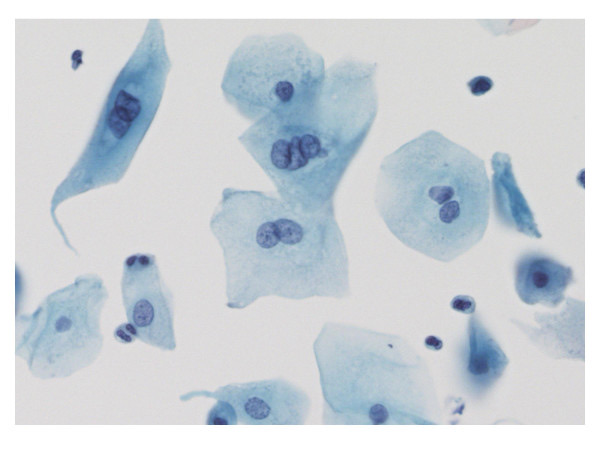
- Bi- and multinucleation. These cells were present in 81% of abnormal samples and 33% of negative samples (P < 0.0001) (Arain et al. 2005).
- Signs of HPV in HIV-positive Japanese MSM. Cytologic signs of HPV infection were present in 96.6% of samples at ASC-US or worse. Compression-positive binucleated cells were the most frequent sign (92.9%), followed by atypical parakeratotic cells (71.4%) (Okayama et al. 2017).
Low-grade squamous intraepithelial lesion in anal cytology
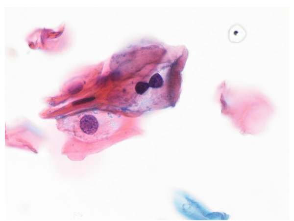
- Most frequent features. LSIL samples most often showed mildly dysplastic cells and bi- or multinucleated squamous cells, followed by parakeratotic cells, atypical parakeratotic cells and koilocytes (Arain et al. 2005).
- Mildly dysplastic cells. In 84% of LSIL samples, three or more cells showed mild dysplasia. Three samples contained typical koilocytes without dysplastic cells (Arain et al. 2005).
- Histologic follow-up of LSIL cytology. Of 36 LSIL samples with follow-up, 5 were negative, 11 showed AIN 1 and 20 showed AIN 2–3 (Arain et al. 2005).
- Condyloma. The WHO Classification of Digestive System Tumours, 6th edition, places condyloma acuminatum within LSIL. Anal condylomas are sampled thoroughly because both LSIL and HSIL can be present (Arends et al. 2026).
High-grade squamous intraepithelial lesion in anal cytology
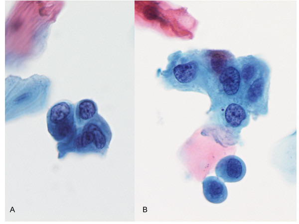
- Diagnostic cells. Every HSIL sample contained three or more squamous cells with moderate or severe dysplasia. HSIL samples also contained many atypical parakeratotic cells (Arain et al. 2005).
- Coexisting low-grade changes. Each HSIL sample also contained mildly dysplastic cells. Seventy-one percent of ASC-H and HSIL samples contained three or more mildly dysplastic cells (Arain et al. 2005).
- Cell size. The HSIL cells illustrated in the Arain series are small to medium and severely dysplastic (Arain et al. 2005).
- Cellularity of HSIL samples. All HSIL samples and 16 of 17 ASC-H samples averaged 8 or more nucleated squamous cells per high-power field (Arain et al. 2005).
- Histologic confirmation. All 4 HSIL samples with follow-up showed AIN 2–3 (1 AIN II, 3 AIN III) (Arain et al. 2005).
Atypical squamous cells in anal cytology
- ASC-US. The ASC-US samples in the Arain series showed parakeratosis, atypical parakeratosis, koilocytes, bi- or multinucleation and mild dysplasia, each in a minority of samples. None showed moderate or severe dysplasia (Arain et al. 2005).
- ASC-H. Five ASC-H samples contained two or fewer cells with features of high-grade dysplasia. The rest contained small cells with dense cytoplasm and atypical nuclei. These cells were interpreted as possible atypical metaplastic or atypical parakeratotic cells (Arain et al. 2005).
- Follow-up of ASC-H. Follow-up within 6 months was available for 15 of 17 ASC-H samples: 13 showed AIN 2–3 and 2 showed AIN 1 (Arain et al. 2005).
- Follow-up of ASC-US. Follow-up of 11 ASC-US samples showed 4 negative results, 2 AIN 1, 1 AIN 2 and 4 AIN 3 (Arain et al. 2005).
Distribution of interpretations in anal cytology series
| Series | Population | Unsatisfactory | NILM | ASC-US | LSIL | ASC-H | HSIL |
|---|---|---|---|---|---|---|---|
| Morency et al. 2019 | 1,417 tests | 17.4% | 27.9% | 19.5% | 24.1% | 3.6% | 7.5% |
| Arain et al. 2005 | 200 SurePath samples from 198 males | 8.5% (17) | 29% (58) | 21% (42) | 29.5% (59) | 8.5% (17) | 3.5% (7) |
- Abnormal rate in the Morency series. Of the 1,417 tests, 54.7% were abnormal (Morency et al. 2019).
- Young MSM with HIV. In 1,255 MSM with HIV aged 18–34 years, the abnormal cytology rate was 65%. Biopsies showed benign findings in 10%, LSIL in 43% and HSIL in 47% (Liu et al. 2024).
- hrHPV in NILM and ASC-US samples. In 642 anal samples, 47% of NILM and 73% of ASC-US samples were hrHPV positive. Non-16, non-18 types were the most common. The relative risk of histologic HSIL or worse was 2.3 times higher with HPV16 or HPV18 than with other high-risk types alone (Chebib et al. 2015).
Sources of error in grading anal squamous intraepithelial lesions
| Source of error | Effect | Source |
|---|---|---|
| Surface maturation and keratinization of high-grade lesions | May obscure sampling of dysplastic cells; AIN 2 and AIN 2/3 were detected less often (P < 0.001) | Russell et al. 2026 |
| Mixed low- and high-grade cells | All HSIL samples also contain mildly dysplastic cells | Arain et al. 2005 |
| Atypical parakeratosis | Atypical parakeratotic cells increase with grade and are absent in negative samples; ordinary parakeratosis is nonspecific | Arain et al. 2005 |
| Absence of a transformation zone component | More frequent in false-negative samples (32.3%) than in true-positive samples (11.0%) | Roberts et al. 2016 |
| Fewer abnormal cells in small lesions | Sensitivity higher with more than 1 anal octant of HSIL (92.9% versus 77.7%) | Jin et al. 2016 |
| Low abnormal-cell count | Two or fewer high-grade cells are reported as ASC-H | Arain et al. 2005 |
- Proficiency testing. CAP program participants classified LSIL correctly in 85% of challenges and HSIL in 57.1%. LSIL concordance improved over 2006–2011, and HSIL concordance did not (Darragh et al. 2013, CAP).
Histologic correlation and terminology for anal squamous intraepithelial lesions
- LAST terminology. Lower Anogenital Squamous Terminology (LAST) applies the same two-tier terms, LSIL and HSIL, to HPV-associated squamous lesions at all lower anogenital sites. It also standardizes the use of biomarkers (Darragh et al. 2012).
- p16. Diffuse p16 immunostaining distinguishes HSIL from reactive changes (Arends et al. 2026).
- WHO Digestive System Tumours, 6th edition (2026). HSIL includes lesions previously classified as moderate dysplasia, severe dysplasia, AIN II, AIN III, carcinoma in situ, Bowen disease and bowenoid papulosis. LSIL includes mild dysplasia, AIN I, anal SIL and condyloma acuminatum. The 6th edition also recognizes HPV-independent subsets analogous to TP53-mutant and TP53-wild-type vulvar intraepithelial neoplasia (Arends et al. 2026). The alignment of anal canal terminology with LAST and the HPV-independent subsets are new in the 6th edition per Arends et al. 2026. These statements come from the published summary of the 6th edition (Arends et al. 2026).
Differential diagnosis of atypical squamous cells in anal cytology
| Finding | Features favoring a benign interpretation | Features favoring SIL | Source |
|---|---|---|---|
| Parakeratotic cells | Small dense orangeophilic cells with nuclei lacking atypia; present in 63% of negative samples | Atypical nuclei (atypical parakeratosis); absent from negative samples | Arain et al. 2005 |
| Binucleated cells | Isolated binucleation, found in 33% of negative samples | Numerous bi- or multinucleated cells; compression-positive binucleated cells | Arain et al. 2005; Okayama et al. 2017 |
| Small cells with dense cytoplasm | Metaplastic cells without nuclear atypia | Atypical nuclei, as in ASC-H or HSIL | Arain et al. 2005 |
| Cells with HSV changes | Multinucleated viral cytopathic effect | Coexisting dysplastic cells, present in 86% of HSV-positive anal samples | Greebon et al. 2014 |
Anal Squamous Cell Carcinoma and Other Anal Malignancies on Cytology
Definition. Anal squamous cell carcinoma is an invasive carcinoma of the anal canal or perianal skin with squamous differentiation. Almost all anal squamous cell carcinomas are caused by human papillomavirus (HPV) and are preceded by high-grade squamous intraepithelial lesion (HSIL) (Stier et al. 2024). Anal cytology is designed to detect HSIL. Digital anal rectal examination (DARE), performed at every screening visit, provides screening for early anal cancers detectable by palpation (Stier et al. 2024). In external proficiency testing, concordance for anal squamous cell carcinoma slide challenges was 56.2% (Darragh et al. 2013, CAP). Cytologic criteria for squamous cell carcinoma in anal samples are those of the Bethesda System for cervical cytology (Bean and Chhieng 2010; Hopp et al. 2023). Malignancies other than squamous cell carcinoma, including mucosal melanoma and adenocarcinoma, are occasionally detected on anorectal cytology (Lau et al. 2017).
Related page: Anal Cancer Screening with Anal Cytology (screening populations, IANS management and adequacy) Related page: Cytomorphology of Anal Cytology Samples (HSIL and other squamous findings in anal cytology) Related page: Squamous Cell Carcinoma in Cervical Cytology (Bethesda cytologic criteria for squamous cell carcinoma) Related page: anal squamous cell carcinoma (histology, staging and treatment of anal squamous cell carcinoma)
Epidemiology of anal squamous cell carcinoma
- Global burden. An estimated 30,416 anal squamous cell carcinomas occurred worldwide in 2020: 19,792 in women and 10,624 in men (Deshmukh et al. 2023).
- Distribution by Human Development Index. Fifty-three percent of male cases and 65% of female cases occurred in countries with a very high Human Development Index (Deshmukh et al. 2023).
- HIV. People with HIV accounted for 21% of the male burden (2,203 cases) and 3% of the female burden (561 cases). The male HIV-associated burden was concentrated in North America, Europe and Africa (Deshmukh et al. 2023).
- Age-standardized incidence by sex and HIV status. The global age-standardized incidence of HIV-negative anal squamous cell carcinoma was 0.55 per 100,000 in women and 0.28 per 100,000 in men. The incidence of HIV-positive anal squamous cell carcinoma was 0.07 per 100,000 in men and 0.02 per 100,000 in women (Deshmukh et al. 2023).
- Incidence by risk group. Anal cancer incidence by risk group and the IANS risk categories are tabulated in Anal Cancer Screening with Anal Cytology.
Progression from HSIL to invasive anal carcinoma
- Progression after HSIL cytology. In 2,804 HIV-infected patients with a median follow-up of 4 years, the estimated 5-year probability of invasive anal cancer after baseline HSIL cytology was 1.7% (upper 95% bound 4.5%). The annual progression risk was 1 in 263 (Cachay et al. 2015).
- Effect of treatment of HSIL. Treatment of biopsy-proven HSIL reduced anal cancer incidence by 57% in the ANCHOR trial (Palefsky et al. 2022). The trial is described in Anal Cancer Screening with Anal Cytology.
- Pathology in ANCHOR. HSIL eligibility required AIN 3 or p16-positive AIN 2 in an anal canal or perianal biopsy. Central review confirmed HSIL in 96.6% of screening participants (Palefsky et al. 2022).
Detection of invasive anal carcinoma by screening
- Digital anal rectal examination. DARE is performed at every screening visit and screens for early cancers detectable by palpation. Where HRA is unavailable, screening is limited to DARE (Stier et al. 2024). IANS published a recommended DARE technique with quality-assurance metrics (Hillman et al. 2019).
- High-resolution anoscopy (HRA) capacity. Most HRA providers practice in large cities in the United States, Western Europe and Australia. Much of the world has very limited access to HRA (Stier et al. 2024).
- Cytology sensitivity for cancer. In a laboratory series with 413 biopsy diagnoses of HSIL and 8 of anal squamous cell carcinoma, cytology at a threshold of possible LSIL or worse had 86.3% sensitivity and 31.9% specificity for high-grade lesions (Russell et al. 2026).
- Reference standard of the screening meta-analysis. The pooled performance figures for anal cytology and HPV tests used AIN2+, including anal cancer, as the reference standard (Clarke et al. 2022).
Cytologic recognition of anal squamous cell carcinoma
- Bethesda criteria. Anal squamous cell carcinoma is reported with the Bethesda category of squamous cell carcinoma, and its criteria are those used in cervical cytology (Bean and Chhieng 2010; Hopp et al. 2023). Keratinizing and nonkeratinizing patterns, tumor diathesis and the related criteria are described in Squamous Cell Carcinoma in Cervical Cytology.
- Interpretive performance. In the CAP interlaboratory comparison program from 2006 to 2011, concordance for anal squamous cell carcinoma slide challenges was 56.2% (Darragh et al. 2013, CAP).
Anal cytology after treatment of anal squamous cell carcinoma
- Standard surveillance. After successful treatment of anal squamous cell carcinoma, surveillance for local recurrence uses anoscopy and digital rectal examination (Brasseur et al. 2022).
- Cytology in surveillance. Of 104 patients successfully treated and followed for 5 years, 23 also had anal cytology. HSIL was found in 5 of them: 2 by cytology, 1 by HRA and 2 by examination under anesthesia with biopsy (Brasseur et al. 2022).
- Proposed role. The authors proposed anal cytology as a surveillance tool that requires fewer specialized resources than HRA (Brasseur et al. 2022). IANS considered guidelines for management after treatment of anal HSIL or cancer to be outside the scope of its screening guidelines (Stier et al. 2024).
Anorectal mucosal melanoma on anal cytology
- Reported features. In the first reported case of primary anorectal melanoma detected on anorectal cytology, the sample showed isolated malignant cells with oval nuclei, prominent nucleoli and elongated, wispy cytoplasmic projections (Lau et al. 2017).
- Absent features. Melanin pigment and squamous dysplasia were absent (Lau et al. 2017).
- Interpretive principle. Malignancies other than squamous cell carcinoma are considered when malignant cells are present without evidence of anal intraepithelial neoplasia (Lau et al. 2017).
Glandular malignancies of the anal region
- Primary perianal adenocarcinoma of intestinal type. This proposed subtype of extramucosal anal adenocarcinoma presents with pagetoid intraepithelial growth. In 14 cases, the median age was 73 years and 9 of 14 patients were women. Underlying invasive carcinoma was found in 9 cases, and no other primary was identified (Bahceci et al. 2026).
- Immunophenotype. All but 1 case expressed cytokeratin 20 and CDX2, with variable cytokeratin 7 (Bahceci et al. 2026).
- Molecular profile. TP53 mutations were present in 86% and ERBB2 alterations in 57%. MYC amplification was present in 36%. APC, KRAS and BRAF alterations typical of rectal adenocarcinoma were absent (Bahceci et al. 2026).
- Contrast with primary extramammary Paget disease. Primary extramammary Paget disease showed recurrent PIK3CA and KMT2C mutations (Bahceci et al. 2026).
- Behavior. Metastases developed in 4 of 14 patients. Survival ranged from 29 to 176 months for localized disease and from 13 to 75 months for metastatic disease (Bahceci et al. 2026).
- Vulvar and perineal Paget disease. Paget cells in vulvar and vaginal cytology are covered under Lower Anogenital Cytology Outside the Cervix and Anus.
Differential diagnosis of malignant cells in anal cytology
| Diagnosis | Cytologic and clinical features | Source |
|---|---|---|
| HSIL with keratinization | Small to medium cells with moderate or severe dysplasia; atypical parakeratotic cells; mildly dysplastic cells in the background | Arain et al. 2005; Russell et al. 2026 |
| Squamous cell carcinoma | Bethesda criteria for squamous cell carcinoma; usually arises from HSIL | Bean and Chhieng 2010; Stier et al. 2024 |
| Anorectal mucosal melanoma | Isolated cells with oval nuclei, prominent nucleoli and wispy cytoplasmic projections; pigment may be absent; no squamous dysplasia | Lau et al. 2017 |
| Perianal intestinal-type adenocarcinoma | Pagetoid intraepithelial growth with or without invasion; cytokeratin 20 and CDX2 positive | Bahceci et al. 2026 |
Reporting malignant cells in anal cytology
- Category. A malignant squamous population is reported as squamous cell carcinoma in the Bethesda System (Bean and Chhieng 2010). In cervical samples, nonsquamous malignant cells, including Paget cells, have been reported under the Bethesda category of other malignant neoplasms (Takahashi et al. 2021).
- Clinical follow-up. A cytologic interpretation of carcinoma leads to examination and biopsy. HRA is the diagnostic procedure for abnormal anal screening results (Stier et al. 2024).
Lower Anogenital Cytology Outside the Cervix and Anus
Definition. Lower anogenital cytology outside the cervix and anus comprises vaginal cytology (usually after hysterectomy), vulvar scraping or brushing cytology, neovaginal cytology, penile cytology, and fine-needle aspiration (FNA) of penile tumors and inguinal lymph nodes. Vaginal cytology is reported with the Bethesda System terminology used for cervical cytology. HPV-associated squamous intraepithelial lesions at all lower anogenital sites are classified with the two-tier Lower Anogenital Squamous Terminology (LAST): low-grade (LSIL) and high-grade (HSIL) squamous intraepithelial lesion (Darragh et al. 2012). The WHO Classification of Female Genital Tumours, 5th edition (2020), separates HPV-associated from HPV-independent squamous neoplasia of the vulva and vagina (Höhn et al. 2021).
Related page: Anal Cancer Screening with Anal Cytology (anal screening after vulvar HSIL or cancer and in men) Related page: Cytomorphology of Anal Cytology Samples (squamous intraepithelial lesion morphology in anal samples) Related page: Anal Squamous Cell Carcinoma and Other Anal Malignancies on Cytology (perianal adenocarcinoma and anal malignancies) Related page: The Bethesda System for Reporting Cervical Cytology (Bethesda System categories) Related page: Metastatic Carcinoma in Lymph Nodes (Cytology) (FNA of metastatic carcinoma in lymph nodes)
Classification of vulvar and vaginal squamous neoplasia in the WHO Female Genital Tumours, 5th edition
- HPV association. The 2020 WHO classification is based on the distinction between HPV-associated and HPV-independent squamous cell carcinoma of the lower female genital organs. This distinction for squamous cell carcinoma is new in the 5th edition per Höhn et al. 2021. HPV status does not replace grading, and no specific grading system is recommended (Höhn et al. 2021).
- HPV-associated vulvar intraepithelial neoplasia. The distinction between HPV-associated and HPV-negative VIN was retained from the previous edition. HPV-associated VIN corresponds to LSIL (VIN 1) and HSIL (VIN 2 and VIN 3) (Höhn et al. 2021).
- HPV-independent vulvar intraepithelial neoplasia. VIN without HPV is termed HPV-independent VIN, a term introduced in the 2020 classification. Höhn et al. allocate differentiated VIN, with its horizontal spread, as the precursor of a more aggressive squamous cell carcinoma than the keratinizing carcinoma linked to DEVIL (Höhn et al. 2021).
- Associated carcinoma patterns. HPV-associated vulvar squamous cell carcinoma is typically nonkeratinizing, with a plump pattern of invasion and p16 block staining. HPV-independent carcinoma is typically keratinizing, with a netlike pattern of invasion and aberrant p53 expression (Höhn et al. 2021).
- Other HPV-independent lesions. Differentiated exophytic vulvar intraepithelial lesion (DEVIL) and vulvar acanthosis with altered differentiation (VAAD) are covered by the umbrella term vulvar aberrant maturation, which is not part of the WHO classification. DEVIL is linked to keratinizing squamous cell carcinoma and VAAD to verrucous carcinoma, and their status as precursors is uncertain (Höhn et al. 2021).
- Vagina. The WHO recommends separating HPV-associated from HPV-independent vaginal squamous cell carcinoma, although HPV status currently has no therapeutic relevance. Most vaginal squamous cell carcinomas are HPV associated; distal (introitus) carcinomas are often keratinizing and HPV independent. The classification of vaginal intraepithelial neoplasia did not change in the 5th edition (Höhn et al. 2021).
- Current WHO volume. The 5th edition is the current WHO volume for female genital tumours. Anal canal neoplasia is classified in the WHO Digestive System Tumours, 6th edition, described in Cytomorphology of Anal Cytology Samples.
- Dual-stain immunocytochemistry on vulvar liquid-based cytology. In a pilot study of 45 vulvar liquid-based specimens, all cases of VIN and vulvar carcinoma were positive for Sec62/Ki-67 and p16/Ki-67 dual staining, and normal and LSIL cases were negative. Cytology detected 22 of 22 VIN+ cases. The authors proposed the dual stains as adjuncts to histology (Takacs et al. 2019).
| Precursor | HPV status | Cytologic correlate | Associated carcinoma | Source |
|---|---|---|---|---|
| LSIL (VIN 1, VaIN 1) | HPV-associated | LSIL | Not stated in the source | Höhn et al. 2021; Darragh et al. 2012 |
| HSIL (VIN 2–3, VaIN 2–3) | HPV-associated | HSIL | Nonkeratinizing HPV-associated squamous cell carcinoma | Höhn et al. 2021; Darragh et al. 2012 |
| Differentiated VIN | HPV-independent | No reliable cytologic category | Keratinizing HPV-independent squamous cell carcinoma | Höhn et al. 2021 |
Vaginal cytology after hysterectomy for benign disease
- Screening after hysterectomy for benign disease. Screening after total hysterectomy for benign disease is generally not recommended in patients with no CIN 2+ in the previous 25 years (Perkins et al. 2020; Khan et al. 2016).
- Low prevalence of abnormalities. In 53,891 vaginal tests from women after hysterectomy for benign disease, the prevalence of ASC was 0.13%, LSIL 0.04% and HSIL or squamous cell carcinoma 0.03%. The prevalence ratio for HSIL or carcinoma, relative to women with a cervix, was 0.13 (Vale et al. 2015).
- Evidence base. Data were too limited for evidence-based recommendations on abnormal vaginal tests. An expert-opinion algorithm was proposed (Khan et al. 2016).
| Vaginal result after hysterectomy for benign disease | ASCCP 2019 management |
|---|---|
| ASC-US, HPV positive | Follow-up in 12 months |
| LSIL | Follow-up in 12 months |
| ASC-H | Immediate vaginal colposcopy |
| HSIL | Immediate vaginal colposcopy |
| Atypical glandular cells | Immediate vaginal colposcopy |
| Benign glandular cells | No further evaluation; reported as NILM |
Source: Perkins et al. 2020, sections G.4 and K.4.
Vaginal cytology after hysterectomy for cervical precancer or cancer
- After treatment for cervical HSIL or adenocarcinoma in situ. When hysterectomy is the treatment, patients have 3 consecutive annual HPV-based tests before long-term surveillance. Long-term surveillance after histologic HSIL or adenocarcinoma in situ uses HPV-based testing every 3 years for 25 years, with or without hysterectomy (Perkins et al. 2020).
- Risk of VaIN. The risk of high-grade VaIN is increased after hysterectomy for histologic HSIL. HPV-based testing appears more sensitive than cytology alone in this setting (Perkins et al. 2020).
- After hysterectomy for invasive cervical carcinoma. In 147 patients followed for a mean of 43.3 months, 15.0% developed VaIN or recurrence. The 8 high-grade VaINs and 2 recurrent carcinomas occurred only after squamous cell carcinoma, within the first 2 years. VaIN was found in 54.5% of patients with a positive hrHPV result and 16.7% of those with a negative result (Li et al. 2013).
- After treatment for vulvar HSIL or cancer. Among 191 women followed for a median of 3.7 years, 37% had abnormal cytology: 25% low grade, 12% high grade and 0.5% carcinoma. The risk of VaIN 2+ was 11% after previous hysterectomy, and the risk of CIN 2+ was 8% with an intact cervix. Immunosuppression was the only significant correlate of high-grade cytology in multivariable analysis (adjusted odds ratio 3.7) (Kuroki et al. 2017).
- Anal screening after vulvar disease. IANS places vulvar HSIL or cancer in Risk Category A for anal cancer screening, beginning within 1 year of diagnosis (Stier et al. 2024). The details are in Anal Cancer Screening with Anal Cytology.
Vaginal cytology after hysterectomy for endometrial carcinoma
| Study | Patients | Finding |
|---|---|---|
| Novetsky et al. 2013 | 433 women; 2,378 vaginal tests | Abnormal cytology in 13% of women; no recurrence diagnosed from isolated abnormal cytology; sensitivity 40% and positive predictive value 7.3% for local recurrence |
| Zhao et al. 2016 | 420 women with vaginal follow-up, 414 of them with cytology | Vaginal recurrence in 11 of 420 women; abnormal cytology led to biopsy diagnosis in 2 of 11 |
- Radiation. Abnormal vaginal cytology after endometrial cancer was strongly associated with prior postoperative radiation therapy (P < 0.001) (Novetsky et al. 2013).
- Colposcopy after low-grade results. No recurrent cancer followed ASC-US or LSIL results, and the authors concluded that colposcopy is not needed after these results (Novetsky et al. 2013).
- Routine surveillance. Vaginal cytology is no longer a recommended routine surveillance option for vaginal cuff recurrence of endometrial carcinoma (Zhao et al. 2016).
- Timing of recurrence. Eight of the 11 vaginal recurrences in the Zhao series occurred within 2 years of hysterectomy (Zhao et al. 2016).
Paget disease in vulvar and vaginal cytology
- Frequency of involvement of Pap tests. Among 40 patients with vulvar or vaginal extramammary Paget disease, 10 of 67 Pap tests (15%) taken concurrently or after diagnosis showed possible involvement (Burk et al. 2026).
- Cytologic features. Suspicious features were hyperchromatic nuclei without visible nucleoli, increased nuclear-to-cytoplasmic ratios and anisonucleosis (Burk et al. 2026). In a separate case, Paget cells formed atypical groups with enlarged nuclei and occasional prominent nucleoli (Gilliland et al. 2013).
- Misinterpretation as squamous or glandular lesions. In a woman with long-standing vulvar and vaginal Paget disease, yearly Pap tests had been interpreted as HSIL, atypical glandular cells and adenocarcinoma (Gilliland et al. 2013).
- Immunocytochemistry. Carcinoembryonic antigen (CEA) and gross cystic disease fluid protein (GCDFP) immunoreactivity distinguished Paget cells from HSIL (Gilliland et al. 2013).
- Standard immunohistochemical panel. Primary extramammary Paget disease is cytokeratin 7 positive, cytokeratin 20 negative, GCDFP-15 positive and GATA3 positive. Secondary disease from urothelial carcinoma is cytokeratin 7 and cytokeratin 20 positive, GCDFP-15 negative, and GATA3 and uroplakin II or III positive. Secondary disease from colorectal carcinoma is cytokeratin 7 negative, cytokeratin 20 positive, GCDFP-15 negative and CDX2 positive (Takahashi et al. 2021).
- Vulvar brushing. Vigorous brushing of an eczematoid vulvar lesion yielded isolated malignant Paget-type cells, and excision confirmed Paget disease. The authors proposed brushing cytology as an initial study for suspicious vulvar lesions (Funamoto et al. 2010).
- Secondary Paget disease from urothelial carcinoma. In a 77-year-old woman with prior urothelial carcinoma, cervical, endometrial and urethral cytology showed malignant cells reported as other malignant neoplasms. Vulvar Paget cells were positive for cytokeratin 7, cytokeratin 20, GATA3 and uroplakin II and negative for gross cystic disease fluid protein 15 (GCDFP-15). In a literature review of extramammary Paget disease with abnormal cervical cytology, primary disease and secondary disease from urothelial carcinoma were the most frequent (Takahashi et al. 2021).
- Perianal intestinal-type disease. Pagetoid perianal adenocarcinoma of intestinal type is cytokeratin 20 and CDX2 positive. It is described in Anal Squamous Cell Carcinoma and Other Anal Malignancies on Cytology (Bahceci et al. 2026).
| Marker | Paget cells | HSIL | Source |
|---|---|---|---|
| CEA | Positive | Negative in the reported comparison | Gilliland et al. 2013 |
| GCDFP and GCDFP-15 | Positive in primary disease; GCDFP-15 negative in secondary disease from urothelial or colorectal carcinoma | Negative in the reported comparison | Gilliland et al. 2013; Takahashi et al. 2021 |
| Uroplakin II | Positive in secondary disease from urothelial carcinoma | Not reported | Takahashi et al. 2021 |
| CDX2 and cytokeratin 20 | Positive in perianal intestinal-type disease and in secondary disease from colorectal carcinoma | Not reported | Bahceci et al. 2026; Takahashi et al. 2021 |
| p16 | Not reported | Diffusely positive (block staining) | Arends et al. 2026; Höhn et al. 2021 |
Neovaginal cytology
- Penile-skin neovagina. In 50 transgender women with an inverted penile-skin neovagina, well-preserved nucleated squamous cells were found in 72% of Pap samples. Four samples showed ASC-US, all negative for hrHPV. One showed hrHPV-positive LSIL, and no HSIL was identified (Weyers et al. 2010).
- Differences from cervicovaginal cytology. A cervicovaginal pattern with superficial, intermediate and parabasal cells and Döderlein bacilli was found in only 4% of samples. Inflammation was present in 30.6% of samples with squamous cells (Weyers et al. 2010).
- Molecular testing. In 16 transgender women with a neovagina, cytology was negative in all 13 sampled participants, while PCR detected hrHPV in 38.5% of those tested. Chlamydia trachomatis, Neisseria gonorrhoeae, Mycoplasma hominis and Ureaplasma urealyticum were also detected (Cabrera López et al. 2026).
Penile and male genital cytology
Penile liquid-based cytology for HPV. In 99 penile samples from men with condyloma or male partners of HPV-infected women, HPV DNA was detected in 93.9%. Koilocytosis was found in 1 sample and nonclassic cytologic signs in 23. The authors concluded that penile cytology does not improve the diagnosis of HPV infection in men (de Lima Rocha et al. 2008).
Limitations of male sampling. Analysis of male HPV infection is limited by inconsistent collection methods, variable adequacy and low sensitivity of cytology (de Lima Rocha et al. 2008).
Diagnostic yield of penile FNA. In 11 patients aged 46–89 years, FNA of the penis yielded material sufficient for diagnosis in every case, with tolerable discomfort and no complications (Skoog et al. 1998).
Diagnoses. The series included 5 squamous cell carcinomas (3 primary, 1 locally recurrent and 1 metastatic from the lung), 3 metastatic carcinomas from colon, prostate and bladder, and 1 each of recurrent melanoma, metastatic testicular mesothelioma and Peyronie disease (Skoog et al. 1998).
Testicular fine-needle aspiration
- Testicular FNA in male infertility. In a comparison of testicular FNA and biopsy in 34 infertile men, the two methods were concordant in 88% of cases. In discordant cases, FNA yielded more diagnostic material, based on the presence of secondary spermatocytes and spermatids. Charcot-Böttcher crystalloids were frequent in Sertoli cell cytoplasm in both normal and abnormal spermatogenesis (Amin et al. 2006).
- Testicular FNA for sperm retrieval in non-obstructive azoospermia. In 89 men with non-obstructive azoospermia, testicular FNA retrieved sperm in 40 (45%), with no significant postoperative complications. The fertilization rate was 16.6% when up to 10 sperm were retrieved. It was 40.1% with dozens of sperm and 47.2% with hundreds to thousands of sperm (P = 0.003) (Sakas et al. 2024).
- Testicular germ cell tumors diagnosed by FNA. In a 49-year-old man, FNA smears suggested a germ cell tumor, and the cell block established seminoma, which histology confirmed (Pandey et al. 2011). Yolk sac tumor of the undescended testis of a 2-year-old child was diagnosed primarily by FNA (Choudhury et al. 2015). The WHO Classification of Urinary and Male Genital Tumours, 5th edition (2022), retains the division of germ cell tumors into those derived from germ cell neoplasia in situ (GCNIS) and those unrelated to it. It keeps the term seminoma, places seminoma in a germinoma family, and leaves yolk sac tumor essentially unchanged (Berney et al. 2022).
FNA of inguinal lymph nodes in penile squamous cell carcinoma
| Setting | Patients | Sensitivity | Specificity | Source |
|---|---|---|---|---|
| Palpable inguinal nodes before treatment of the primary tumor | 16 men; 25 nodes | 93% | 91% | Saisorn et al. 2006 |
| Clinically node-negative groins, ultrasound-guided FNA | 43 patients; 83 groins | 39% | 100% | Kroon et al. 2005 |
| Follow-up after treatment, ultrasound-guided FNA | 247 patients | 87.3% | 99.9% | Djajadiningrat et al. 2014 |
- Palpable nodes. FNA of palpable nodes allowed early inguinal lymphadenectomy without prolonged initial antibiotic treatment. No needle-tract implantation occurred (Saisorn et al. 2006).
- Clinically node-negative groins. A positive ultrasound-guided FNA allowed earlier therapeutic inguinal dissection and reduced the number of groins requiring dynamic sentinel node biopsy by 11%. A negative FNA does not exclude metastasis because sensitivity was 39% (Kroon et al. 2005).
- Follow-up. Ultrasound-guided FNA detected 12 of 15 nonpalpable inguinal recurrences (80%). Physical examination detected 73% of recurrent groins (Djajadiningrat et al. 2014).
Differential diagnosis of atypical cells in vulvar and vaginal cytology
| Finding | Key features | Source |
|---|---|---|
| HSIL (VaIN or VIN extension) | Dysplastic squamous cells; p16 block positivity on tissue | Höhn et al. 2021; Darragh et al. 2012 |
| Paget cells | Clusters of cells with enlarged hyperchromatic nuclei; CEA positive | Burk et al. 2026; Gilliland et al. 2013 |
| Secondary Paget disease from urothelial carcinoma | Uroplakin II and GATA3 positive; GCDFP-15 negative | Takahashi et al. 2021 |
| Recurrent endometrial carcinoma at the vaginal cuff | Neoplastic glandular cells; most recurrences within 2 years | Zhao et al. 2016 |
| Post-radiation change | Abnormal cytology more frequent after radiation; no recurrence found from isolated abnormal cytology | Novetsky et al. 2013 |
References
- Amin A, Monabati A, Tadayon A, et al. Testicular fine needle aspiration cytology in male infertility. Acta Cytol. 2006;50(2):147–150. doi:10.1159/000325923. PMID 16610681
- Arain S, Walts AE, Thomas P, Bose S. The anal Pap smear: cytomorphology of squamous intraepithelial lesions. Cytojournal. 2005;2(1):4. doi:10.1186/1742-6413-2-4. PMID 15715910
- Arends MJ, Esposito I, Gill AJ, et al. Changes in the 6th edition of the World Health Organization classification of tumours of the digestive system. Histopathology. 2026;88(7):1295–1314. doi:10.1111/his.70116. PMID 41724188
- Bahceci D, Saoud C, Isidro RA, et al. Perianal intestinal-type Paget disease with and without invasion, unassociated with internal malignancy: a distinct form of primary perianal adenocarcinoma. Mod Pathol. 2026;39(1):100917. doi:10.1016/j.modpat.2025.100917. PMID 41106477
- Bean SM, Chhieng DC. Anal-rectal cytology: a review. Diagn Cytopathol. 2010;38(7):538–546. doi:10.1002/dc.21242. PMID 19941374
- Benevolo M, Rollo F, Latini A, et al. Interobserver agreement in the interpretation of anal cytology. Cancer Cytopathol. 2024;132(7):419–424. doi:10.1002/cncy.22807. PMID 38451011
- Berney DM, Cree I, Rao V, Moch H. An introduction to the WHO 5th edition 2022 classification of testicular tumours. Histopathology. 2022;81(4):459–466. doi:10.1111/his.14675. PMID 35502823
- Brasseur B, Subillaga O, Vrees M, et al. Can anal cytology be a tool in following patients treated for squamous cell carcinoma of the anus? Am Surg. 2022;88(7):1621–1625. doi:10.1177/00031348221080426. PMID 35258352
- Brouwer OR, Albersen M, Parnham A, et al. European Association of Urology-American Society of Clinical Oncology Collaborative Guideline on Penile Cancer: 2023 Update. Eur Urol. 2023;83(6):548–560. doi:10.1016/j.eururo.2023.02.027. PMID 36906413
- Burk JL, Crumley S, Duan X, et al. Cervicovaginal cytologic features in patients with vulvar Paget disease. J Am Soc Cytopathol. 2026;15(5):361–368. doi:10.1016/j.jasc.2026.06.001. PMID 42364948
- Cabrera López TJ, Rodríguez MNT, Nieto EV, et al. Neovaginal cytologic and molecular screening findings for human papillomavirus and sexually transmitted infections among transgender women. Open Forum Infect Dis. 2026;13(9):ofag516. doi:10.1093/ofid/ofag516. PMID 42703621
- Cachay E, Agmas W, Mathews C. Five-year cumulative incidence of invasive anal cancer among HIV-infected patients according to baseline anal cytology results: an inception cohort analysis. HIV Med. 2015;16(3):191–195. doi:10.1111/hiv.12190. PMID 25197003
- Chebib I, Tetreault D, Tambouret RH. Non-16/non-18 high-risk human papillomavirus types predominate in anal cytology categories of negative for intraepithelial lesion and atypical squamous cell of undetermined significance. J Am Soc Cytopathol. 2015;4(6):344–351. doi:10.1016/j.jasc.2015.06.004. PMID 31051749
- Chopra S, Kakar A, Bakshi P. Anal cytology screening: an aid to diagnose tuberculosis infection in HIV/AIDS. J Cytol. 2020;37(3):147–148. doi:10.4103/JOC.JOC_167_19. PMID 33088034
- Choudhury M, Sehgal S, Nangia A, Chadha R. Yolk sac tumor of cryptorchid testis diagnosed by fine needle aspiration cytology. J Cytol. 2015;32(1):53–55. doi:10.4103/0970-9371.155239. PMID 25948949
- Clarke MA, Deshmukh AA, Suk R, et al. A systematic review and meta-analysis of cytology and HPV-related biomarkers for anal cancer screening among different risk groups. Int J Cancer. 2022;151(11):1889–1901. doi:10.1002/ijc.34199. PMID 35793241
- Clifford GM, Georges D, Shiels MS, et al. A meta-analysis of anal cancer incidence by risk group: toward a unified anal cancer risk scale. Int J Cancer. 2021;148(1):38–47. doi:10.1002/ijc.33185. PMID 32621759
- Darragh TM, Colgan TJ, Cox JT, et al. The Lower Anogenital Squamous Terminology Standardization Project for HPV-associated lesions: background and consensus recommendations from the College of American Pathologists and the American Society for Colposcopy and Cervical Pathology. Arch Pathol Lab Med. 2012;136(10):1266–1297. doi:10.5858/arpa.LGT200570. PMID 22742517
- Darragh TM, Tokugawa D, Castle PE, et al. Interrater agreement of anal cytology. Cancer Cytopathol. 2013;121(2):72–78. doi:10.1002/cncy.21218. PMID 22811048
- Darragh TM, Winkler B, Souers RJ, et al. Room for improvement: initial experience with anal cytology: observations from the College of American Pathologists interlaboratory comparison program in nongynecologic cytology. Arch Pathol Lab Med. 2013;137(11):1550–1554. doi:10.5858/arpa.2012-0574-CP. PMID 24168493
- Darragh TM, Winkler B. Anal cancer and cervical cancer screening: key differences. Cancer Cytopathol. 2011;119(1):5–19. doi:10.1002/cncy.20126. PMID 21319310
- de Lima Rocha MG, Faria FL, Souza Mdo C, et al. Detection of human papillomavirus infection in penile samples through liquid-based cytology and polymerase chain reaction. Cancer. 2008;114(6):489–493. doi:10.1002/cncr.23947. PMID 18973183
- Deshmukh AA, Damgacioglu H, Georges D, et al. Global burden of HPV-attributable squamous cell carcinoma of the anus in 2020, according to sex and HIV status: a worldwide analysis. Int J Cancer. 2023;152(3):417–428. doi:10.1002/ijc.34269. PMID 36054026
- Djajadiningrat RS, Teertstra HJ, van Werkhoven E, et al. Ultrasound examination and fine needle aspiration cytology: useful for followup of the regional nodes in penile cancer? J Urol. 2014;191(3):652–655. doi:10.1016/j.juro.2013.08.046. PMID 23994372
- Dyer CEF, Jin F, Hillman RJ, et al. Self-collected versus clinician-collected anal swabs for anal cancer screening: a systematic review and meta-analysis. Int J Cancer. 2025;156(1):79–90. doi:10.1002/ijc.35133. PMID 39140279
- European Institute of Oncology. Anal cancer and/or precancer screening: performance analysis of the BD Onclarity HPV assay on anal specimens. ClinicalTrials.gov identifier NCT06327568. First submitted 2024-03-18. https://clinicaltrials.gov/study/NCT06327568
- Funamoto Y, Haba R, Kobayashi TK, et al. Cytologic diagnosis of vulvar Paget's disease by means of brushing smear: report of a case. Diagn Cytopathol. 2010;38(2):127–131. doi:10.1002/dc.21160. PMID 19691090
- Gilliland K, Knapik J, Wilkinson EJ. Cytology of vulvar/vaginal Paget disease: report of a case and review of the literature. J Low Genit Tract Dis. 2013;17(4):e26–e30. doi:10.1097/LGT.0b013e31828487a2. PMID 23609593
- Greebon LJ, Avery DL, Prihoda TJ, et al. The frequency of herpes simplex virus changes in anal Pap smear and its association with squamous intraepithelial lesions in high-risk male patients. Diagn Cytopathol. 2014;42(6):487–490. doi:10.1002/dc.23006. PMID 24692314
- Guzmán-Arocho YD, Helland TL, VanderLaan PA, et al. Multi-institutional 10-year retrospective review of amoeba diagnosed on cytologic evaluation of anal pap tests: what is the significance? J Am Soc Cytopathol. 2023;12(3):197–205. doi:10.1016/j.jasc.2023.01.004. PMID 36935302
- Hillman RJ, Berry-Lawhorn JM, Ong JJ, et al. International Anal Neoplasia Society guidelines for the practice of digital anal rectal examination. J Low Genit Tract Dis. 2019;23(2):138–146. doi:10.1097/LGT.0000000000000458. PMID 30907777
- Höhn AK, Brambs CE, Hiller GGR, et al. 2020 WHO classification of female genital tumors. Geburtshilfe Frauenheilkd. 2021;81(10):1145–1153. doi:10.1055/a-1545-4279. PMID 34629493
- Hopp AM, Puzyrenko A, Giorgadze T. Comprehensive review of anal cytology. Acta Cytol. 2023;67(2):185–194. doi:10.1159/000528532. PMID 36626881
- Jin F, Grulich AE, Poynten IM, et al. The performance of anal cytology as a screening test for anal HSILs in homosexual men. Cancer Cytopathol. 2016;124(6):415–424. doi:10.1002/cncy.21702. PMID 26915346
- Jin F, Poynten IM, Hillman RJ, et al. Does use of anal cytology as a triage test improve the performance of high-risk human papillomavirus screening in gay and bisexual men for anal cancer prevention? Int J Cancer. 2025;156(3):575–586. doi:10.1002/ijc.35185. PMID 39279187
- Khan MJ, Massad LS, Kinney W, et al. A common clinical dilemma: management of abnormal vaginal cytology and human papillomavirus test results. J Low Genit Tract Dis. 2016;20(2):119–125. doi:10.1097/LGT.0000000000000185. PMID 26901279
- Khattab R, McMeekin E, Taege AJ, et al. Unsatisfactory exfoliative anal cytology samples, 15-year experience with histologic, cytologic, and molecular follow-up. Diagn Cytopathol. 2018;46(2):117–121. doi:10.1002/dc.23858. PMID 29124900
- Kroon BK, Horenblas S, Deurloo EE, et al. Ultrasonography-guided fine-needle aspiration cytology before sentinel node biopsy in patients with penile carcinoma. BJU Int. 2005;95(4):517–521. doi:10.1111/j.1464-410X.2005.05330.x. PMID 15705071
- Kuroki LM, Frolova AI, Wu N, et al. Yield of cytology surveillance after high-grade vulvar intraepithelial neoplasia or cancer. J Low Genit Tract Dis. 2017;21(3):193–197. doi:10.1097/LGT.0000000000000321. PMID 28644191
- Lau RP, Chiaffarano J, Alexander M, et al. Primary anorectal mucosal melanoma detected by anorectal cytology. Diagn Cytopathol. 2017;45(5):452–455. doi:10.1002/dc.23675. PMID 28160456
- Leijte JA, Graafland NM, Valdés Olmos RA, et al. Prospective evaluation of hybrid 18F-fluorodeoxyglucose positron emission tomography/computed tomography in staging clinically node-negative patients with penile carcinoma. BJU Int. 2009;104(5):640–644. doi:10.1111/j.1464-410X.2009.08450.x. PMID 19281465
- Li Z, Barron S, Hong W, et al. Surveillance for recurrent cancers and vaginal epithelial lesions in patients with invasive cervical cancer after hysterectomy: are vaginal cytology and high-risk human papillomavirus testing useful? Am J Clin Pathol. 2013;140(5):708–714. doi:10.1309/AJCPH4AFSZHU8EKK. PMID 24124151
- Liu Y, Bhardwaj S, Sigel K, et al. Anal cancer screening results from 18-to-34-year-old men who have sex with men living with HIV. Int J Cancer. 2024;154(1):21–27. doi:10.1002/ijc.34734. PMID 37728489
- Mohamed A, Gonzalez RS, Lawson D, Wang J, Cohen C. SOX10 expression in malignant melanoma, carcinoma, and normal tissues. Appl Immunohistochem Mol Morphol. 2013;21(6):506–510. doi:10.1097/PAI.0b013e318279bc0a. PMID 23197006
- Morency EG, Harbert T, Fatima N, et al. Anal cytology: institutional statistics, correlation with histology, and development of multidisciplinary screening program with review of the current literature. Arch Pathol Lab Med. 2019;143(1):23–29. doi:10.5858/arpa.2017-0242-RA. PMID 29652190
- Novetsky AP, Kuroki LM, Massad LS, et al. The utility and management of vaginal cytology after treatment for endometrial cancer. Obstet Gynecol. 2013;121(1):129–135. doi:10.1097/aog.0b013e31827499a9. PMID 23262937
- Okayama K, Okodo M, Kitamura H, Itoda I. Significance of the cytological signs of human papillomavirus infection in anal Pap smears of human immunodeficiency virus-infected Japanese men who have sex with men. Asian Pac J Cancer Prev. 2017;18(11):3173–3178. doi:10.22034/APJCP.2017.18.11.3173. PMID 29172296
- Palefsky JM, Lee JY, Jay N, et al. Treatment of anal high-grade squamous intraepithelial lesions to prevent anal cancer. N Engl J Med. 2022;386(24):2273–2282. doi:10.1056/NEJMoa2201048. PMID 35704479
- Pandey A, Nandini N, Jha A, Manjunath G. Fine needle aspiration cytology and cell block in the diagnosis of seminoma testis. J Cytol. 2011;28(1):39–41. doi:10.4103/0970-9371.76950. PMID 21552408
- Pedreira C, Prado Escobar D, Memari K, et al. Silent invasion of the rectum: aggressive anorectal melanoma with pelvic mass effect, venous compression, and hepatic metastases. Cureus. 2026;18(4):e106339. doi:10.7759/cureus.106339. PMID 42083691
- Perkins RB, Guido RS, Castle PE, et al. 2019 ASCCP risk-based management consensus guidelines for abnormal cervical cancer screening tests and cancer precursors. J Low Genit Tract Dis. 2020;24(2):102–131. doi:10.1097/LGT.0000000000000525. PMID 32243307
- Revollo B, Videla S, Llibre JM, et al. Routine screening of anal cytology in persons with human immunodeficiency virus and the impact on invasive anal cancer: a prospective cohort study. Clin Infect Dis. 2020;71(2):390–399. doi:10.1093/cid/ciz831. PMID 31504329
- Roberts JM, Jin F, Thurloe JK, et al. The value of a transformation zone component in anal cytology to detect HSIL. Cancer Cytopathol. 2016;124(8):596–601. doi:10.1002/cncy.21730. PMID 27176896
- Russell S, Bentel J, Laycock A. Evaluating the performance of anal cytology and human papillomavirus testing for detection of anal cancer and pre-cancerous lesions. Diagn Cytopathol. 2026;54(8):597–609. doi:10.1002/dc.70144. PMID 42135996
- Saisorn I, Lawrentschuk N, Leewansangtong S, Bolton DM. Fine-needle aspiration cytology predicts inguinal lymph node metastasis without antibiotic pretreatment in penile carcinoma. BJU Int. 2006;97(6):1225–1228. doi:10.1111/j.1464-410X.2006.06159.x. PMID 16686716
- Sakas J, Radin O, Abrahami N, et al. Testicular fine-needle aspiration in infertile men with absolute non-obstructive azoospermia: a single-centre cohort study. Aust N Z J Obstet Gynaecol. 2024;64(3):258–263. doi:10.1111/ajo.13783. PMID 38078572
- Skoog L, Collins BT, Tani E, Ramos RR. Fine needle aspiration cytology of penile tumors. Acta Cytol. 1998;42(6):1336–1340. doi:10.1159/000332164. PMID 9850639
- Stier EA, Clarke MA, Deshmukh AA, et al. International Anal Neoplasia Society's consensus guidelines for anal cancer screening. Int J Cancer. 2024;154(10):1694–1702. doi:10.1002/ijc.34850. PMID 38297406
- Takacs FZ, Radosa JC, Bochen F, et al. Sec62/Ki67 and p16/Ki67 dual-staining immunocytochemistry in vulvar cytology for the identification of vulvar intraepithelial neoplasia and vulvar cancer: a pilot study. Arch Gynecol Obstet. 2019;299(3):825–833. doi:10.1007/s00404-018-5021-0. PMID 30607586
- Takahashi K, Kikuchi A, Horiuchi A, et al. Vulvar extramammary Paget disease detected by cytology for cervical cancer screening: a case report and literature review. Am J Case Rep. 2021;22:e933655. doi:10.12659/AJCR.933655. PMID 34679073
- Templeton DJ, Roberts JM, Poynten IM, et al. Prevalence and predictors of unsatisfactory anal cytology tests in a cohort of gay and bisexual men in Sydney, Australia: baseline findings from the Study of the Prevention of Anal Cancer (SPANC). Eur J Cancer Prev. 2017;26(3):212–216. doi:10.1097/CEJ.0000000000000321. PMID 28350724
- Vale DB, Bragança JF, Xavier-Junior JC, et al. Usefulness of vaginal cytology tests in women with previous hysterectomy for benign diseases: assessment of 53,891 tests. Gynecol Oncol. 2015;137(2):270–273. doi:10.1016/j.ygyno.2015.02.011. PMID 25703672
- Weyers S, Lambein K, Sturtewagen Y, et al. Cytology of the 'penile' neovagina in transsexual women. Cytopathology. 2010;21(2):111–115. doi:10.1111/j.1365-2303.2009.00663.x. PMID 19735277
- Wiley DJ, Hsu H, Bolan R, et al. Comparison of 2 anal cytology protocols to predict high-grade anal intraepithelial neoplasia. J Low Genit Tract Dis. 2013;17(4):414–424. doi:10.1097/LGT.0b013e318281d36e. PMID 23595039
- Zaccarini DJ, Khurana KK. Histopathologic and cytologic follow-up in high risk male patients with unsatisfactory anal cytology. Patholog Res Int. 2017;2017:9780213. doi:10.1155/2017/9780213. PMID 29075548
- Zhao C, Karunamurthy A, Jain S, et al. Vaginal cytology results in follow-up of endometrial carcinoma after hysterectomy. Am J Clin Pathol. 2016;146(2):244–247. doi:10.1093/ajcp/aqw100. PMID 27371362
Image credits
- Anal LSIL. Image: Arain Shehla et al., PMC open access (Cytojournal. 2005 Feb 16;2:4. doi: 10.1186/1742-6413-2-4) Figure 2, CC BY.
- Anal LSIL. Image: Arain Shehla et al., PMC open access (Cytojournal. 2005 Feb 16;2:4. doi: 10.1186/1742-6413-2-4) Figure 3, CC BY.
- Anal HSIL. A, a small cluster of immature squamous cells with high nuclear-to-cytoplasmic ratios and hyperchromatic, irregular nuclei. B, a group of similar dysplastic cells with dense cyanophilic cytoplasm and enlarged nuclei with coarse chromatin and irregular membranes. Papanicolaou stain, 40× objective. Image: Arain Shehla et al., PMC open access (Cytojournal. 2005 Feb 16;2:4. doi: 10.1186/1742-6413-2-4) Figure 5, CC BY.