Last reviewed 2026-11-04 by a board-certified pathologist. 79 references.
Normal Liver, Indications for Fine-Needle Aspiration and Reactive Changes on Liver FNA
Definition. Liver fine-needle aspiration (FNA) is image-guided sampling of a focal liver lesion with a fine needle for smears, liquid-based preparations and a cell block. Its main tasks are separation of benign hepatocytic nodules from reactive hepatocytes, separation of well-differentiated hepatocellular carcinoma (HCC) from benign hepatocellular nodules, separation of poorly differentiated HCC from intrahepatic cholangiocarcinoma and metastatic carcinoma, and identification of the primary site of a metastasis (Wee 2005; Wee 2011). Interpretation of a liver aspirate starts with recognition of the normal cellular components and of the reactive changes that occur in chronic liver disease, because both are present in aspirates of most liver lesions.
Indications for liver FNA when HCC can be diagnosed by imaging
Imaging diagnosis without tissue. In patients at risk for HCC, the diagnosis can be established by noninvasive imaging criteria. Arterial phase hyperenhancement and washout have high specificity and positive predictive value in lesions ≥1 cm. An LR-5 observation has a 95–99% probability of being HCC (Singal et al. 2023, AASLD).
Observations that require tissue. Biopsy is advised for LR-M observations. In these observations 93–100% of cases are malignant on tissue sampling, but only 29–44% are HCC. Biopsy is also recommended for tumor in vein (LR-TIV), for nodules in patients without cirrhosis or without hepatitis B virus (HBV) infection, and for nodules with noncharacteristic imaging, independent of the alpha-fetoprotein (AFP) level (Singal et al. 2023).
LR-4 observations. Biopsy or follow-up imaging at 3 months is advised, depending on the clinical scenario, after multidisciplinary discussion (Singal et al. 2023).
Clinical trials and precision treatment. The AASLD guidance advises biopsy of all LR-4 and LR-5 lesions in clinical trials. HCC biopsies inform molecular and immune classes and predictive gene signatures (Singal et al. 2023).
Serum AFP. AFP at 400 ng/mL was previously used as a diagnostic criterion for HCC. More than 40% of HCCs have normal AFP levels, and AFP is no longer recommended for the diagnosis of HCC (Singal et al. 2023).
Biopsy versus imaging criteria in earlier guidance. The European Association for the Study of the Liver (EASL) criteria of 2000 allowed a diagnosis of HCC in cirrhosis without biopsy for lesions >2 cm with arterial hypervascularization and concordant imaging. Punctures were limited to nodules <2 cm (as summarized by Wee 2005).
Clinical and imaging setting
Recommendation
Source
Cirrhosis, LR-5 observation
Diagnosis by imaging; tissue not required
Singal et al. 2023
LR-4 observation
Biopsy or repeat imaging at 3 months after multidisciplinary review
Singal et al. 2023
LR-M observation
Biopsy
Singal et al. 2023
LR-TIV
Biopsy
Singal et al. 2023
Nodule without cirrhosis or chronic HBV infection
Pathological diagnosis
Singal et al. 2023
Noncharacteristic imaging
Biopsy, independent of AFP
Singal et al. 2023
Clinical trial enrollment
Biopsy of LR-4 and LR-5 lesions
Singal et al. 2023
Practical advantages of FNA. FNA is suited to patients with cirrhosis, poor liver function and bleeding risk, to patients with obstructive jaundice or a risk of bile leakage, to lesions near large vessels or reached through bowel, to small (<2 cm) deep nodules and to multiple redirected passes through large lesions. It allows on-site assessment of adequacy and triage for ancillary studies (Wee 2005).
FNA, cell block and core needle biopsy of the liver
Needle types. Cutting aspiration needles of 21 gauge yield both smears and microbiopsy-like cores (Wee 2005).
Comparative accuracy. FNA and core needle biopsy (CNB) each reached an accuracy of 78% alone and 88% in combination (as summarized by Wee 2005). When a biopsy is indicated for a clinically suspected HCC, CNB has been favored over FNA (Wee 2005).
Value of the cell block. In a series of 936 liver aspirates, 72.1% of 796 concurrent cell blocks contained enough tissue for diagnosis. Sensitivity for liver tumors rose from 78.4% with smears alone to 85.1% with smears and cell blocks, and specificity rose from 97.4% to 98.7% (Kuo et al. 2004).
Reticulin stain on the cell block. All HCCs in the Kuo series had a decreased or absent reticulin pattern, and benign hepatocellular lesions usually had a normal trabecular reticulin framework (Kuo et al. 2004). In a cell block and thin-core series, all 40 HCCs had an abnormal reticulin pattern, and all 25 benign hepatocytic lesions had a normal trabecular pattern (de Boer et al. 2000).
Diagnostic sensitivity of liver biopsy. The sensitivity of liver biopsy ranges from 70% to 93% for most tumors and has been reported as low as about 60% for tumors <2 cm. A negative biopsy does not exclude HCC (Singal et al. 2023).
Specificity of liver FNA. Specificity and positive predictive value of liver FNA for malignancy approach 100% in most studies (as summarized by Wee 2005).
Requirements for optimal results. Optimal results depend on a dedicated radiologist-cytopathologist team, on-site cytology, a combined cytohistological approach, immunohistochemistry and clinicopathological correlation (Wee 2011).
Complications of percutaneous liver sampling
Needle tract seeding after biopsy of HCC. A meta-analysis of eight studies gave a pooled seeding rate of 2.7% (95% confidence interval 1.8–4.0%) overall, or 0.9% per year (Silva et al. 2008).
Combined complication rate. The risk of complications after liver biopsy, including tumor seeding and bleeding, has been reported at about 3%. The rate has decreased substantially with coaxial needle technique (Singal et al. 2023).
Underestimated seeding rate in older literature. A commonly cited seeding rate of 0.006% has been regarded by many authors as a gross underestimate (as summarized by Wee 2005).
Transplant candidates. Needle biopsy has usually been avoided in patients considered for liver transplantation because of the possibility of seeding (Wee 2005).
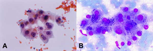
Monolayered clusters of loosely cohesive, well-defined polygonal cells with ample dense granular cytoplasm, round central nuclei and a low nuclear-to-cytoplasmic (N/C) ratio; binucleation and some variation in nuclear size
Wee 2005
Bile duct and ductular epithelium
Small cells with scant cytoplasm in a double-layered row
Wee 2005
Kupffer cells
Cells with elongated nuclei
Wee 2005
Endothelial cells
Normal component of the aspirate
Wee 2005
Mesothelial cells
Normal component of the aspirate
Wee 2005
Tissue fragments of nonneoplastic liver. In a comparison of 14 nonneoplastic liver aspirates with 14 well-differentiated HCCs, all nonneoplastic aspirates contained large tissue fragments that resisted smearing (Yang, Yang and Tao 2004).
Bile duct cells in benign liver and HCC. Bile duct cells are scarce in HCC, and their absence favors HCC (Wee 2005).
Normal N/C ratio. The N/C ratio of benign hepatocytes is about one third or less (Wee 2005).
Pigments and inclusions in hepatocytes on liver FNA
Finding
Papanicolaou stain
Romanowsky (May-Grünwald-Giemsa) stain
Significance
Source
Lipofuscin
Brown granules
Black granules
Normal wear-and-tear pigment
Wee 2005
Bile
Dark brown blobs
Black
Within cells, canaliculi or pseudoacini; in a malignant hepatocytic tumor it supports HCC
Wee 2005
Iron (hemosiderin)
Dark brown
Black
Confirmed by Perls stain; iron accumulation favors a benign process
Wee 2005
Fat vacuoles
Clear vacuoles
Clear vacuoles
Occur in benign hepatocytes and in HCC
Wee 2005
Mallory-Denk bodies and hyaline bodies
Cytoplasmic inclusions
Cytoplasmic inclusions
Occur in benign hepatocytes and in HCC
Wee 2005
Intranuclear inclusions
Nuclear inclusions
Nuclear inclusions
Not specific for HCC
Wee 2005
Pigments of similar color. Lipofuscin, bile and iron overlap in color on both stains. The Perls stain separates iron from the other two (Wee 2005).
Fatty change in neoplasms. Fatty change occurs in well-differentiated HCC, hepatocellular adenoma, focal nodular hyperplasia and dysplastic nodules (Wee 2005). It therefore does not separate these lesions from each other.
Reactive and regenerative hepatocyte changes in chronic liver disease
Large cell change. Hepatocytes show cell and nuclear enlargement with mild atypia, and the N/C ratio stays at about one third or less. Large cell change is a low-grade lesion (Wee 2005).
Small cell change. Small, monotonous hepatocytes show a subtle increase in N/C ratio and nuclear crowding. Small cell change is a high-grade lesion and a putative precursor of HCC. Both large and small cell change are common in cirrhosis (Wee 2005).
Dysplastic foci and dysplastic nodules. Dysplastic foci are <1 mm, and dysplastic nodules are >1 mm (Wee 2005). Low-grade and high-grade dysplastic nodules and large regenerative nodules are the benign and premalignant nodules of cirrhosis (Di Tommaso et al. 2007).
Frequency of HCC in cirrhosis. HCC occurs in 5–40% of patients with cirrhosis, and foci of cancer are found in about a third of dysplastic nodules (as summarized by Wee 2005).
Feature
Large cell change
Small cell change
Well-differentiated HCC
Cell size
Enlarged
Small
Small, monotonous
N/C ratio
About one third or less
Slightly increased
Increased
Nuclear crowding
Absent
Present
Present
Grade of lesion
Low grade
High grade, putative precursor
Malignant
Trabeculae and endothelium
Normal plates
Normal plates
Trabeculae more than two cells thick, wrapped or crossed by endothelium
Reticulin on cell block
Intact
Intact
Decreased or absent
Sources: Wee 2005; Wee and Nilsson 2003; Kuo et al. 2004.
Separation of reactive hepatocytes from well-differentiated HCC on FNA
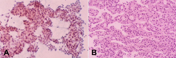
Architectural criteria. In 95 aspirates reviewed against histology, the most useful criteria for highly well-differentiated HCC were architectural: hypercellularity, arborescent cohesive clusters, broad trabeculae, and transgressing and peripheral endothelium (Wee and Nilsson 2003).
Cytologic criteria. Small monotonous hepatocytes with nuclear crowding, decreased cytoplasm, increased N/C ratio, atypical naked nuclei and tumor giant cells supported well-differentiated HCC (Wee and Nilsson 2003).
Unreliable criteria. Well-defined cell borders, abundant cytoplasm, eccentric nuclei, thick nuclear membranes, irregular nuclear contours, dense chromatin and macronucleoli were not always present in highly well-differentiated HCC. Some of these features occurred in dysplastic hepatocytes (Wee and Nilsson 2003).
Microtrabecular and microacinar HCC. In 14 of 123 HCCs (11.4%), aspirates showed small hepatocytes with minimal atypia in microacini of five or more cells and microtrabeculae of irregular thickness without apparent peripheral endothelium. Transgressing capillaries were found on careful inspection, and all cases had a deficient reticulin framework (Yang, Yang and Tao 2004).
CD34. CD34 is negative in normal sinusoids and stains sinusoids diffusely in HCC. Benign lesions show focal staining confined to periportal and periseptal areas. Diffuse CD34 staining was present in 82.5% of HCCs and in 8% of benign lesions in cell block and thin-core material (de Boer et al. 2000).
Hep Par 1. Hep Par 1 stains macroregenerative nodules, dysplastic nodules, focal nodular hyperplasia and hepatocellular adenoma. It therefore does not separate benign from malignant hepatocytic nodules (Wee 2005).
The spectrum of benign aspirates depends on the population. In a series of 1,741 ultrasound-guided liver aspirates from northern India, 372 aspirates were benign (Nigam et al. 2026).
Benign diagnosis
Number of cases
Cytologic findings reported
Related page
Abscess
178
Degenerated inflammatory cells and karyorrhectic debris
Source: Nigam et al. 2026. Inflammatory lesions formed 69.3% of benign diagnoses in this series.
Granulomas. Epithelioid granulomas are reported as benign. A Ziehl-Neelsen stain identified acid-fast bacilli in tuberculous granulomas in the Nigam series (Nigam et al. 2026).
The proposed WHO Reporting System for Liver Cytopathology
Status. The International Academy of Cytology (IAC), the International Agency for Research on Cancer (IARC) and the WHO have published reporting systems for lung, pancreaticobiliary, lymph node, thymus and spleen, and soft tissue cytopathology. In 2026 the liver system was one of four systems described as nearing completion (Field et al. 2026).
Common design. Each WHO system links a diagnostic category to a hierarchical risk of malignancy (ROM) and gives management recommendations, diagnostic criteria and guidance on ancillary testing and specimen handling (Field et al. 2026).
Proposed categories. One single-institution series reclassified 1,741 liver aspirates into the five categories of the proposed WHO Reporting System for Liver Cytology: nondiagnostic/inadequate, benign, atypical, suspicious for malignancy and malignant (Nigam et al. 2026). The WHO liver system itself was not yet published in 2026 (Field et al. 2026).
Proposed category
Aspirates, n (%)
With histological follow-up
Malignant on follow-up
ROM
Nondiagnostic/inadequate
81 (4.7%)
33
2
6.1%
Benign
372 (21.4%)
225
11
4.9%
Atypical
34 (2.0%)
27
11
40.7%
Suspicious for malignancy
39 (2.2%)
28
27
96%
Malignant
1,215 (69.8%)
667
667
100%
Source: Nigam et al. 2026 (1,741 aspirates; single institution; histology in 980). Sensitivity for malignancy was 98.46%, specificity 92.64% and accuracy 97.04%. The ROM ranges that the authors quote for the proposed WHO system were 15–67% (nondiagnostic), 3–16% (benign), 21.4–100% (atypical), 77–100% (suspicious) and 100% (malignant).
Adequacy in that series. At least two passes were made, and a pathologist confirmed adequacy on site. The nondiagnostic rate was 4.65% (Nigam et al. 2026).
Limits of the data. The ROM values come from one retrospective series with a high proportion of malignant aspirates. The published WHO system will define the final ROM values.
Sources of diagnostic error in nonneoplastic liver aspirates
Sampling of the lesion. A hepatocytic aspirate raises four questions: whether the sample is representative, whether the cells are benign or malignant, whether they are neoplastic or nonneoplastic, and whether they are intralesional or extralesional (Wee 2005).
Well-differentiated HCC. The lower accuracy of cytology alone in the Kuo series was mainly due to difficulty separating well-differentiated HCC from benign lesions (Kuo et al. 2004).
Small nodules in surveillance programs. Surveillance of cirrhotic patients detects smaller nodules, and tissue characterization of well-differentiated nodules ≤2 cm is difficult (Wee 2011).
Reactive changes as false positives. Atypia secondary to inflammation or regeneration can be mistaken for malignancy (Nigam et al. 2026, discussion). In that series, 1 of 28 suspicious aspirates with histology was chronic hepatitis C.
Situation
Interpretation
Reason
Benign hepatocytes with bile duct cells from a 3-cm solid mass
Nonrepresentative unless imaging explains the finding
Extralesional hepatocytes cannot be excluded (Wee 2005)
Enlarged hepatocytes with N/C ratio ≤1/3 in cirrhosis
Large cell change
Low-grade change common in cirrhosis (Wee 2005)
Small monotonous hepatocytes in broad trabeculae with endothelial wrapping
Well-differentiated HCC
Architectural criteria of HCC (Wee and Nilsson 2003)
Small hepatocytes in microacini, reticulin deficient
Well-differentiated HCC
Microacinar pattern of HCC (Yang, Yang and Tao 2004)
Iron-laden benign hepatocytes
Benign
Iron accumulation favors a benign process (Wee 2005)
Benign hepatocytes on FNA. A, a loose cluster of polygonal cells with ample granular cytoplasm, round central nuclei, small nucleoli and occasional binucleation; golden-brown lipofuscin granules sit in the cytoplasm (Papanicolaou stain). B, lipofuscin appears as dark blue-black granules (Romanowsky-type stain). Magnification not stated.Image: Wee Aileen, PMC open access (Cytojournal. 2005 Jun 8;2:7. doi: 10.1186/1742-6413-2-7) Figure 2, CC BY.
Benign Liver Lesions on Fine-Needle Aspiration: Abscess, Hydatid Cyst, Focal Nodular Hyperplasia, Hepatocellular Adenoma and Hemangioma
Definition. Benign focal liver lesions sampled by fine-needle aspiration (FNA) fall into three groups. Inflammatory and infectious lesions include pyogenic abscess, amebic abscess and cystic echinococcosis (hydatid cyst). Benign hepatocellular nodules include focal nodular hyperplasia (FNH) and hepatocellular adenoma (HCA). Vascular lesions include cavernous hemangioma. Inflammatory lesions formed 69.3% of benign diagnoses in a series of 1,741 liver aspirates, and abscess was the most common benign diagnosis (Nigam et al. 2026). The benign hepatocellular nodules are the most difficult group on FNA. Their separation from normal liver and from well-differentiated hepatocellular carcinoma (HCC) depends on architecture, which is often not preserved in aspirates (Balitzer and Greenland 2024).
Frequency. Abscess accounted for 178 of 372 benign liver aspirates in a northern Indian series. The smears showed degenerated inflammatory cells and karyorrhectic debris (Nigam et al. 2026).
Amebic abscess: clinical setting. In one series, seven patients treated for amebic dysentery had multiple liver lesions that were clinically suspected to be malignant. Ultrasound showed multiple well-defined hypoechoic cystic lesions of varying size (Mokhtari and Kumar 2014).
Amebic abscess: cytology. Smears showed necrotic material with mixed inflammatory cells and Entamoeba histolytica trophozoites. The trophozoites were round, with well-defined borders, a single eccentric nucleus with a central karyosome and ingested red blood cells in the cytoplasm. Charcot-Leyden crystals were present (Mokhtari and Kumar 2014).
Amebic abscess: value of FNA. FNA established the diagnosis and allowed early treatment (Mokhtari and Kumar 2014).
Pyogenic abscess: management. Pyogenic liver abscess has an increasing incidence and no international management guideline. Percutaneous drainage appeared superior to needle aspiration alone in a 2026 evidence synthesis. Colonoscopy is recommended after a diagnosis of cryptogenic pyogenic abscess (Gaillard et al. 2026).
Necrotic tumor as a mimic of abscess. Colorectal metastases show abundant tumor diathesis (Wee 2005). Necrotic debris alone from a liver mass therefore does not exclude a necrotic carcinoma, and the aspirate is examined for viable tumor cells.
Feature
Pyogenic abscess
Amebic abscess
Necrotic metastatic carcinoma
Background
Degenerated neutrophils, karyorrhectic debris
Necrotic material, mixed inflammatory cells
Necrotic tumor diathesis
Diagnostic element
Inflammatory cells without organisms or tumor cells
Trophozoites with a single eccentric nucleus, central karyosome and ingested red cells
Viable malignant epithelial cells
Other findings
—
Charcot-Leyden crystals
—
Source
Nigam et al. 2026; Gaillard et al. 2026
Mokhtari and Kumar 2014
Wee 2005
Cystic echinococcosis (hydatid cyst) on FNA
Organisms. Two Echinococcus species are of medical importance. Echinococcus granulosus causes cystic echinococcosis, and Echinococcus multilocularis causes alveolar echinococcosis (Brunetti, Kern and Vuitton 2010).
Management of cystic echinococcosis. The WHO Informal Working Group on Echinococcosis (WHO-IWGE) expert consensus recommends an image-based, stage-specific approach. The options are percutaneous treatment, surgery, anti-infective drug treatment and watch-and-wait (Brunetti, Kern and Vuitton 2010).
Management of alveolar echinococcosis. Early diagnosis and radical, tumor-like surgery followed by albendazole prophylaxis are the main elements of management. Most patients present at a stage when radical surgery is no longer possible (Brunetti, Kern and Vuitton 2010).
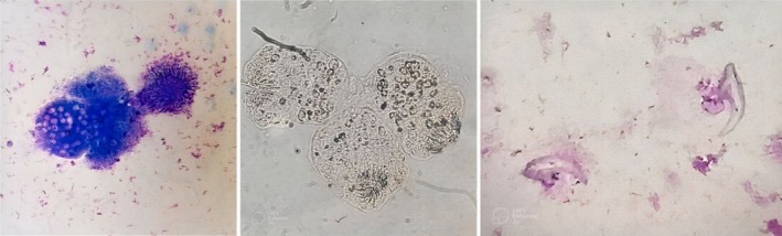
Cytologic elements. May-Grünwald-Giemsa-stained smears are examined for protoscolices, laminated membranes, scolices, hooklets and the background inflammatory response (Sadiku et al. 2026). Hooklets and protoscolices identified the hydatid cysts in the Nigam series (Nigam et al. 2026).
Diagnostic yield. In 14 aspirates of hepatic hydatid disease, seven (50%) showed diagnostic features of Echinococcus. The other seven yielded only histiocytes, necrotic debris or nonspecific inflammation (Sadiku et al. 2026).
Correlation with imaging and serology. A specific cytologic diagnosis requires identification of the characteristic elements. Imaging, serology and histopathology are correlated with the aspirate for safe and accurate management (Sadiku et al. 2026).
Mucinous cystic neoplasm in the differential. Mucinous cystic neoplasm is far more frequent in the liver than in the extrahepatic bile ducts and is an epithelial neoplasm in the differential diagnosis of a cystic liver lesion (Arends et al. 2026).
Risk of anaphylaxis. A systematic review of 5,943 percutaneous procedures on 5,517 echinococcal cysts found lethal anaphylaxis in 0.03% of procedures and reversible allergic reactions in 1.7%. Lethal anaphylaxis after percutaneous treatment was no more frequent than drug-related anaphylaxis (Neumayr et al. 2011).
Difficulty on FNA. Separation of well-differentiated HCC from FNH and large regenerative nodules requires an assessment of architectural features. This is difficult on FNA when intact tissue fragments are not sampled (Balitzer and Greenland 2024).
Glutamine synthetase. In 79 resected FNHs, glutamine synthetase (GS) staining formed large anastomosing areas of positive hepatocytes in a "map-like" pattern, often around hepatic veins. Hepatocytes next to fibrous bands containing arteries and ductules were negative (Bioulac-Sage et al. 2009, Liver Int).
Glutamine synthetase in the differential. HCA showed a GS pattern different from the map-like pattern of FNH, even when fibrous bands were present. β-catenin-mutated HCA and well-differentiated HCC showed diffuse GS staining without a map-like pattern (Bioulac-Sage et al. 2009, Liver Int).
Hep Par 1 and reticulin. Hep Par 1 stains FNH. The reticulin framework may be poorly developed in HCA and FNH (Wee 2005).
Other stains. Some FNHs show deficient canaliculi on polyclonal carcinoembryonic antigen (pCEA) staining, and some show substantial CD34 reactivity (Wee 2005). These findings overlap with HCC and do not establish malignancy in a hepatocytic nodule.
Hybrid lesions. Single nodules with distinct HCA and FNH components are exceptionally rare. In two cases, the HCA component was HNF1A-inactivated with loss of FABP1, the FNH component retained FABP1 and showed map-like GS, and the molecular findings supported a collision of two separate lesions (Zen and Schirmacher 2026).
Hepatocellular adenoma on FNA
Clinical setting. The most frequent setting is solitary or multiple adenomas in the normal liver of women of childbearing age exposed to oral contraceptives. HCA also occurs with steatohepatitis, vascular liver diseases and alcohol-related cirrhosis, in men and in infants (Bioulac-Sage, Sempoux and Balabaud 2017).
Complications. The two major complications are bleeding and malignant transformation (Bioulac-Sage, Sempoux and Balabaud 2017). In 411 patients, symptomatic bleeding occurred in 14%, 7% of nodules were borderline between HCA and HCC, and 3% of patients developed HCC. Of the patients, 85% were female, and the median age was 38 years (Nault et al. 2017).
β-catenin activation and HCC. In a surgical series of 128 HCAs, all six patients who developed HCC had β-catenin-activated tumors, with or without inflammatory features (Bioulac-Sage et al. 2009, Hepatology).
Morphologic correlates. Tumor steatosis and microadenomas were more frequent in HNF1A-inactivated HCA. Body mass index >25, peliosis and steatosis of the background liver were more frequent in inflammatory HCA (Bioulac-Sage et al. 2009, Hepatology).
Subtypes of hepatocellular adenoma in the WHO classification
The 6th edition of the WHO classification of digestive system tumours (2026) lists HNF1A-inactivated HCA, inflammatory HCA, β-catenin-activated HCA, β-catenin-activated inflammatory HCA and sonic hedgehog HCA. Sonic hedgehog HCA is newly defined in the 6th edition (Arends et al. 2026). In the 5th edition (2019), the HCA classification was based on biological and molecular factors (Nagtegaal et al. 2020). Before sonic hedgehog HCA was defined, about 10% of HCAs remained unclassified (Sala et al. 2020). With immunohistochemistry and molecular testing, fewer than 5% of HCAs now remain unclassified (Sempoux and Park 2026).
Subtype
Molecular basis
Immunohistochemical surrogate
Clinical association
Sources
HNF1A-inactivated
Inactivating HNF1A mutations
Loss of liver fatty acid-binding protein (LFABP)
Tumor steatosis, microadenomas
Margolskee et al. 2016; Bioulac-Sage et al. 2009, Hepatology
Inflammatory
Mutations that activate JAK/STAT3 signaling
Serum amyloid A (SAA) overexpression
Obesity, peliosis, background steatosis
Bioulac-Sage, Sempoux and Balabaud 2017; Margolskee et al. 2016
β-catenin-activated
Activating CTNNB1 mutations
GS upregulation and nuclear β-catenin
Higher risk of HCC
Bioulac-Sage et al. 2009, Hepatology; Margolskee et al. 2016
β-catenin-activated inflammatory
Both pathways
SAA with GS
Higher risk of HCC
Arends et al. 2026; Bioulac-Sage et al. 2009, Hepatology
Bland, mostly small hepatocytes; high risk of hemorrhage; no significant risk of malignant transformation
Arends et al. 2026; Nault et al. 2017; Sala et al. 2020
Frequency of sonic hedgehog HCA. The WHO 6th edition gives 5–10% of HCAs (Arends et al. 2026). In the 2017 molecular series that defined the group, it formed 4% of HCAs and was associated with obesity and bleeding (Nault et al. 2017).
Limits of immunohistochemistry. LFABP loss reliably identified HNF1A-mutated adenomas. GS and β-catenin staining did not consistently identify β-catenin-mutated adenomas, and mutational analysis improved their recognition (Margolskee et al. 2016).
Findings on FNA. Hep Par 1 stains HCA. The reticulin framework may be poorly developed, some HCAs show deficient canaliculi on pCEA staining, and CD34 can show substantial reactivity. Serum AFP is not elevated (Wee 2005). These findings overlap with HCC. Subtyping requires a cell block with immunohistochemistry and, for β-catenin-mutated adenomas, mutational analysis (Margolskee et al. 2016).
Hepatic hemangioma on FNA
Aspirate content. In 33 hemangiomas sampled by ultrasound-guided FNA, the aspirate was blood alone in 24. In the other nine, endothelial cells or clusters of capillaries were present and were considered diagnostic (Solbiati et al. 1985).
Diagnostic yield. In a second series of 10 FNAs from eight patients, capillary vessels or endothelial cells established the diagnosis in six. The authors gave a true-positive yield of 60% (Caturelli et al. 1986).
Cytology in a recent series. Hemangiomas showed spindle cells in a hemorrhagic background (Nigam et al. 2026).
Complications. One of 33 hemangiomas showed intratumoral bleeding on ultrasound that required no treatment (Solbiati et al. 1985). No complication followed 10 FNAs in the Caturelli series (Caturelli et al. 1986). A fatality after FNA of a hepatic hemangioma has been reported (Terriff, Gibney and Scudamore 1990).
Technique. A possible hemangioma was not considered an absolute contraindication to FNA when a fine needle and an optimal route were used (Solbiati et al. 1985).
Interpretation of a bloody aspirate. Blood alone from a lesion with imaging features of hemangioma is consistent with hemangioma but is not diagnostic. Endothelial cells or capillary fragments support the diagnosis (Solbiati et al. 1985).
Differential diagnosis of benign liver lesions on FNA
Lesion
Cytologic findings
Ancillary studies
Main mimic
Source
Pyogenic abscess
Degenerated neutrophils, karyorrhectic debris
—
Necrotic metastasis
Nigam et al. 2026; Wee 2005
Amebic abscess
Necrotic material, trophozoites with ingested red cells, Charcot-Leyden crystals
—
Pyogenic abscess
Mokhtari and Kumar 2014
Cystic echinococcosis
Protoscolices, hooklets, laminated membrane
Serology, imaging stage
Simple cyst; abscess
Sadiku et al. 2026; Brunetti, Kern and Vuitton 2010
Focal nodular hyperplasia
Benign hepatocytes; ductules and arteries in fibrous bands on cell block
Map-like GS pattern
HCA; well-differentiated HCC
Bioulac-Sage et al. 2009, Liver Int
Hepatocellular adenoma
Bland hepatocytes; reticulin may be poorly developed
LFABP, SAA, GS, β-catenin; mutational analysis
Well-differentiated HCC
Margolskee et al. 2016; Wee 2005
Hemangioma
Blood; endothelial cells or capillary fragments in a minority
—
Angiosarcoma, which also has a bloody background
Solbiati et al. 1985; Caturelli et al. 1986; Boucher et al. 2000
Reporting benign liver aspirates
Specific benign diagnosis. In the proposed WHO system, an aspirate with diagnostic benign findings is reported as benign.
Nonspecific findings. Hemorrhagic, necrotic or inflammatory aspirates without diagnostic elements are interpreted with imaging (Sadiku et al. 2026).
Benign hepatocytes from a mass. A sample of benign hepatocytes from a mass raises the question of whether the sampled cells are intralesional (Wee 2005). The report states whether the findings explain the imaging lesion.
Aspirate
Report
Reason
Hooklets and laminated membrane
Benign, cystic echinococcosis
Diagnostic parasitic elements (Sadiku et al. 2026)
Histiocytes and debris only from a cyst with imaging features of hydatid disease
Nonspecific; correlation with serology and imaging
Half of hydatid aspirates lack parasitic elements (Sadiku et al. 2026)
Trophozoites with ingested red cells
Benign, amebic abscess
Diagnostic organism (Mokhtari and Kumar 2014)
Blood only from a lesion with hemangioma imaging
Nondiagnostic or consistent with hemangioma, by imaging correlation
Blood alone in 24 of 33 hemangiomas (Solbiati et al. 1985)
Bland hepatocytes with map-like GS on cell block
Benign, consistent with FNH
Characteristic GS pattern (Bioulac-Sage et al. 2009, Liver Int)
Echinococcal (hydatid) cyst contents on FNA. Left, a Giemsa-stained smear with scolices. Middle, an unstained wet preparation with scolices bearing radially arranged hooklets. Right, suckers and detached hooklets, each pointed at one end and bifid at the other. The aspirate came from a thyroid hydatid cyst; scolices and hooklets have the same appearance in hepatic cysts.Image: Ghimire Prashant et al., PMC open access (Clin Case Rep. 2025 Apr 9;13(4):e70337. doi: 10.1002/ccr3.70337) FIGURE 2, CC BY.
Hepatocellular Carcinoma, Fibrolamellar Carcinoma and Hepatoblastoma on Fine-Needle Aspiration, with Immunohistochemistry
Definition. Hepatocellular carcinoma (HCC) is a malignant epithelial tumor with hepatocellular differentiation. It accounts for about 90% of primary liver cancers (EASL 2025). Fine-needle aspiration (FNA) is used for HCC when imaging is not diagnostic, in noncirrhotic livers and for tissue-based treatment selection (Normal Liver, FNA Indications and Reactive Changes on Liver FNA). The two main problems of HCC on FNA are the separation of well-differentiated HCC from benign hepatocellular nodules and the separation of poorly differentiated HCC from intrahepatic cholangiocarcinoma and metastatic carcinoma (Wee 2011). Fibrolamellar carcinoma is a subtype of HCC defined by the DNAJB1::PRKACA fusion (Nagtegaal et al. 2020). Some fibrolamellar carcinomas carry biallelic inactivating PRKAR1A mutations, which activate the same protein kinase A pathway (Rhee and Park 2026). Hepatoblastoma is the most common malignant liver tumor of children (Subramanian et al. 2019).
6th edition (2026). The 6th edition recognizes nine HCC subtypes, which account for about 35% of HCCs. Sarcomatoid HCC is added as a distinct subtype to the eight subtypes of the 5th edition. Two histological patterns are newly introduced: the vessels encapsulating tumor clusters (VETC) pattern and the CTNNB1-mutated pattern. The steatohepatitic subtype can evolve into a "burned-out" form with less fat and more stromal fibrosis (Rhee and Park 2026; Sempoux and Park 2026). The published summary of changes lists only the new hepatocellular adenoma subtype and the separation of small and large duct intrahepatic cholangiocarcinoma for the liver (Arends et al. 2026).
Subtypes in the 5th edition (2019; previous edition). Several rarer subtypes, together 20–30% of HCCs, are defined by consistent morphological, molecular and clinical features (Nagtegaal et al. 2020). Up to 35% of HCCs can be assigned to a subtype (Choi and Thung 2023). Eight subtypes are listed: steatohepatitic, clear cell, macrotrabecular massive, scirrhous, chromophobe, fibrolamellar, neutrophil-rich and lymphocyte-rich (Choi and Thung 2023).
Subtypes in the 4th edition (2010). The 2010 classification recognized five morphological subtypes: fibrolamellar, scirrhous, undifferentiated, lymphoepithelioma-like and sarcomatoid HCC. Architectural and cytologic variants were not separate subtypes (El Jabbour, Lagana and Lee 2019).
Grading. The WHO system is three-tiered (well, moderately and poorly differentiated). Both the worst and the predominant grades are reported, and the worst grade generally determines prognosis (Choi and Thung 2023).
Growth patterns. The predominant pattern is trabecular in about 70% of HCCs, solid (compact) in about 20%, pseudoglandular in about 10% and macrotrabecular in about 1%. Any pattern can occur in any subtype (Torbenson 2021).
Driver mutations.CTNNB1 mutations (40% of 343 resected HCCs) and TP53 mutations (21%) were mutually exclusive. CTNNB1-mutated tumors were well differentiated and cholestatic, with microtrabecular and pseudoglandular patterns. TP53-mutated tumors were poorly differentiated, with a compact pattern and pleomorphic cells (Calderaro et al. 2017).
Subtype (WHO 6th edition)
Approximate frequency
Defining feature
Prognosis compared with conventional HCC
Steatohepatitic
5–20%
Steatohepatitic features in ≥50% of the tumor
Similar
Clear cell
3–7%
>80% clear cells, due to glycogen
Better
Macrotrabecular massive
5–20%
Macrotrabecular pattern (≥6–10 cells thick) in ≥50% of the tumor
Worse
Scirrhous
4%
Dense intratumoral fibrosis in ≥50% of the tumor
Variable
Chromophobe
3%
Clear to eosinophilic cytoplasm, mostly bland nuclei; alternative lengthening of telomeres
Similar
Fibrolamellar
1%
Large eosinophilic cells, lamellar fibrosis, DNAJB1::PRKACA fusion
Similar to HCC in noncirrhotic liver
Neutrophil-rich
<1%
Numerous intratumoral neutrophils; granulocyte colony-stimulating factor production
Worse
Lymphocyte-rich
<1%
Lymphocytes outnumber tumor cells in most areas
Better
Sarcomatoid (new in the 6th edition)
<1%
Predominantly spindle cells
Worse
Sources: Choi and Thung 2023; Rhee and Park 2026. Sarcomatoid HCC was not a subtype in the 5th edition.
Molecular correlates of subtypes. The scirrhous subtype was associated with TSC1/TSC2 mutations and a progenitor expression profile. The steatohepatitic subtype showed IL-6/JAK/STAT activation without CTNNB1, TERT or TP53 alterations. The macrotrabecular massive subtype was associated with poor survival, high serum alpha-fetoprotein (AFP), vascular invasion, TP53 mutations and FGF19 amplification (Calderaro et al. 2017).
Histological patterns new in the 6th edition. The VETC pattern consists of tumor clusters encapsulated by continuous sinusoid-like endothelial vessels. It is present in 30–40% of resected HCCs and is associated with larger tumors, raised serum AFP and an unfavorable prognosis. The CTNNB1-mutated pattern, seen in about 60% of CTNNB1-mutated HCCs, is a well-differentiated tumor with microtrabecular, solid or acinar growth, frequent bile formation, scant inflammation and increased glutamine synthetase expression. Its prognosis is favorable (Rhee and Park 2026).
Keratin 19. Keratin 19 expression is an immunohistochemical marker of poor prognosis in HCC (Sempoux and Park 2026).
Macrotrabecular thickness. Macrotrabeculae have been defined as at least 6 or at least 10 cells thick, and published studies have used thresholds of 30–50% of the tumor (Torbenson 2021).
Cytologic criteria for HCC on FNA
Feature
Finding in HCC
Source
Trabeculae
Cords more than two cells thick, wrapped by peripheral endothelium
Wee 2005
Transgressing endothelium
Capillaries crossing cell aggregates (sinusoidal capillarization), best seen on Giemsa stain
Wee 2005
Naked nuclei
Atypical naked hepatocytic nuclei, often abundant
Wee 2005
N/C ratio
Increased, with monomorphism and nuclear crowding in well-differentiated tumors
Wee 2005
Bile
Within cells, canaliculi or pseudoacini
Wee 2005
Inclusions
Hyaline bodies, pale bodies, Mallory-Denk bodies; intranuclear inclusions are not specific
Wee 2005
Cytoplasmic contents
Fat and glycogen are common
Wee 2005
Bile duct cells
Scarce; their absence favors HCC
Wee 2005
Giant cells
Osteoclast-like or pleomorphic; their presence does not raise the grade
Poorly differentiated HCC. Pleomorphism, nuclear contour irregularity and prominent nucleoli are seen. The hepatocellular nature may be inapparent, and immunohistochemistry is required (Wee 2005).
Reticulin on the cell block. Reticulin staining increased accuracy, particularly for well-differentiated HCC (Kuo et al. 2004).
Cytologic variants of HCC on FNA
Fatty change. Fatty change can occur in HCC without background steatosis, especially in small lesions. It may mimic focal fatty change or signet ring cell adenocarcinoma (Wee 2005).
Clear cell change. Clear cells result from glycogen or lipid (Wee 2005).
Small cell pattern. Dissociated small cells with microacinar formation resemble a neuroendocrine tumor (Wee 2005).
Undifferentiated, spindle cell and giant cell patterns. These are rare and lack obvious hepatocytic features (Wee 2005).
Immunohistochemistry for hepatocellular differentiation on liver FNA
Marker
Pattern in HCC
Performance
Source
Arginase-1
Cytoplasmic
No nonhepatic tumor positive on tissue microarrays; positive in 81–84% of HCCs on FNA cell blocks; rare positivity in pancreatic, colorectal and breast adenocarcinomas
Timek et al. 2012; McKnight et al. 2012; Fujiwara et al. 2012
Hep Par 1
Cytoplasmic
Positive in 2.7% of nonhepatic tumors; staining falls from 100% in well-differentiated HCC to <5% in some poorly differentiated HCC
Timek et al. 2012; Wee 2005
Glypican-3
Cytoplasmic
Positive in 3.1% of nonhepatic tumors; negative in benign hepatocytic lesions
Timek et al. 2012; Chikhale et al. 2021
Polyclonal CEA
Canalicular
Sensitivity 95.2%; canalicular staining is specific for hepatocellular differentiation in a carcinoma
Wee 2005
CD10
Canalicular
Sensitivity 68.3%
Wee 2005
AFP
—
Sensitivity about 50% (range 20–75%)
Wee 2005
Albumin mRNA in situ hybridization
—
Positive in >95% of HCCs and in 80–95% of intrahepatic cholangiocarcinomas; negative in metastases
El Jabbour, Lagana and Lee 2019
CK7 and CK19
Focal in high-grade HCC
CK7 in 28% of non-fibrolamellar HCCs at a >15% threshold; diffuse CK7 in fibrolamellar carcinoma
Wee 2005; El Jabbour, Lagana and Lee 2019; Garg et al. 2018
CK20 and CDX2
Usually negative
CK20 in 14% and CDX2 in 5.2% of HCCs
El Jabbour, Lagana and Lee 2019
Recommended panel. On 62 liver FNA specimens, 19 of 29 HCCs were positive for arginase-1, Hep Par 1 and glypican-3, 9 for one or two of them, and 1 for none. A three-marker panel is recommended to separate HCC from metastatic carcinoma (Timek et al. 2012).
Performance in a second FNA series. Hep Par 1 stained 11 of 13 HCCs and glypican-3 stained 8 of 13. Arginase-1 showed cytoplasmic staining in only 3 of 13. All 55 metastatic adenocarcinomas and the cholangiocarcinoma were negative for Hep Par 1, arginase-1, glypican-3 and TTF-1. A panel of arginase-1, glypican-3, Hep Par 1, TTF-1 and CK19 had a sensitivity of 100% and a specificity of 88.9% (Chikhale et al. 2021).
Arginase-1 sensitivity across FNA series. Arginase-1 stained 37 of 44 HCC cell blocks (84.1%), more than Hep Par 1 (72.7%) and glypican-3 (56.8%) (McKnight et al. 2012). It stained 81% of 37 HCCs, more than Hep Par 1 (70%) and glypican-3 (54%) (Fujiwara et al. 2012). The 3 of 13 rate in the Chikhale series comes from a small series. Arginase-1 was diffusely positive in 2 of 15 pancreatic adenocarcinomas (Fujiwara et al. 2012).
Benign hepatocytes. Benign hepatic lesions expressed arginase-1 and Hep Par 1 and were negative for glypican-3 (Chikhale et al. 2021). Arginase-1 and Hep Par 1 therefore confirm hepatocellular origin but do not separate HCC from benign hepatocytes.
Panels applied to smears. A panel of Hep Par 1, AFP, BerEP4, CD31, CD68 and Ki-67 applied to smears separated HCC from metastatic carcinoma and regenerative nodules in 23 HCCs with 100% accuracy (Onofre et al. 2007).
Markers of malignancy in well-differentiated hepatocellular nodules
Glypican-3, HSP70 and glutamine synthetase. For early and grade 1 HCC, sensitivity and specificity were 59% and 86% for glutamine synthetase, 69% and 91% for glypican-3, and 78% and 95% for HSP70. With at least two of the three markers positive, sensitivity was 72% and specificity was 100% (Di Tommaso et al. 2007).
Staining of benign and dysplastic nodules. All three markers were negative in 100% of large regenerative and low-grade dysplastic nodules, in 73% of high-grade dysplastic nodules and in 3% of early or grade 1 HCCs (Di Tommaso et al. 2007).
Four-marker rule. Positive staining for two of four markers (glypican-3, glutamine synthetase, HSP70 and clathrin heavy chain) is highly specific for HCC (Singal et al. 2023).
CD34 and reticulin. Reticulin separated benign from malignant hepatocytic lesions more consistently than diffuse sinusoidal CD34 staining (de Boer et al. 2000).
Histologic features of HCC. The major histologic features are stromal invasion, increased cell density, intratumoral portal tracts, unpaired arteries, a pseudoglandular pattern and diffuse fatty change (Singal et al. 2023).
Lesion
Glypican-3
HSP70
Glutamine synthetase
CD34
Reticulin
Large regenerative nodule
Negative
Negative
Negative
Periportal and periseptal only
Intact
Low-grade dysplastic nodule
Negative
Negative
Negative
Periportal and periseptal only
Intact
High-grade dysplastic nodule
Negative in 73%
Negative in 73%
Negative in 73%
Periportal and periseptal only
Intact
Well-differentiated HCC
Positive in 69%
Positive in 78%
Positive in 59%
Diffuse in 82.5%
Decreased or absent
Sources: Di Tommaso et al. 2007; de Boer et al. 2000; Wee 2005.
Molecular testing for HCC on liver FNA material
Separation of HCC from intrahepatic cholangiocarcinoma.TERT promoter and CTNNB1 mutations are characteristic of HCC. BAP1, IDH1/IDH2 and PBRM1 mutations favor intrahepatic cholangiocarcinoma (Balitzer and Greenland 2024).
Yield on cell blocks. In 12 FNAs with cell blocks from suspected primary liver masses, next-generation sequencing supported HCC in one well-differentiated neoplasm and refined the diagnosis in six of eight poorly differentiated carcinomas. Two tumors remained unclassified (Balitzer and Greenland 2024).
Fibrolamellar carcinoma on FNA
Clinical features. Fibrolamellar carcinoma affects adolescents and young adults without primary liver disease or cirrhosis (Honeyman et al. 2014). The average age is 26 years, and hilar lymph node metastasis is more frequent than in conventional HCC (Torbenson 2021).
Molecular basis. A chimeric DNAJB1::PRKACA transcript results from a deletion of about 400 kilobases on chromosome 19. It was present in all 15 tumors examined (Honeyman et al. 2014).
Cytology. In six aspirates, the mean age was 19 years and serum AFP was normal in all patients. Smears showed large polygonal cells with abundant cytoplasm, vesicular nuclei and prominent nucleoli, with variably cellular fibrous stromal fragments. Intranuclear inclusions, cytoplasmic eosinophilic inclusions and bile were present. The tumor cells were 2.19 times the size of conventional HCC cells (Garg et al. 2018).
Pale bodies and oncocytic cells. Large discohesive oncocytic cells, lamellar fibrosis and pale bodies are characteristic (Wee 2005).
Immunohistochemistry. Fibrolamellar carcinoma showed strong membranous and cytoplasmic CK7 and granular cytoplasmic CD68 staining on cell blocks. Conventional HCC showed weak focal CK7 and canalicular CD68 only (Garg et al. 2018). In histologic material, CD68 showed granular dot-like staining in 31 of 32 fibrolamellar carcinomas, with a sensitivity of 96% and a specificity of 80% (Ross et al. 2011).
CK7 in conventional HCC. With a >15% threshold, 28% of non-fibrolamellar HCCs were CK7-positive and 32% focally positive, and all fibrolamellar carcinomas were positive (El Jabbour, Lagana and Lee 2019).
Hepatoblastoma on FNA
Epidemiology. Hepatoblastoma accounts for less than 1% of pediatric malignancies and is the most common malignant tumor of the liver in children (Subramanian et al. 2019).
Clinical findings. In one cytology series all children presented with an abdominal mass and raised serum AFP (Subramanian et al. 2019).
Cytology of epithelial hepatoblastoma. In 26 aspirates from children aged 4 months to 9 years, epithelial fragments showed a trabecular arrangement and acinar formation, and extramedullary hematopoiesis was present in 20 of 25 epithelial cases (Iyer et al. 2005).
Fetal component. Fetal cells had abundant cytoplasm, a small round nucleus resembling that of a fetal hepatocyte, finely granular chromatin and a single central nucleolus. Pleomorphism and mitoses were absent, and the N/C ratio was ≤1/3 (Iyer et al. 2005).
Embryonal component. Embryonal cells had scant cytoplasm, a pleomorphic nucleus, a high N/C ratio, coarsely granular chromatin, two to four angulated nucleoli and mitoses (Iyer et al. 2005).
Mesenchymal component. Eight of 14 tumors with histology were of mixed epithelial and mesenchymal type, but mesenchymal tissue was not seen in the aspirates (Iyer et al. 2005).
Subtyping on FNA. Fetal and embryonal subtypes can be recognized on smears, and cell blocks support ancillary studies. Extramedullary hematopoiesis supports the diagnosis (Subramanian et al. 2019).
β-catenin. Pathogenic CTNNB1 alterations, mainly involving exon 3, were found in 10 of 17 malignant pediatric liver tumors. Nuclear and cytoplasmic accumulation of β-catenin was present in all hepatoblastomas, including those without a detectable CTNNB1 alteration (Yamaoka et al. 2006).
Small cell undifferentiated tumors. Tumors classified as small cell undifferentiated hepatoblastoma show loss of INI1 expression and SMARCB1 deletion and are regarded as malignant rhabdoid tumors (Vokuhl et al. 2016). Hepatoblastoma with focal small cell histology responds to hepatoblastoma chemotherapy, whereas small cell undifferentiated tumors respond poorly (Fuchs et al. 2022).
Pediatric HCC as a mimic. Pediatric HCC (median age 10 years in one series) closely mimics hepatoblastoma, and serum AFP and imaging do not separate the two (Nalwa et al. 2023).
Feature
Fetal cells
Embryonal cells
Small cell undifferentiated tumor
Cytoplasm
Abundant
Scant
Round cell tumor without differentiation
N/C ratio
≤1/3
High
—
Chromatin
Finely granular
Coarsely granular
—
Nucleoli
Single, central
Two to four, angulated
—
Mitoses
Absent
Present
—
Ancillary finding
Nuclear β-catenin
Nuclear β-catenin
INI1 loss, SMARCB1 deletion
Source
Iyer et al. 2005; Yamaoka et al. 2006
Iyer et al. 2005; Yamaoka et al. 2006
Iyer et al. 2005; Vokuhl et al. 2016
Differential diagnosis of HCC on liver FNA
Differential
Features favoring the mimic
Features favoring HCC
Source
Benign hepatocytic nodule
Normal plates, bile duct cells, intact reticulin; glypican-3 negative
Broad trabeculae, endothelial wrapping, reticulin loss; two of glypican-3, HSP70 and glutamine synthetase positive
Wee and Nilsson 2003; Di Tommaso et al. 2007
Intrahepatic cholangiocarcinoma
Ductular or biliary features, desmoplasia, CK7 and CK19
Hepatocellular markers; TERT promoter or CTNNB1 mutation
Wee 2005; Balitzer and Greenland 2024
Metastatic adenocarcinoma
Arginase-1, Hep Par 1 and glypican-3 negative; site-specific markers
Arginase-1, Hep Par 1 and glypican-3 positive
Timek et al. 2012; Chikhale et al. 2021
Metastatic renal cell carcinoma
Transgressing and papillary endothelium without peripheral endothelium
Peripheral endothelium around trabeculae
Wee 2005
Neuroendocrine tumor
Dissociated cells with neuroendocrine features
Microacinar HCC with transgressing capillaries and reticulin loss
CAP protocol. The College of American Pathologists (CAP) publishes a protocol for the examination of specimens from patients with HCC (version 4.3.0.0) (CAP 2022).
Grade and subtype. The report states the subtype when the sample allows it, and the worst and predominant grades (Choi and Thung 2023).
Proposed WHO category. In the proposed WHO system, an unequivocal HCC is reported as malignant (Nigam et al. 2026).
Material for ancillary testing. Cell blocks support reticulin staining, immunohistochemistry and sequencing (Kuo et al. 2004; Balitzer and Greenland 2024).
hepatocellular nodule immunostain lookup
Well-differentiated hepatocellular carcinoma. A, FNA: thick, branching trabeculae of monotonous hepatocytes with crowded nuclei and round pseudoacinar spaces (Papanicolaou stain). B, matching histology with trabecular and pseudoglandular growth (H&E). Magnification not stated.Image: Wee Aileen, PMC open access (Cytojournal. 2005 Jun 8;2:7. doi: 10.1186/1742-6413-2-7) Figure 5, CC BY.
Intrahepatic Cholangiocarcinoma on Fine-Needle Aspiration and Its Separation from Metastatic Adenocarcinoma
Definition. Intrahepatic cholangiocarcinoma (iCCA) is a malignant epithelial tumor of the liver with biliary differentiation. All intrahepatic carcinomas with a ductal or tubular phenotype are classified as iCCA (Nagtegaal et al. 2020). The 6th edition of the WHO classification of digestive system tumours (2026) recognizes small duct iCCA and large duct iCCA as two separate entities (Arends et al. 2026). On fine-needle aspiration (FNA), iCCA is an adenocarcinoma without specific cytologic features. Its diagnosis depends on the clinical and imaging setting, the immunoprofile, albumin in situ hybridization and molecular testing, because the liver is one of the most common sites of metastatic adenocarcinoma (Shi et al. 2026).
Classification of iCCA in the WHO 5th and 6th editions
5th edition (2019). iCCA was one entity with two major subtypes, large duct and small duct. The large duct type resembles extrahepatic cholangiocarcinoma. The small duct type shares etiological, pathogenetic and imaging features with hepatocellular carcinoma (HCC). The subtypes differ in etiology, molecular alterations, growth pattern and clinical behavior (Nagtegaal et al. 2020).
6th edition (2026). The small duct and large duct types are separate entities (SD-ICCA and LD-ICCA). The change recognizes differences in etiology, pathogenesis, epidemiology, macroscopy, histopathology, molecular alterations, and clinical and therapeutic profile (Arends et al. 2026).
Cholangiolocarcinoma. In the 5th edition, cholangiolocellular carcinoma was removed from combined hepatocellular-cholangiocarcinoma, renamed cholangiolocarcinoma and classified as a subtype of small duct iCCA (Nagtegaal et al. 2020).
Intraductal precursors. Intraductal papillary neoplasm of bile duct, intraductal tubulopapillary neoplasm and intraductal oncocytic papillary neoplasm are separate entities in the 6th edition. They are described in the extrahepatic bile duct chapter and referred to in the liver chapter, together with mucinous cystic neoplasm, which is far more frequent in the liver (Arends et al. 2026).
Point
WHO 4th edition (2010)
WHO 5th edition (2019)
WHO 6th edition (2026)
Large duct and small duct iCCA
—
Two subtypes of one entity
Two separate entities
Cholangiolocellular carcinoma
Subtype of combined hepatocellular-cholangiocarcinoma
Renamed cholangiolocarcinoma; subtype of small duct iCCA
—
Carcinomas with ductal or tubular phenotype
—
All classified as iCCA
—
Sources: Nagtegaal et al. 2020; Arends et al. 2026. A dash indicates a point not addressed in the cited summary.
Small duct and large duct iCCA
Feature
Small duct iCCA
Large duct iCCA
Source
Resemblance
Shares etiology and imaging with HCC
Resembles extrahepatic cholangiocarcinoma
Nagtegaal et al. 2020
Mucin
Little mucin
Mucin production
Hayashi et al. 2016
Immunophenotype
N-cadherin and/or NCAM (CD56); C-reactive protein (CRP); albumin in situ hybridization positive
Diffuse S100P; strong diffuse polyclonal CEA (pCEA)
Hayashi et al. 2016; Gong, Chen and Matsukuma 2025
Chronic liver disease
More frequent
Less frequent
Hayashi et al. 2016
Serum CEA and CA 19-9
Lower
Higher
Hayashi et al. 2016
Perineural invasion and nodal metastasis
Less frequent
More frequent
Hayashi et al. 2016
Characteristic alterations
IDH mutations, FGFR2 translocations
KRAS mutations
Hayashi et al. 2016
Hayashi classification. In 102 consecutive iCCAs classified by mucin and immunophenotype, 42 were type 1 (mucin-producing, diffuse S100P) and 56 were type 2 (little mucin, N-cadherin and/or NCAM). KRAS mutation was significantly more frequent in type 1, and IDH mutation and FGFR2 translocation were restricted to type 2. Type 1 had worse survival on log-rank analysis but was not an independent prognostic factor (Hayashi et al. 2016).
Subclassification in a Western cohort. In 29 North American iCCAs, morphology alone classified 55% as small or large duct type. A panel of pCEA, CRP, N-cadherin and albumin in situ hybridization classified 93%. CRP, N-cadherin and albumin in situ hybridization were specific for small duct iCCA, and diffuse strong pCEA was specific for large duct tumors (Gong, Chen and Matsukuma 2025).
Geographic variation. In the same cohort, 88% of iCCAs were small duct type, and hepatolithiasis was absent, unlike East Asian cohorts (Gong, Chen and Matsukuma 2025).
Separation from extrahepatic cholangiocarcinoma. CRP, albumin in situ hybridization, N-cadherin and pCEA helped separate iCCA from extrahepatic cholangiocarcinoma when the primary site within the biliary tract was unclear (Gong, Chen and Matsukuma 2025).
Cytology of iCCA on FNA
Morphology. iCCA is usually a well to moderately differentiated adenocarcinoma, often with desmoplasia, minimal mucin and clusters of proliferating ductules (Wee 2005).
Diagnostic basis. iCCA is separated from HCC and metastatic carcinoma by its ductular or biliary features and its immunoprofile (Wee 2005).
Overlap with poorly differentiated HCC. Poorly differentiated HCC and iCCA may show overlapping features on FNA, and a subset of primary liver tumors remains unclassifiable after molecular testing (Balitzer and Greenland 2024).
Frequency in an FNA series. In a northern Indian series of 1,254 suspicious or malignant liver aspirates, cholangiocarcinoma formed 54 (4.8%) of the 1,132 diagnoses classed as metastatic, and 13 (10.6%) of the 122 primary liver malignancies were intrahepatic cholangiocarcinomas (Nigam et al. 2026).
Albumin in situ hybridization and immunohistochemistry for iCCA on cell blocks
Albumin in situ hybridization. Albumin messenger RNA in situ hybridization was positive in >95% of HCCs and in 80–95% of iCCAs and negative in metastatic tumors (El Jabbour, Lagana and Lee 2019).
Performance in a large cohort. In 153 iCCAs and 885 other adenocarcinomas, albumin in situ hybridization had a specificity of 98.8% and a sensitivity of 69.5% for iCCA. It was almost completely negative in perihilar cholangiocarcinoma (Albrecht et al. 2025).
C-reactive protein. CRP immunohistochemistry had a sensitivity of 84.1% and a specificity of 86.4% for iCCA. Combined with albumin in situ hybridization, sensitivity rose to 88.0% with a specificity of 86.1% (Albrecht et al. 2025).
Albumin expression and genotype. Albumin in situ hybridization was strongly positive in 93% of iCCAs with FGFR fusions or IDH1/IDH2 mutations. Tumors with BAP1, ARID1A or PBRM1 alterations were less often positive and stained more weakly (Shi et al. 2026). In the European cohort, 16 of 17 IDH1, IDH2 and FGFR2 alterations were in albumin-positive tumors, and the single KRAS mutation was in an albumin-negative tumor (Albrecht et al. 2025).
Hepatocellular markers. Arginase-1, Hep Par 1 and glypican-3 were negative in all metastatic adenocarcinomas and in the cholangiocarcinoma in a liver FNA series (Chikhale et al. 2021). Arginase-1 stained no nonhepatic tumor in 1,240 surgical specimens (Timek et al. 2012).
CK19. CK19 stained the cholangiocarcinoma and 21 of 55 metastatic adenocarcinomas in the same FNA series (Chikhale et al. 2021). CK19 therefore does not separate iCCA from metastatic adenocarcinoma.
Test
iCCA
HCC
Metastatic colorectal adenocarcinoma
Source
Albumin in situ hybridization
Positive in 69.5–95%, mainly small duct type
Positive in >95%
Negative or rarely positive
El Jabbour, Lagana and Lee 2019; Albrecht et al. 2025
CRP
Sensitivity 84.1%; specificity 86.4%
—
—
Albrecht et al. 2025
Arginase-1, Hep Par 1, glypican-3
Negative
Positive
Negative
Chikhale et al. 2021; Timek et al. 2012
CK19
Positive in the single case in the series
Focal in high-grade HCC
Positive in a subset
Chikhale et al. 2021; Wee 2005
SATB2
—
—
Positive in 92.2% of liver biopsies
Zhang et al. 2018
CDX2
—
Positive in 5.2%
Positive in 100% of liver biopsies; specificity 85.4%
Zhang et al. 2018; El Jabbour, Lagana and Lee 2019
CK20
—
Positive in 14%
Positive in 95.1% of liver biopsies
Zhang et al. 2018; El Jabbour, Lagana and Lee 2019
Separation of iCCA from metastatic adenocarcinoma
iCCA as a diagnosis of exclusion. iCCA remains a diagnosis of exclusion after a thorough clinical workup. None of the immunohistochemical markers proposed for it has reached clinical practice (Albrecht et al. 2025).
Colorectal and other metastatic adenocarcinomas. Markers of colorectal origin, smear panels for the primary site and carcinoma of unknown primary are given in Metastases, Lymphoma and Angiosarcoma in Liver FNA.
Regional patterns of metastasis. In the northern Indian FNA series, the leading primary sites of liver metastases were gallbladder (40.6%), pancreas (11.4%) and breast (6.0%) (Nigam et al. 2026).
Molecular alterations of iCCA tested on FNA material
Therapeutic targets.FGFR fusions and IDH1/IDH2 mutations are characteristic of iCCA, and inhibitors of these alterations improve survival (Shi et al. 2026).
Alterations that favor iCCA over HCC. In liver FNAs with cell blocks, IDH1, CDKN2A/CDKN2B and BRAF alterations supported iCCA in three patients (Balitzer and Greenland 2024).
Albumin in situ hybridization as a screen. Strong albumin positivity identified most iCCAs with FGFR fusions or IDH mutations and has been proposed as a screening tool for actionable alterations (Shi et al. 2026).
Combined hepatocellular-cholangiocarcinoma on FNA
Definition. Combined hepatocellular-cholangiocarcinoma contains two distinct cell populations, each confirmed by immunostains (Torbenson 2021).
Detection by biopsy. Histology can identify combined hepatocellular-cholangiocarcinoma among LR-5 observations (Singal et al. 2023).
FNA and sequencing. Next-generation sequencing of FNA cell blocks identified two combined tumors among eight poorly differentiated primary liver carcinomas (Balitzer and Greenland 2024).
Limits of FNA. The diagnosis requires two cell populations that are distinct on H&E, each confirmed by immunostains (Torbenson 2021). An aspirate that contains one component therefore cannot establish a combined tumor.
Differential diagnosis of adenocarcinoma in a liver FNA
Tumor
Clinical and imaging setting
Morphology
Supportive ancillary findings
Source
Small duct iCCA
Mass in the liver, often with chronic liver disease
Little mucin; ductular clusters
Albumin in situ hybridization, CRP, N-cadherin; IDH1/IDH2, FGFR2
Hayashi et al. 2016; Gong, Chen and Matsukuma 2025
Large duct iCCA
Mass near large intrahepatic ducts
Mucin-producing glands
Diffuse S100P and pCEA; albumin negative; KRAS
Hayashi et al. 2016; Gong, Chen and Matsukuma 2025
Perihilar or extrahepatic cholangiocarcinoma
Hilar stricture
Biliary adenocarcinoma
Albumin in situ hybridization almost always negative
Albrecht et al. 2025
Metastatic colorectal adenocarcinoma
Multiple masses; colorectal history
Glands with abundant tumor diathesis
SATB2, CK20, CDX2
Zhang et al. 2018; Wee 2005
Poorly differentiated HCC
Cirrhosis or chronic liver disease
Polygonal cells, trabeculae
Arginase-1, Hep Par 1, glypican-3; TERT promoter, CTNNB1
Timek et al. 2012; Balitzer and Greenland 2024
Combined hepatocellular-cholangiocarcinoma
Any; may present as LR-5
Two populations
Hepatocellular and biliary markers in separate components
Torbenson 2021; Singal et al. 2023
Reporting iCCA on liver FNA
CAP protocol. The College of American Pathologists publishes a protocol for the examination of specimens from patients with carcinoma of the intrahepatic bile ducts (version 4.4.0.0, September 2026) (CAP 2026).
Wording of the diagnosis. An adenocarcinoma with a biliary immunoprofile and positive albumin in situ hybridization supports iCCA. Without albumin positivity, the report states the differential with metastatic biliary-type adenocarcinoma from the gallbladder, pancreas or extrahepatic bile ducts (Albrecht et al. 2025).
Material for molecular testing. Cell blocks from liver FNAs support next-generation sequencing (Balitzer and Greenland 2024). Testing for FGFR2 fusions and IDH1/IDH2 mutations identifies patients for targeted therapy (Shi et al. 2026).
Proposed WHO category. A definite adenocarcinoma is reported in the malignant category of the proposed WHO system (Nigam et al. 2026).
Metastatic Tumors, Lymphoma and Angiosarcoma in Liver Fine-Needle Aspiration
Definition. Malignant tumors in liver fine-needle aspiration (FNA) other than hepatocellular carcinoma (HCC) and intrahepatic cholangiocarcinoma include metastatic carcinoma, melanoma and sarcoma, lymphoma, and primary hepatic angiosarcoma. Metastases formed 90.3% of suspicious and malignant liver aspirates in a large Indian series (Nigam et al. 2026). The proportion depends on the population: in a Taiwanese series, HCC was the most common malignancy in liver aspirates (Kuo et al. 2004). The 6th edition of the WHO classification of digestive system tumours (2026) describes liver metastases in a dedicated section of its chapter on other tumours and metastases, together with a new section on carcinoma of unknown primary (Arends et al. 2026).
Frequency of metastatic tumors in liver FNA series
Series
Aspirates
Leading malignant diagnoses
Source
Northern India, 8 years
1,741
Metastases 90.3% of suspicious or malignant aspirates; primary sites gallbladder 40.6%, pancreas 11.4%, breast 6.0%; primary liver malignancies 9.7%
Nigam et al. 2026
Taiwan, 1 year
936
HCC 45.6% of aspirates; metastatic adenocarcinoma 5.6%
Kuo et al. 2004
Tumor types among metastases. In the Indian series, adenocarcinoma formed 75.6% of the 1,132 metastatic malignancies, ductal carcinoma of the breast 6.0% and neuroendocrine tumor 4.7% (Nigam et al. 2026).
Common primary sites of metastatic adenocarcinoma. Most metastatic adenocarcinomas in liver FNA come from the stomach, colorectum, pancreas, breast and lung (Wee 2005).
Primary liver malignancies in the same series. HCC formed 86.9% of the 122 primary liver malignancies, intrahepatic cholangiocarcinoma 10.6%, hepatoblastoma 1.7% and angiosarcoma 0.8% (Nigam et al. 2026).
Cytologic patterns of metastatic tumors on liver FNA
Pattern
Common tumors
Diagnostic features
Source
Glandular
Adenocarcinoma of the colorectum, stomach, pancreas, breast, lung
Colorectal metastases show abundant tumor diathesis
Wee 2005
Squamous
Squamous cell carcinoma, often from the pancreaticobiliary tract
Tadpole-shaped cells, keratinization, necrosis
Wee 2005
Small round cell
Neuroendocrine tumor, small cell carcinoma, nasopharyngeal carcinoma, lymphoma
Microacinar HCC is a mimic
Wee 2005
Clear cell
Renal cell carcinoma
Transgressing and papillary endothelium without peripheral endothelium
Hepatoid and alpha-fetoprotein-producing carcinomas, mainly of the lung and gastrointestinal tract
Mimic HCC
Wee 2005
Melanocytic
Melanoma
SOX10 and other melanocytic markers
Saad et al. 2024
Immunohistochemistry for the primary site of liver metastases
Panels applied to smears. A panel of CK5/6, CK7, CK20, CA125, TTF-1 and CDX2 applied to the FNA smear identified the primary site of 85 metastatic carcinomas with an accuracy of 90.3%. The primary remained clinically unknown in 23 cases (Onofre et al. 2007).
Colorectal adenocarcinoma. In 192 liver biopsies, SATB2 had a sensitivity of 92.2% and a specificity of 97.8%, CK20 95.1% and 91.0%, and CDX2 100% and 85.4%. All three markers were positive in 89.3% of colorectal and 2.2% of noncolorectal liver metastases (Zhang et al. 2018).
SATB2 with CK7 and CK20. SATB2 had a sensitivity of 93% and a specificity of 77% for colorectal origin, rising to a specificity of 100% in combination with CK7 and CK20 (Dragomir et al. 2014).
GIST. A spindle cell metastasis in the liver requires CD117 (c-kit) staining to identify GIST (Wee 2005).
Suspected primary
Markers
Performance
Source
HCC
Arginase-1, Hep Par 1, glypican-3
Arginase-1: no nonhepatic tumor positive on tissue microarrays; rare adenocarcinomas positive on FNA, diffusely in 2 of 15 pancreatic adenocarcinomas
Timek et al. 2012; Fujiwara et al. 2012
Intrahepatic cholangiocarcinoma
Albumin in situ hybridization, C-reactive protein
Albumin specificity 98.8%
Albrecht et al. 2025
Colorectal adenocarcinoma
SATB2, CK20, CDX2
Triple positive in 89.3%
Zhang et al. 2018
Melanoma
SOX10, PRAME, HMB45, Melan-A, S100
SOX10 sensitivity 100% in cytology
Saad et al. 2024
Angiosarcoma
CD31, FLI-1, CD34
Endothelial differentiation confirmed by immunocytochemistry
Gupta et al. 2023; Boucher et al. 2000
GIST
CD117
—
Wee 2005
Carcinoma of unknown primary in the WHO 6th edition
New section. Carcinoma of unknown primary is included in a separate section for the first time (Arends et al. 2026).
Definition. Carcinoma of unknown primary is a metastatic epithelial malignancy whose primary site has not been identified despite comprehensive clinical, radiological and pathological investigation, including immunohistochemical and molecular testing. It is an operational term (Arends et al. 2026).
Subdivision. Tumors are first divided by whether the clinical, radiological, immunohistochemical and molecular profile suggests origin within or outside the gastrointestinal tract, and then into favorable and unfavorable groups. The favorable group generally benefits from site-specific therapy, and the optimal therapy for the unfavorable group is not defined (Arends et al. 2026).
Colon-like subtype. The only favorable subtype with a gastrointestinal-like profile is the colon-like subtype: CDX2 and CK20 positive and CK7 negative, or with a molecular profile similar to colorectal carcinoma (Arends et al. 2026).
Metastatic melanoma in liver FNA
SOX10. SOX10 had a sensitivity of 100% and a specificity of 93% for melanoma in histologic material. It stained 12% of breast carcinomas and no ovarian, endometrial, lung or colon carcinomas (Mohamed et al. 2013).
Marker sensitivity in cytology specimens. In 93 melanomas sampled by cytology, PRAME stained 86%, SOX10 100%, HMB45 84%, Melan-A 82% and S100 71% (Saad et al. 2024).
PRAME limitations. Spindle cell melanoma was PRAME-negative in 4 of 10 cases. PRAME also stained 23% of carcinomas, 33% of sarcomas and 11% of hematologic malignancies, giving a specificity of 82% (Saad et al. 2024).
Separation from HCC. A large-cell malignant tumor negative for arginase-1, Hep Par 1 and glypican-3 is tested with melanocytic markers when the history or morphology suggests melanoma.
Marker
Sensitivity in cytology specimens
Notes
Source
SOX10
100%
Also stains 12% of breast carcinomas
Saad et al. 2024; Mohamed et al. 2013
PRAME
86%
Negative in 40% of spindle cell melanomas; stains some carcinomas and sarcomas
Saad et al. 2024
HMB45
84%
—
Saad et al. 2024
Melan-A
82%
—
Saad et al. 2024
S100
71%
Lowest sensitivity
Saad et al. 2024
Lymphoma involving the liver
Primary hepatic lymphoma. Primary hepatic lymphoma is a biopsy-proven liver lesion without lymphomatous involvement of other solid organs. It accounts for 0.4% of primary extranodal non-Hodgkin lymphomas (Ma et al. 2025).
Subtypes. In an institutional cohort of 30 patients, diffuse large B-cell lymphoma (DLBCL) formed 50%, marginal zone lymphoma 17%, chronic lymphocytic leukemia/small lymphocytic lymphoma 13%, follicular lymphoma 10% and T-cell lymphoma 7%. In 192 published cases, DLBCL formed 40%, marginal zone lymphoma 34%, T-cell lymphoma 13% and Burkitt lymphoma 7% (Ma et al. 2025).
Associations. Hepatitis C was present in 16% and hepatitis B in 17% of published cases (Ma et al. 2025).
Presentation. On imaging, primary hepatic lymphoma presents as solitary or multiple nodules that resemble carcinoma or benign inflammatory conditions (Choi and Gill 2018).
Flow cytometry on FNA. Flow cytometry is part of the FNA assessment of lymphoid lesions. Adequacy criteria for flow cytometry, assessment of blood contamination and TRBC1/TRBC2 analysis of T-cell populations are part of the evaluation (Sewell et al. 2026).
Hepatosplenic T-cell lymphoma
Cell of origin and setting. Hepatosplenic T-cell lymphoma most commonly arises from γδ T cells and is more common in adolescents and young adults, with a median age of 34 years and a strong male predominance. About 20% of cases arise with chronic immunosuppression or immune dysregulation (Pro, Allen and Behdad 2020).
Inflammatory bowel disease. Risk factors in inflammatory bowel disease are young age, concomitant use of anti-tumor necrosis factor (TNF) agents and thiopurines, Crohn disease, male sex and thiopurine use for 2 years or more (Pro, Allen and Behdad 2020).
Pattern of involvement. The liver, spleen and bone marrow are involved in a sinusoidal pattern. Cytopenias are common, and lymphadenopathy is typically absent (Pro, Allen and Behdad 2020).
Immunophenotype. The cells are CD3, CD2 and CD7 positive, usually CD56 positive and CD57 negative, and TIA-1 positive. They are typically CD4 and CD8 double negative, although CD8 may be expressed. Perforin and granzyme B are typically negative. About 20% express the αβ T-cell receptor (Pro, Allen and Behdad 2020).
Cytogenetics. Isochromosome 7q and trisomy 8 occur in up to 63% and 50% of cases and frequently coexist (Pro, Allen and Behdad 2020).
Hepatosplenic involvement in about 0.2% of cases; diffuse or sinusoidal and portal infiltration
Similar to the tumor at other sites
Zhang et al. 2023
Primary hepatic angiosarcoma on FNA
Etiology. Vinyl chloride is one of only two environmental agents definitely linked to a type of liver cancer, and it causes angiosarcoma of the liver (McGlynn et al. 2025).
Clinical features. In 44 patients from four institutions, the median age was 63.7 years, 81.4% had bilobar disease and 37.2% had metastases at presentation. Only 10 patients underwent resection. Median overall survival was 5.8 months, with 1-, 3- and 5-year survival of 30.0%, 8.1% and 5.6% (Wilson et al. 2019).
Imaging. Imaging is often nondiagnostic (Wilson et al. 2019).
Cytology. Smears showed singly scattered, large, markedly pleomorphic epithelioid to elongated cells with oval to elongated nuclei, irregular nuclear contours, coarse chromatin, macronucleoli and abundant finely vacuolated cytoplasm in a bloody background (Gupta et al. 2023).
Vasoformative features. In 15 angiosarcomas from various sites, intracellular red blood cells, well-formed vessels, microacinar or lumen formation and intracytoplasmic lumens were variably present. The background was bloody in all specimens. Atypia ranged from relatively bland to highly pleomorphic (Boucher et al. 2000).
Immunocytochemistry. The diagnosis can be suggested by FNA when vasoformative features are present and established with immunocytochemical confirmation of endothelial differentiation (Boucher et al. 2000). In a hepatic case, the cell block was diffusely positive for vimentin and CD31 with nuclear FLI-1 (Gupta et al. 2023).
Feature
Hemangioma
Angiosarcoma
Source
Aspirate
Blood alone in most cases
Bloody background in all cases
Solbiati et al. 1985; Boucher et al. 2000
Cells
Endothelial cells or capillary fragments in a minority
Pleomorphic epithelioid or spindled cells, singly or in groups
Solbiati et al. 1985; Gupta et al. 2023
Vasoformative features
Capillary fragments
Intracytoplasmic lumens and intracellular red cells
Boucher et al. 2000
Endothelial markers
—
CD31, FLI-1, CD34
Gupta et al. 2023; Boucher et al. 2000
Epithelioid hemangioendothelioma of the liver on FNA
Classification. The 6th edition of the WHO classification of digestive system tumours (2026) describes mesenchymal tumors in a dedicated chapter aligned with other WHO tumour volumes (Arends et al. 2026).
Behavior. Epithelioid hemangioendothelioma (EHE) is a rare vascular tumor of intermediate malignancy, often with indolent behavior (Bourgeau et al. 2021). Hepatic EHE has a more favorable prognosis than HCC (Gupta et al. 2010).
Imaging. Multiple liver nodules resemble metastatic carcinoma on imaging (Gupta et al. 2010).
Cytology. The cytologic features of hepatic EHE include discohesive atypical cells with intracytoplasmic lumina and intranuclear inclusions. Intracytoplasmic lumina are rarely seen in aspirates. In one hepatic aspirate, paucicellular smears showed single cells and small tissue fragments, with small bland epithelioid cells and larger pleomorphic cells with hyperchromatic nuclei. Many cells contained sharply defined intranuclear pseudoinclusions. The smears were interpreted as poorly differentiated carcinoma, and the diagnosis was made on core biopsy (Gupta et al. 2010). In another case, FNA showed a vascular proliferation with an epithelioid component, and a small biopsy established the diagnosis (Campione et al. 2015).
Endothelial markers and keratin. On core biopsy, the tumor cells were strongly and diffusely positive for CD31 and CD34 and focally positive for pancytokeratin (Gupta et al. 2010). Keratin staining was present in frozen sections of 1 of 2 EHEs and in an epithelioid angiosarcoma (Gray et al. 1990). ERG was expressed in 42 of 43 EHEs and in 96 of 100 angiosarcomas (Miettinen et al. 2011).
CAMTA1. Diffuse nuclear CAMTA1 staining was present in 51 of 59 EHEs (86%), including 92% of conventional tumors. Six of the eight CAMTA1-negative tumors were TFE3-positive. Other epithelioid vascular and nonvascular mesenchymal tumors were negative, except one tumor diagnosed as epithelioid angiosarcoma on core biopsy (Doyle, Fletcher and Hornick 2016). In a second series, CAMTA1 was positive in 14 of 16 EHEs and in 1 of 276 other tumors, a ductal carcinoma of the breast with focal weak staining (Shibuya et al. 2015). In 34 hepatic EHEs and 25 hepatic angiosarcomas, CAMTA1 was positive in 91.2% of EHEs and negative in all angiosarcomas. Aberrant p53 expression, high Ki-67 labeling and high mitotic activity were associated with angiosarcoma (Yoo et al. 2024).
WWTR1::CAMTA1 fusion. The t(1;3)(p36.3;q25) translocation produces the WWTR1::CAMTA1 fusion. Rearrangement of both genes was present in all 17 EHEs tested, including tumors of the liver, and in none of the other epithelioid vascular tumors examined (Errani et al. 2011). The fusion is present in about 90% of EHEs (Doyle, Fletcher and Hornick 2016).
YAP1::TFE3 subset. Fewer than 5% of EHEs carry a YAP1::TFE3 fusion (Doyle, Fletcher and Hornick 2016). These tumors occurred in young adults (mean age 30 years), showed at least focal well-formed vascular channels with variably solid architecture, and expressed endothelial markers and strong nuclear TFE3 (Antonescu et al. 2013). A hepatic YAP1::TFE3 EHE was diagnosed by FNA and core biopsy in an 18-year-old male with synchronous lung and liver involvement (Bourgeau et al. 2021).
Feature
Epithelioid hemangioendothelioma
Angiosarcoma
Metastatic carcinoma
Smears
Paucicellular; single bland and pleomorphic epithelioid cells; intranuclear pseudoinclusions; intracytoplasmic lumina rare
Bloody background; intracellular red blood cells
Usually hypercellular; pleomorphic cells with prominent nucleoli; mucin-containing vacuoles in some cases
ERG
42 of 43
96 of 100
30 of 66 prostatic adenocarcinomas and 1 of 42 large cell undifferentiated lung carcinomas; other carcinomas negative
Keratin
Focal in some cases
Present in epithelioid angiosarcoma
Positive in ≥98% of samples in 62 of 75 epithelial tumor entities; lower rates in renal cell, neuroendocrine and poorly differentiated carcinomas
CAMTA1
Nuclear in 86–91%
Negative in hepatic angiosarcomas
Focal weak staining in 1 ductal carcinoma of the breast; other non-EHE tumors negative
Sources
Gupta et al. 2010; Gray et al. 1990; Miettinen et al. 2011; Doyle, Fletcher and Hornick 2016; Yoo et al. 2024
Boucher et al. 2000; Gray et al. 1990; Miettinen et al. 2011; Yoo et al. 2024
Gupta et al. 2010; Miettinen et al. 2011; Menz et al. 2023; Shibuya et al. 2015
Differential diagnosis of malignant tumors other than HCC and cholangiocarcinoma in liver FNA
Cytologic finding
Leading diagnoses
First-line ancillary studies
Source
Glands with dirty necrosis
Metastatic colorectal adenocarcinoma
SATB2, CK20, CDX2
Zhang et al. 2018; Wee 2005
Large malignant cells without glandular differentiation
Melanoma; poorly differentiated HCC
SOX10; arginase-1, Hep Par 1, glypican-3
Saad et al. 2024; Timek et al. 2012
Lymphoid cells
Lymphoma
Flow cytometry; cell block immunohistochemistry
Sewell et al. 2026
Pleomorphic cells in a bloody background
Angiosarcoma
CD31, FLI-1, CD34
Gupta et al. 2023; Boucher et al. 2000
Spindle cells
GIST; leiomyosarcoma
CD117
Wee 2005
Clear cells
Renal cell carcinoma; clear cell HCC
Hepatocellular markers
Wee 2005; Timek et al. 2012
Reporting metastatic tumors, lymphoma and angiosarcoma on liver FNA
Malignant category. Unequivocal malignant tumors are reported in the malignant category of the proposed WHO system (Nigam et al. 2026).
Statement of the primary site. The report names the tumor type and, when the immunoprofile allows it, the likely primary site. Carcinomas whose primary site is not identified after comprehensive clinical, radiological and pathological investigation are classified as carcinoma of unknown primary (Arends et al. 2026).
Triage for lymphoma. A lymphoid aspirate is triaged for flow cytometry and cell block (Sewell et al. 2026).
References
Albrecht T, Rossberg A, Rose F, et al. Combined analysis of albumin in situ hybridisation and C reactive protein immunohistochemistry for the diagnosis of intrahepatic cholangiocarcinoma: towards a molecular classification paradigm. J Clin Pathol. 2025;78(5):307–316. doi:10.1136/jcp-2024-209429. PMID 38749660
Antonescu CR, Le Loarer F, Mosquera JM, et al. Novel YAP1-TFE3 fusion defines a distinct subset of epithelioid hemangioendothelioma. Genes Chromosomes Cancer. 2013;52(8):775–784. doi:10.1002/gcc.22073. PMID 23737213
Arends MJ, Esposito I, Gill AJ, et al. Changes in the 6th edition of the World Health Organization classification of tumours of the digestive system. Histopathology. 2026;88(7):1295–1314. doi:10.1111/his.70116. PMID 41724188
Balitzer DJ, Greenland NY. The utility of next-generation sequencing in challenging liver FNA biopsies. Cancer Cytopathol. 2024;132(11):714–722. doi:10.1002/cncy.22893. PMID 39097802
Bioulac-Sage P, Laumonier H, Couchy G, et al. Hepatocellular adenoma management and phenotypic classification: the Bordeaux experience. Hepatology. 2009;50(2):481–489. doi:10.1002/hep.22995. PMID 19585623
Bioulac-Sage P, Laumonier H, Rullier A, et al. Over-expression of glutamine synthetase in focal nodular hyperplasia: a novel easy diagnostic tool in surgical pathology. Liver Int. 2009;29(3):459–465. doi:10.1111/j.1478-3231.2008.01849.x. PMID 18803590
Bioulac-Sage P, Sempoux C, Balabaud C. Hepatocellular adenoma: classification, variants and clinical relevance. Semin Diagn Pathol. 2017;34(2):112–125. doi:10.1053/j.semdp.2016.12.007. PMID 28131467
Boucher LD, Swanson PE, Stanley MW, Silverman JF, Raab SS, Geisinger KR. Cytology of angiosarcoma. Findings in fourteen fine-needle aspiration biopsy specimens and one pleural fluid specimen. Am J Clin Pathol. 2000;114(2):210–219. doi:10.1309/PXMU-LF05-3894-W29F. PMID 10941336
Bourgeau M, Martinez A, Deeb KK, et al. Cytologic features of hepatic YAP1-TFE3 rearranged epithelioid hemangioendothelioma. Diagn Cytopathol. 2021;49(12):E447–E452. doi:10.1002/dc.24853. PMID 34411462
Brunetti E, Kern P, Vuitton DA; Writing Panel for the WHO-IWGE. Expert consensus for the diagnosis and treatment of cystic and alveolar echinococcosis in humans. Acta Trop. 2010;114(1):1–16. doi:10.1016/j.actatropica.2009.11.001. PMID 19931502
Calderaro J, Couchy G, Imbeaud S, et al. Histological subtypes of hepatocellular carcinoma are related to gene mutations and molecular tumour classification. J Hepatol. 2017;67(4):727–738. doi:10.1016/j.jhep.2017.05.014. PMID 28532995
Campione S, Cozzolino I, Mainenti P, et al. Hepatic epithelioid hemangioendothelioma: pitfalls in the diagnosis on fine needle cytology and "small biopsy" and review of the literature. Pathol Res Pract. 2015;211(9):702–705. doi:10.1016/j.prp.2015.06.009. PMID 26187370
Caturelli E, Rapaccini GL, Sabelli C, et al. Ultrasound-guided fine-needle aspiration biopsy in the diagnosis of hepatic hemangioma. Liver. 1986;6(6):326–330. doi:10.1111/j.1600-0676.1986.tb00299.x. PMID 3553820
Chikhale M, Toi PC, Siddaraju N, Ananthakrishnan R. The strength of cytomorphology and efficacy of immuno-cytochemistry in distinguishing hepatocellular carcinoma from its mimics on fine-needle aspiration cytology. Diagn Cytopathol. 2021;49(7):864–875. doi:10.1002/dc.24759. PMID 33929782
Choi JH, Thung SN. Advances in histological and molecular classification of hepatocellular carcinoma. Biomedicines. 2023;11(9):2582. doi:10.3390/biomedicines11092582. PMID 37761023
de Boer WB, Segal A, Frost FA, Sterrett GF. Can CD34 discriminate between benign and malignant hepatocytic lesions in fine-needle aspirates and thin core biopsies? Cancer. 2000;90(5):273–278. PMID 11038423
Di Tommaso L, Franchi G, Park YN, et al. Diagnostic value of HSP70, glypican 3, and glutamine synthetase in hepatocellular nodules in cirrhosis. Hepatology. 2007;45(3):725–734. doi:10.1002/hep.21531. PMID 17326147
Doyle LA, Fletcher CD, Hornick JL. Nuclear expression of CAMTA1 distinguishes epithelioid hemangioendothelioma from histologic mimics. Am J Surg Pathol. 2016;40(1):94–102. doi:10.1097/PAS.0000000000000511. PMID 26414223
Dragomir A, de Wit M, Johansson C, Uhlen M, Pontén F. The role of SATB2 as a diagnostic marker for tumors of colorectal origin: results of a pathology-based clinical prospective study. Am J Clin Pathol. 2014;141(5):630–638. doi:10.1309/AJCPWW2URZ9JKQJU. PMID 24713733
El Jabbour T, Lagana SM, Lee H. Update on hepatocellular carcinoma: pathologists' review. World J Gastroenterol. 2019;25(14):1653–1665. doi:10.3748/wjg.v25.i14.1653. PMID 31011252
Errani C, Zhang L, Sung YS, et al. A novel WWTR1-CAMTA1 gene fusion is a consistent abnormality in epithelioid hemangioendothelioma of different anatomic sites. Genes Chromosomes Cancer. 2011;50(8):644–653. doi:10.1002/gcc.20886. PMID 21584898
European Association for the Study of the Liver. EASL Clinical Practice Guidelines on the management of hepatocellular carcinoma. J Hepatol. 2025;82(2):315–374. doi:10.1016/j.jhep.2024.08.028. PMID 39690085
Field AS, Pitman MB, Rekhi B, Cree IA, Lokuhetty D, Schmitt F. WHO Reporting Systems in cytopathology: a review of current status and the future. Acta Cytol. 2026 (online ahead of print). doi:10.1159/acy/abjag010. PMID 42814652
Fuchs J, Murtha-Lemekhova A, Kessler M, et al. A systematic review and meta-analysis of malignant rhabdoid and small cell undifferentiated liver tumors: a rational for a uniform classification. Cancers (Basel). 2022;14(2):272. doi:10.3390/cancers14020272. PMID 35053437
Fujiwara M, Kwok S, Yano H, Pai RK. Arginase-1 is a more sensitive marker of hepatic differentiation than HepPar-1 and glypican-3 in fine-needle aspiration biopsies. Cancer Cytopathol. 2012;120(4):230–237. doi:10.1002/cncy.21190. PMID 22434791
Gaillard C, Costa AC, Uldry É, et al. Clinical management of pyogenic liver abscess: a point-by-point synthesis of the available evidence and existing gaps. Infect Dis Now. 2026 (online ahead of print):105379. doi:10.1016/j.idnow.2026.105379. PMID 42967575
Garg R, Srinivasan R, Dey P, et al. Utility of cytokeratin7 immunocytochemistry in the cytopathological diagnosis of fibrolamellar hepatocellular carcinoma. J Cytol. 2018;35(2):75–78. doi:10.4103/JOC.JOC_130_17. PMID 29643652
Gong R, Chen ZE, Matsukuma K. Morphologic and immunohistochemical characterization of small and large duct cholangiocarcinomas in a Western cohort: a panel of pCEA, CRP, N-cadherin, and albumin in situ hybridization aids in subclassification. Ann Diagn Pathol. 2025;75:152437. doi:10.1016/j.anndiagpath.2025.152437. PMID 39832462
Gupta P, Singh B, Chaluvashetty SB, et al. Cytomorphologic and immunocytochemical diagnosis of primary hepatic angiosarcoma in a young adult: challenging diagnosis of a rare, aggressive malignancy. Diagn Cytopathol. 2023;51(3):E82–E88. doi:10.1002/dc.25076. PMID 36409510
Gupta R, Mathur SR, Gupta SD, et al. Hepatic epithelioid hemangioendothelioma: a diagnostic pitfall in aspiration cytology. Cytojournal. 2010;6:25. doi:10.4103/1742-6413.58951. PMID 20165548
Hayashi A, Misumi K, Shibahara J, et al. Distinct clinicopathologic and genetic features of 2 histologic subtypes of intrahepatic cholangiocarcinoma. Am J Surg Pathol. 2016;40(8):1021–1030. doi:10.1097/PAS.0000000000000670. PMID 27259014
Honeyman JN, Simon EP, Robine N, et al. Detection of a recurrent DNAJB1-PRKACA chimeric transcript in fibrolamellar hepatocellular carcinoma. Science. 2014;343(6174):1010–1014. doi:10.1126/science.1249484. PMID 24578576
Iyer VK, Kapila K, Agarwala S, Verma K. Fine needle aspiration cytology of hepatoblastoma. Recognition of subtypes on cytomorphology. Acta Cytol. 2005;49(4):355–364. doi:10.1159/000326166. PMID 16124162
Kuo FY, Chen WJ, Lu SN, Wang JH, Eng HL. Fine needle aspiration cytodiagnosis of liver tumors. Acta Cytol. 2004;48(2):142–148. doi:10.1159/000326307. PMID 15085744
Ma J, Daou R, Bou Eid J, et al. Management approaches for primary hepatic lymphoma: 10 year institutional experience with comprehensive literature review. Front Oncol. 2025;15:1475118. doi:10.3389/fonc.2025.1475118. PMID 40182049
Margolskee E, Bao F, de Gonzalez AK, et al. Hepatocellular adenoma classification: a comparative evaluation of immunohistochemistry and targeted mutational analysis. Diagn Pathol. 2016;11:27. doi:10.1186/s13000-016-0475-5. PMID 26961851
McGlynn KA, Watling CZ, Hernandez BY, Groopman JD. Environmental risk factors for liver cancer. Hepatology. 2025 (online ahead of print). doi:10.1097/HEP.0000000000001463. PMID 40622863
McKnight R, Nassar A, Cohen C, Siddiqui MT. Arginase-1: a novel immunohistochemical marker of hepatocellular differentiation in fine needle aspiration cytology. Cancer Cytopathol. 2012;120(4):223–229. doi:10.1002/cncy.21184. PMID 22298472
Menz A, Gorbokon N, Viehweger F, et al. Pan-keratin immunostaining in human tumors: a tissue microarray study of 15,940 tumors. Int J Surg Pathol. 2023;31(6):927–938. doi:10.1177/10668969221117243. PMID 35946088
Miettinen M, Wang ZF, Paetau A, et al. ERG transcription factor as an immunohistochemical marker for vascular endothelial tumors and prostatic carcinoma. Am J Surg Pathol. 2011;35(3):432–441. doi:10.1097/PAS.0b013e318206b67b. PMID 21317715
Mohamed A, Gonzalez RS, Lawson D, Wang J, Cohen C. SOX10 expression in malignant melanoma, carcinoma, and normal tissues. Appl Immunohistochem Mol Morphol. 2013;21(6):506–510. doi:10.1097/PAI.0b013e318279bc0a. PMID 23197006
Mokhtari M, Kumar PV. Amebic liver abscess: fine needle aspiration diagnosis. Acta Cytol. 2014;58(3):225–228. doi:10.1159/000358865. PMID 24662424
Nagtegaal ID, Odze RD, Klimstra D, et al. The 2019 WHO classification of tumours of the digestive system. Histopathology. 2020;76(2):182–188. doi:10.1111/his.13975. PMID 31433515
Nalwa A, Nakra T, Yadav R, et al. Cytomorphology of paediatric hepatocellular carcinoma: a useful diagnostic adjunct. Cytopathology. 2023;34(5):479–488. doi:10.1111/cyt.13266. PMID 37357840
Nault JC, Couchy G, Balabaud C, et al. Molecular classification of hepatocellular adenoma associates with risk factors, bleeding, and malignant transformation. Gastroenterology. 2017;152(4):880–894.e6. doi:10.1053/j.gastro.2016.11.042. PMID 27939373
Neumayr A, Troia G, de Bernardis C, et al. Justified concern or exaggerated fear: the risk of anaphylaxis in percutaneous treatment of cystic echinococcosis—a systematic literature review. PLoS Negl Trop Dis. 2011;5(6):e1154. doi:10.1371/journal.pntd.0001154. PMID 21695106
Nigam N, Kumari N, Singh J, et al. Fine-needle aspiration cytology of hepatic masses: a retrospective observational study and assessment of risk of malignancy based on the proposed World Health Organization Reporting System for Liver Cytology. J Cytol. 2026;43(3):157–166. doi:10.4103/joc.joc_117_25. PMID 42592409
Onofre AS, Pomjanski N, Buckstegge B, Böcking A. Immunocytochemical diagnosis of hepatocellular carcinoma and identification of carcinomas of unknown primary metastatic to the liver on fine-needle aspiration cytologies. Cancer. 2007;111(4):259–268. doi:10.1002/cncr.22768. PMID 17567831
Pro B, Allen P, Behdad A. Hepatosplenic T-cell lymphoma: a rare but challenging entity. Blood. 2020;136(18):2018–2026. doi:10.1182/blood.2019004118. PMID 32756940
Rhee H, Park YN. Comprehensive radiopathological and molecular review of HCC: expanding on the WHO, 6th edition. Clin Mol Hepatol. 2026 (online ahead of print). doi:10.3350/cmh.2026.0431. PMID 42576691
Ross HM, Daniel HD, Vivekanandan P, et al. Fibrolamellar carcinomas are positive for CD68. Mod Pathol. 2011;24(3):390–395. doi:10.1038/modpathol.2010.207. PMID 21113139
Saad M, Cantley R, Hao W, et al. Performance of preferentially expressed antigen in melanoma (PRAME) immunohistochemistry for metastatic melanoma in cytology specimens. Diagn Cytopathol. 2024;52(7):362–368. doi:10.1002/dc.25313. PMID 38558495
Sadiku F, Qerimi A, Bunjaku G, Maloku I, Pajaziti A. Fine-needle aspiration cytology of hepatic hydatid disease: a five-year case series with emphasis on diagnostic limitations. Acta Cytol. 2026;70(4):387–392. doi:10.1159/000550489. PMID 41553949
Sala M, Gonzales D, Leste-Lasserre T, et al. ASS1 overexpression: a hallmark of sonic hedgehog hepatocellular adenomas; recommendations for clinical practice. Hepatol Commun. 2020;4(6):809–824. doi:10.1002/hep4.1514. PMID 32490318
Sempoux C, Park YN. Integration of histopathology and molecular features for the classification of benign and malignant hepatocellular neoplasms. Virchows Arch. 2026 (online ahead of print). doi:10.1007/s00428-026-04685-x. PMID 42678415
Sewell WA, Al-Abbadi MA, Geddie WR, et al. The power of flow cytometry in the cytopathologic diagnosis of lymphoid lesions. Acta Cytol. Published online January 8, 2026:1–10. doi:10.1159/000550290. PMID 41505355
Shi J, Hosseini S, Lang E, et al. Comparison of albumin in situ hybridization in genetically characterized cholangiocarcinoma and tumors of other origins. Mod Pathol. 2026;39(11):101078. doi:10.1016/j.modpat.2026.101078. PMID 42660428
Shibuya R, Matsuyama A, Shiba E, Harada H, Yabuki K, Hisaoka M. CAMTA1 is a useful immunohistochemical marker for diagnosing epithelioid haemangioendothelioma. Histopathology. 2015;67(6):827–835. doi:10.1111/his.12713. PMID 25879300
Silva MA, Hegab B, Hyde C, Guo B, Buckels JA, Mirza DF. Needle track seeding following biopsy of liver lesions in the diagnosis of hepatocellular cancer: a systematic review and meta-analysis. Gut. 2008;57(11):1592–1596. doi:10.1136/gut.2008.149062. PMID 18669577
Singal AG, Llovet JM, Yarchoan M, et al. AASLD Practice Guidance on prevention, diagnosis, and treatment of hepatocellular carcinoma. Hepatology. 2023;78(6):1922–1965. doi:10.1097/HEP.0000000000000466. PMID 37199193
Solbiati L, Livraghi T, De Pra L, Ierace T, Masciadri N, Ravetto C. Fine-needle biopsy of hepatic hemangioma with sonographic guidance. AJR Am J Roentgenol. 1985;144(3):471–474. doi:10.2214/ajr.144.3.471. PMID 3881893
Subramanian KS, Chikhale M, Barwad A, Gochhait D, Toi PC, Siddaraju N. Fine-needle aspiration cytology and cell block in diagnosis and subtyping of hepatoblastoma. Diagn Cytopathol. 2019;47(7):706–710. doi:10.1002/dc.24167. PMID 31009172
Terriff BA, Gibney RG, Scudamore CH. Fatality from fine-needle aspiration biopsy of a hepatic hemangioma. AJR Am J Roentgenol. 1990;154(1):203–204. doi:10.2214/ajr.154.1.2104717. PMID 2104717
Timek DT, Shi J, Liu H, Lin F. Arginase-1, HepPar-1, and glypican-3 are the most effective panel of markers in distinguishing hepatocellular carcinoma from metastatic tumor on fine-needle aspiration specimens. Am J Clin Pathol. 2012;138(2):203–210. doi:10.1309/AJCPK1ZC9WNHCCMU. PMID 22904131
Torbenson MS. Hepatocellular carcinoma: making sense of morphological heterogeneity, growth patterns, and subtypes. Hum Pathol. 2021;112:86–101. doi:10.1016/j.humpath.2020.12.009. PMID 33387587
Vokuhl C, Oyen F, Häberle B, von Schweinitz D, Schneppenheim R, Leuschner I. Small cell undifferentiated (SCUD) hepatoblastomas: all malignant rhabdoid tumors? Genes Chromosomes Cancer. 2016;55(12):925–931. doi:10.1002/gcc.22390. PMID 27356182
Wee A, Nilsson B. Highly well differentiated hepatocellular carcinoma and benign hepatocellular lesions. Can they be distinguished on fine needle aspiration biopsy? Acta Cytol. 2003;47(1):16–26. doi:10.1159/000326470. PMID 12585026
Wee A. Fine needle aspiration biopsy of hepatocellular carcinoma and hepatocellular nodular lesions: role, controversies and approach to diagnosis. Cytopathology. 2011;22(5):287–305. doi:10.1111/j.1365-2303.2011.00882.x. PMID 21762411
Wee A. Fine needle aspiration biopsy of the liver: algorithmic approach and current issues in the diagnosis of hepatocellular carcinoma. Cytojournal. 2005;2:7. doi:10.1186/1742-6413-2-7. PMID 15941489
Wilson GC, Lluis N, Nalesnik MA, et al. Hepatic angiosarcoma: a multi-institutional, international experience with 44 cases. Ann Surg Oncol. 2019;26(2):576–582. doi:10.1245/s10434-018-7062-9. PMID 30456677
Yamaoka H, Ohtsu K, Sueda T, Yokoyama T, Hiyama E. Diagnostic and prognostic impact of beta-catenin alterations in pediatric liver tumors. Oncol Rep. 2006;15(3):551–556. PMID 16465411
Yang GC, Yang GY, Tao LC. Cytologic features and histologic correlations of microacinar and microtrabecular types of well-differentiated hepatocellular carcinoma in fine-needle aspiration biopsy. Cancer. 2004;102(1):27–33. doi:10.1002/cncr.20000. PMID 14968415
Yoo Y, Shin J, Jun E, et al. Proposed diagnostic and prognostic markers of primary malignant hepatic vascular neoplasms. Diagn Pathol. 2024;19(1):68. doi:10.1186/s13000-024-01482-5. PMID 38741104
Zen Y, Schirmacher P. Hybrid lesions of hepatocellular adenoma and focal nodular hyperplasia: a clinicopathological and molecular study of two cases. Histopathology. 2026 (online ahead of print). doi:10.1111/his.70305. PMID 42899380
Zhang YH, Li Z, Zhao S, et al. Extranodal natural killer/T-cell lymphoma with hepatosplenic involvement: a retrospective study of a consecutive 14-year case series. Ann Hematol. 2023;102(10):2803–2813. doi:10.1007/s00277-023-05358-4. PMID 37434096
Zhang YJ, Chen JW, He XS, et al. SATB2 is a promising biomarker for identifying a colorectal origin for liver metastatic adenocarcinomas. EBioMedicine. 2018;28:62–69. doi:10.1016/j.ebiom.2018.01.001. PMID 29396302
Image credits
Benign hepatocytes on FNA. A, a loose cluster of polygonal cells with ample granular cytoplasm, round central nuclei, small nucleoli and occasional binucleation; golden-brown lipofuscin granules sit in the cytoplasm (Papanicolaou stain). B, lipofuscin appears as dark blue-black granules (Romanowsky-type stain). Magnification not stated. Image: Wee Aileen, PMC open access (Cytojournal. 2005 Jun 8;2:7. doi: 10.1186/1742-6413-2-7) Figure 2, CC BY.
Echinococcal (hydatid) cyst contents on FNA. Left, a Giemsa-stained smear with scolices. Middle, an unstained wet preparation with scolices bearing radially arranged hooklets. Right, suckers and detached hooklets, each pointed at one end and bifid at the other. The aspirate came from a thyroid hydatid cyst; scolices and hooklets have the same appearance in hepatic cysts. Image: Ghimire Prashant et al., PMC open access (Clin Case Rep. 2025 Apr 9;13(4):e70337. doi: 10.1002/ccr3.70337) FIGURE 2, CC BY.