Library › Papillary thyroid carcinoma
Papillary thyroid carcinoma
Last reviewed 2026-11-04 by a board-certified pathologist. 57 references.
Papillary Thyroid Carcinoma (PTC)
Definition. A malignant epithelial tumor of follicular cells, diagnosed by its characteristic nuclear features, with or without papillae (WHO 2022). It is the most common thyroid malignancy. In US SEER-9 data from 1974 to 2013, PTC accounted for 64,625 of 77,276 thyroid cancers (84%) (Lim 2017). Imaging, treatment and follow-up are covered in Papillary Thyroid Carcinoma: Imaging, Treatment and Follow-up, and cytology in Papillary Thyroid Carcinoma (Cytology).
Epidemiology of papillary thyroid carcinoma
- Incidence: US thyroid cancer incidence nearly tripled from 1975 to 2009, from 4.9 to 14.3 per 100,000. PTC accounted for almost the entire increase, from 3.4 to 12.5 per 100,000 (Davies 2014).
- Detection and true increase: much of the rise reflects detection of small tumors (Davies 2014). PTC incidence also rose for regional and distant stages, and incidence-based mortality for distant-stage PTC increased 2.9% per year from 1994 to 2013 (Lim 2017).
- Sex and age: women accounted for 75% of patients with thyroid cancer in SEER-9, and the mean age at diagnosis was 48 years (Lim 2017).
- Children: in a 715-patient multicenter pediatric cohort, PTC accounted for 94% of differentiated thyroid carcinomas, and 16.5% of children with PTC had distant metastasis (Wasserman 2026).
Clinical features of papillary thyroid carcinoma
- Main risk factor: ionizing radiation, especially in childhood. After the Chornobyl accident, the frequency of fusion-positive PTC increased with thyroid radiation dose (Karyadi 2025).
- Presentation: a painless nodule, often detected incidentally.
- Spread: mainly through lymphatics to central (level VI) and lateral neck nodes. Lymph node metastases are present in 30–80% of patients with PTC, and the central neck is the most common compartment involved (Stack 2012). Blood-borne spread to lung and bone is uncommon.
- Prognosis: in SEER data for 2016–2022, five-year relative survival for thyroid cancer was 98.3% overall, 99.9% for localized, 98.1% for regional and 48.3% for distant disease (SEER 2026). Age at diagnosis (AJCC cutoff 55 years), distant metastasis and gross extrathyroidal extension are the main determinants of stage.
Pathogenesis of papillary thyroid carcinoma: MAPK pathway activation
- Driver alterations: BRAF and RAS mutations and RET and NTRK1 fusions all activate the MAPK pathway and are almost always mutually exclusive (TCGA 2014).
- TCGA 2014 (496 tumors):
- BRAF mutations in 61.7%, mostly V600E
- gene fusions in 15.3%, mutually exclusive with BRAF, RAS and EIF1AX mutations
- TERT promoter mutations in 9.4%
- new drivers (EIF1AX, PPM1D, CHEK2 and further fusions) reduced the fraction of tumors with no known driver from 25% to 3.5%
- Signaling output: BRAF V600E signals as a monomer and escapes negative feedback from ERK, which produces high MAPK output. RAS mutations and kinase fusions signal through RAF dimers that respond to ERK feedback, which produces lower output (TCGA 2014).
- BRAF-like and RAS-like groups: the BRAF V600E–RAS score (BRS) places each tumor on a scale between the two expression profiles (TCGA 2014).
- BRAF-like: BRAF V600E and BRAF fusions; RET fusions are weakly BRAF-like. These tumors are mostly classic and tall cell PTC and have lower thyroid differentiation scores.
- RAS-like: RAS mutations, BRAF K601E and PAX8::PPARG fusion. These tumors are mostly follicular-patterned and have higher thyroid differentiation scores.
- TERT promoter mutations occur in less-differentiated PTC and are associated with a high risk of recurrence (TCGA 2014). BRAF V600E with a TERT promoter mutation identifies the PTC with the highest recurrence rate (Xing 2014).
- BRAF V600E and mortality: PTC-related mortality was 5.3% with the mutation and 1.1% without it. After adjustment for lymph node metastasis, extrathyroidal extension and distant metastasis, the association was no longer significant (Xing 2013).
- MicroRNA: miR-146b, miR-155, miR-187, miR-197, miR-221, miR-222 and miR-224 are overexpressed in thyroid carcinomas compared with hyperplastic nodules, and this set detected cancer in FNA samples (Nikiforova 2008).
Familial papillary thyroid carcinoma and inherited syndromes
Familial non-medullary thyroid carcinoma falls into two groups. In the first group, non-thyroid tumors predominate. In the second, non-medullary thyroid tumors predominate, as in pure familial PTC, familial PTC with papillary renal neoplasia and familial PTC with multinodular goiter. Familial tumors present at a younger age and show more lymph node metastasis and extrathyroidal invasion (Guilmette 2018).
| Syndrome | Gene | Thyroid findings |
|---|---|---|
| Familial adenomatous polyposis | APC | Cribriform-morular thyroid carcinoma, classified separately from PTC in WHO 2022 (Baloch 2022) |
| PTEN hamartoma tumor syndrome (Cowden syndrome) | PTEN | Thyroid cancer standardized incidence ratio of 72 in PTEN mutation carriers; follicular carcinoma is overrepresented. SDHx variants and KLLN epimutation in Cowden-like patients are associated with PTC (Ngeow 2011) |
| Carney complex | PRKAR1A | Follicular cell-derived thyroid tumors (Guilmette 2018) |
| DICER1 syndrome | DICER1 | Multinodular goiter, with PTC arising in hyperplastic nodules in children without prior chemotherapy (Rutter 2016) |
| Werner syndrome | WRN | In Japanese patients, 48% follicular, 35% papillary and 13% anaplastic carcinoma, compared with 14%, 78% and 2% in the general Japanese population (Ishikawa 1999) |
Gross findings and multifocality in papillary thyroid carcinoma
- Appearance: firm, white, infiltrative or encapsulated mass, often with gritty calcification. Cystic change is common in nodal metastases.
- Papillary microcarcinoma: a tumor ≤1 cm. WHO 2022 no longer treats it as a subtype, and the CAP protocol states that the term is not used alone without a subtype designation (CAP 2023).
- Encapsulated tumors: the entire capsule is submitted, because NIFTP requires exclusion of capsular and vascular invasion (Nikiforov 2016).
- Multifocality: PTC is frequently multifocal. Separate foci arise either by intraglandular spread from one tumor or as independent primary tumors. In an X-chromosome inactivation study, separate foci showed discordant clonal patterns, indicating independent origin, in 5 of 10 informative patients (Shattuck 2005).
- Staging of multifocal tumors: the tumor with the highest T category (usually the largest) determines the T category, and the suffix (m) records multiple tumors (Amin 2017; CAP 2023).
Microscopic findings in papillary thyroid carcinoma
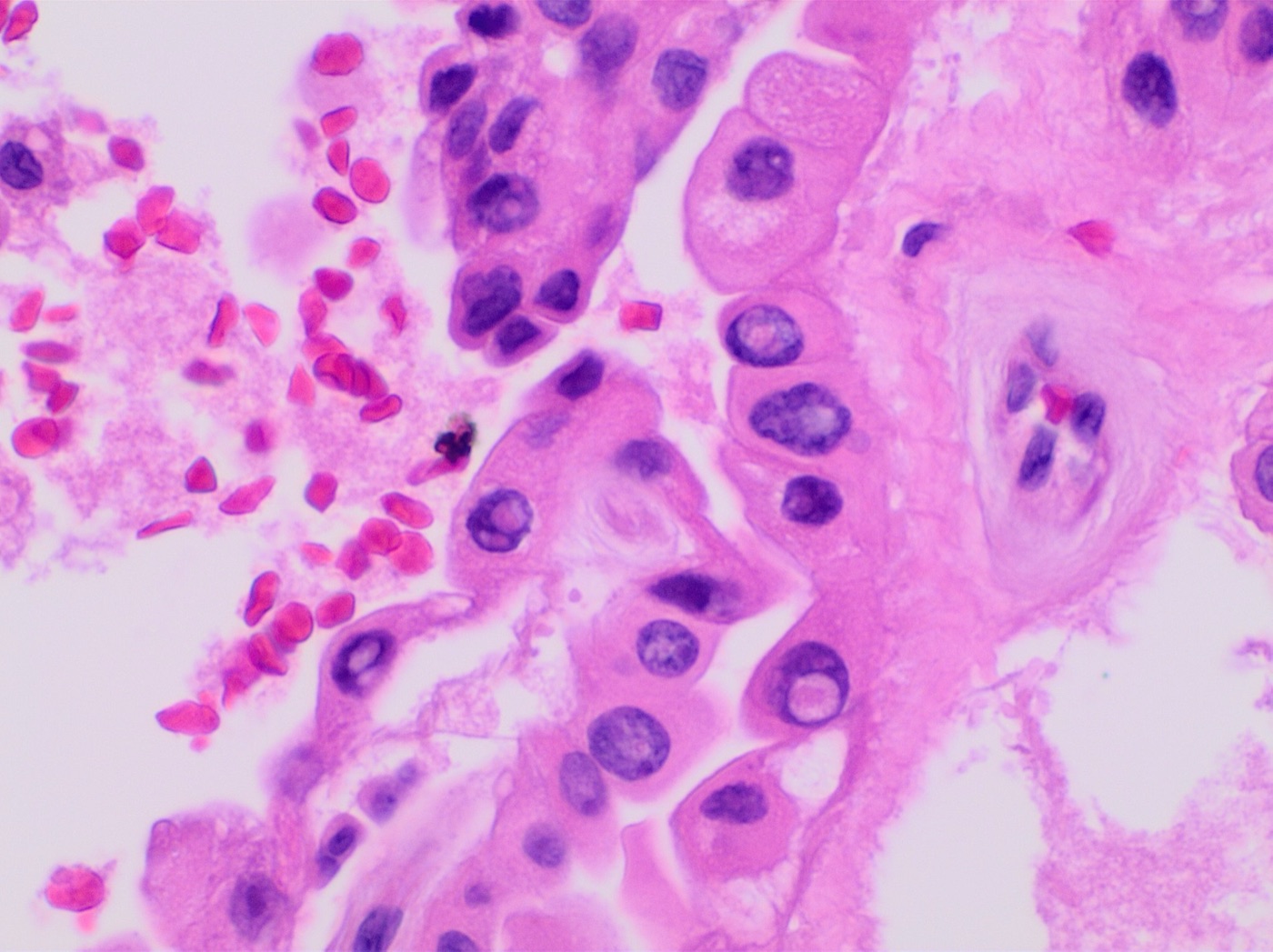
Nuclear features (these define the diagnosis):
- enlargement, overlapping and crowding
- elongation, with irregular contours and nuclear grooves
- intranuclear pseudoinclusions (cytoplasmic invaginations)
- chromatin clearing, giving "Orphan Annie eye" nuclei (most prominent in formalin-fixed tissue)
Architecture and other features:
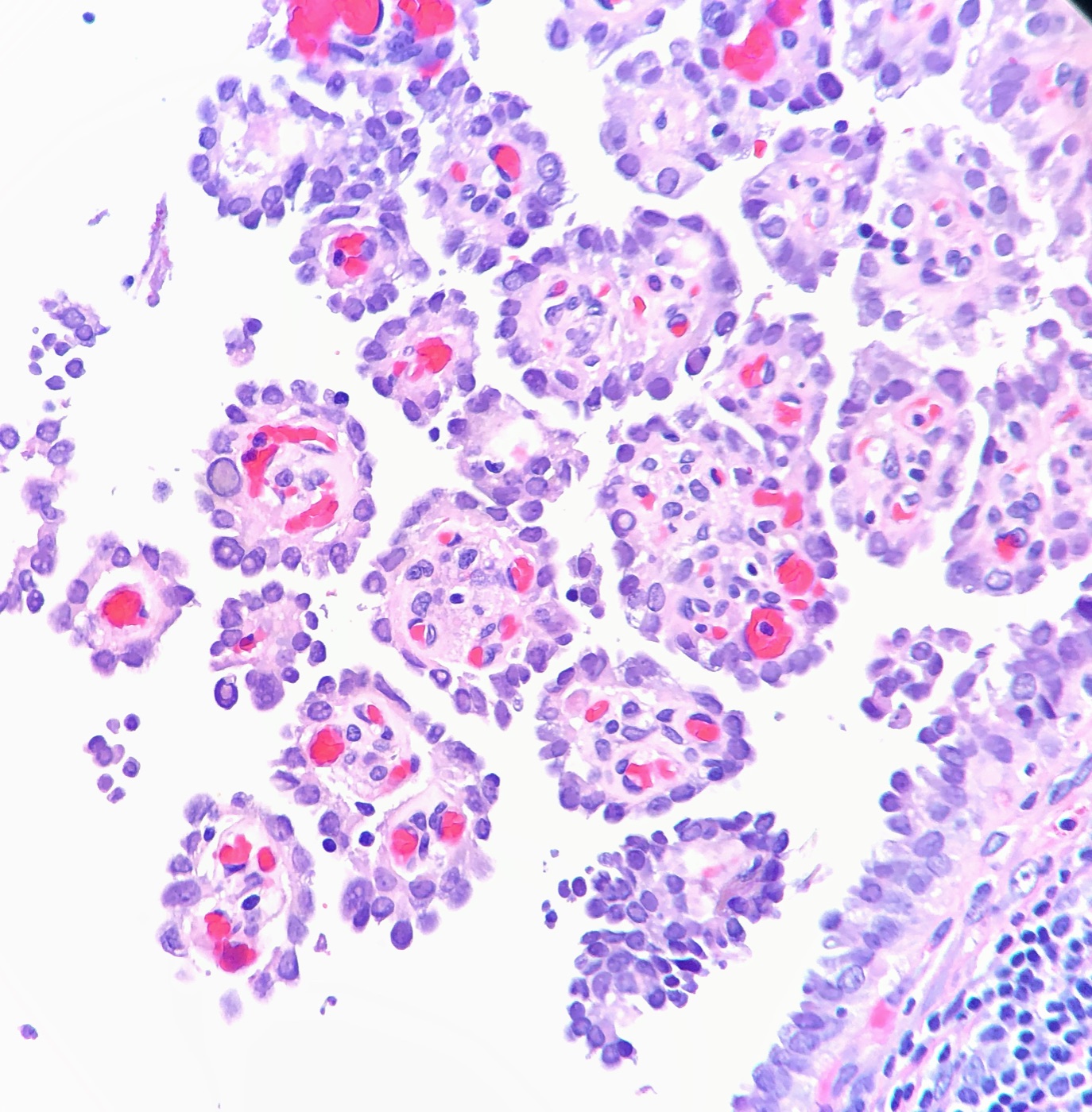
- true papillae with fibrovascular cores
- psammoma bodies (lamellated calcifications)
- stromal fibrosis
Three-point nuclear score for papillary-type nuclei
The consensus nuclear features are grouped into three classes. Each class scores 0 when absent and 1 when present (Nikiforov 2016).
| Class | Features included | Points |
|---|---|---|
| Size and shape | Nuclear enlargement, overlapping or crowding, elongation | 0 or 1 |
| Nuclear membrane irregularities | Irregular contours, grooves, pseudoinclusions | 0 or 1 |
| Chromatin characteristics | Clearing with margination, glassy nuclei | 0 or 1 |
- A total of 0–1 indicates nuclei that are not papillary type. A total of 2–3 indicates papillary-type nuclei and is required for NIFTP.
- The score was developed for encapsulated follicular-patterned tumors. Its sensitivity for NIFTP was 98.6% and its specificity 90.1% (Nikiforov 2016).
WHO 5th edition (2022) subtypes of papillary thyroid carcinoma
| Subtype | Key features | Behavior |
|---|---|---|
| Classic | Papillae with PTC nuclear features | Favorable |
| Encapsulated classic | Classic PTC surrounded by a complete capsule | Favorable |
| Infiltrative follicular | Follicular pattern, infiltrative edge | Similar to classic |
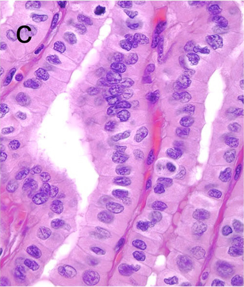
| Tall cell | ≥30% of tumor cells at least 3 times as tall as wide, with dense eosinophilic cytoplasm and distinct cell borders (WHO 2022; Turchini 2023); BRAF V600E | Aggressive |
| Columnar cell | Stratified nuclei, subnuclear vacuoles (endometrial-like) | Aggressive when invasive |
| Hobnail | ≥30% hobnail cells, loss of polarity | Aggressive |
| Diffuse sclerosing | Young patients, diffuse involvement, many psammoma bodies, squamous morules, lymphocytes | More nodal and lung metastasis |
| Solid/trabecular | ≥50% solid; children, radiation, RET fusions | Less favorable than classic PTC |
| Warthin-like | Oncocytic papillae with lymphoid stroma; background thyroiditis | Similar to classic |
| Oncocytic | Oncocytic cells with PTC nuclei | Similar to classic |
| Fibromatosis/fasciitis-like stroma | PTC with abundant myofibroblastic stroma | Similar to classic |
| Clear cell | Clear cytoplasm in >50% of tumor cells, with PTC nuclei; NCOA4::RET fusion in 2 of 4 reported cases (Xu 2026) | Rare; long-term outcome undefined |
| Spindle cell | Bland spindle cells of follicular lineage (TTF-1 and thyroglobulin positive) within PTC; fewer than 20 reported cases (Leong 2022) | Rare; poorly characterized |
The CAP protocol lists each of these subtypes in its histologic type element (CAP 2023).
Related entities classified separately from PTC in WHO 5th edition:
- Invasive encapsulated follicular variant PTC (IEFVPTC): RAS-like, and behaves like follicular carcinoma.
- Noninvasive follicular thyroid neoplasm with papillary-like nuclear features (NIFTP): a low-risk neoplasm, classified outside the carcinomas.
- Cribriform-morular thyroid carcinoma: a separate tumor with APC or CTNNB1 alterations, nuclear β-catenin staining and an association with familial adenomatous polyposis.
- Differentiated high-grade thyroid carcinoma: PTC-type or follicular-type carcinoma with ≥5 mitoses per 2 mm² and/or tumor necrosis, without anaplastic features. Poorly differentiated carcinoma is a separate entity defined by the Turin criteria: solid, trabecular or insular growth; absence of the conventional nuclear features of PTC; and at least one of convoluted nuclei, ≥3 mitoses per 10 high-power fields or tumor necrosis (Volante 2007). WHO 2022 expresses the mitotic criterion as ≥3 mitoses per 2 mm², and the CAP protocol records mitotic rate in the same units (CAP 2023).
NIFTP criteria (WHO 2022):
- Essential:
- encapsulated or clearly demarcated
- follicular growth pattern with no well-formed papillae (2018 revised criteria, which replaced the original <1% papillae threshold) (Nikiforov 2018)
- no psammoma bodies
- <30% solid, trabecular or insular growth
- nuclear score 2–3
- no capsular or vascular invasion
- no tumor necrosis and fewer than 3 mitoses per 2 mm²
- Desirable: absence of BRAF V600E and of other high-risk alterations (TERT promoter, TP53). Molecular testing is not required for the diagnosis. BRAF V600E is evidence against NIFTP.
Papillary thyroid carcinoma in thyroglossal duct cysts
Carcinoma arising in a thyroglossal duct cyst is rare. In a systematic review of 484 cases, 94.2% were PTC, and a synchronous carcinoma in the thyroid gland was present in 34.6%. The median age was 40 years, the recurrence rate 7.4% and overall survival 99.1%. The Sistrunk procedure is the primary operation (Thimsen 2025).
Ancillary testing in papillary thyroid carcinoma
- IHC:
- TTF-1, PAX8 and thyroglobulin positive
- CK19, HBME-1 and galectin-3 favor PTC in the distinction from benign lesions; none is specific
- BRAF V600E (VE1) IHC serves as a surrogate marker for the mutation
- Molecular:
- BRAF V600E in about 60% (TCGA 2014), typical of the classic and tall cell subtypes
- RAS in follicular-patterned tumors
- RET fusions (CCDC6::RET = RET/PTC1, NCOA4::RET = RET/PTC3) in radiation-associated and pediatric tumors
- NTRK and ALK fusions, mainly in pediatric tumors and potentially targetable
- TERT promoter mutations, especially together with BRAF V600E, indicate more aggressive behavior
- Molecular testing of indeterminate FNA samples is covered in Papillary Thyroid Carcinoma: Imaging, Treatment and Follow-up and Thyroid Molecular Testing.
Differential diagnosis of papillary thyroid carcinoma
| Entity | Distinguishing features |
|---|---|
| Papillary hyperplasia (Graves disease, adenomatous nodule) | Lacks the full nuclear features; papillae often point toward the center of a cystic space |
| Hashimoto thyroiditis | Patchy nuclear clearing in reactive cells; no mass or invasion |
| Follicular adenoma or carcinoma | Round nuclei with dark chromatin; no grooves or pseudoinclusions |
| Hyalinizing trabecular tumor | Trabeculae with hyaline material; PAX8::GLIS3 fusion; membranous MIB-1 staining |
| NIFTP | Encapsulated, no invasion, no papillae or psammoma bodies (NIFTP criteria, WHO 2022) |
AJCC 8th edition staging of papillary thyroid carcinoma
The same T, N and M definitions apply to all differentiated thyroid carcinomas (Amin 2017; CAP 2023).
| Category | Definition |
|---|---|
| T1a | Tumor ≤1 cm, limited to the thyroid |
| T1b | Tumor >1 cm and ≤2 cm, limited to the thyroid |
| T2 | Tumor >2 cm and ≤4 cm, limited to the thyroid |
| T3a | Tumor >4 cm, limited to the thyroid |
| T3b | Gross extrathyroidal extension invading only strap muscles (sternohyoid, sternothyroid, thyrohyoid or omohyoid), from a tumor of any size |
| T4a | Gross extrathyroidal extension invading subcutaneous soft tissues, larynx, trachea, esophagus or recurrent laryngeal nerve, from a tumor of any size |
| T4b | Gross extrathyroidal extension invading prevertebral fascia or encasing the carotid artery or mediastinal vessels, from a tumor of any size |
| N0a | One or more cytologically or histologically confirmed benign lymph nodes |
| N0b | No radiologic or clinical evidence of locoregional lymph node metastasis |
| N1a | Metastasis to level VI or VII (pretracheal, paratracheal, prelaryngeal/Delphian or upper mediastinal) lymph nodes, unilateral or bilateral |
| N1b | Metastasis to unilateral, bilateral or contralateral lateral neck lymph nodes (levels I, II, III, IV or V) or retropharyngeal lymph nodes |
| M0 | No distant metastasis |
| M1 | Distant metastasis |
- Minimal (microscopic) extrathyroidal extension no longer changes the T category.
- Upper mediastinal (level VII) nodes are regional nodes (N1a). Their involvement does not count as distant metastasis.
AJCC 8th edition prognostic stage groups for papillary thyroid carcinoma
| Age at diagnosis | Stage | T | N | M |
|---|---|---|---|---|
| <55 years | I | Any T | Any N | M0 |
| <55 years | II | Any T | Any N | M1 |
| ≥55 years | I | T1 or T2 | N0 or NX | M0 |
| ≥55 years | II | T1 or T2 | N1 | M0 |
| ≥55 years | II | T3a or T3b | Any N | M0 |
| ≥55 years | III | T4a | Any N | M0 |
| ≥55 years | IVA | T4b | Any N | M0 |
| ≥55 years | IVB | Any T | Any N | M1 |
CAP synoptic reporting elements for papillary thyroid carcinoma
The CAP thyroid protocol, version 4.4.0.0 (March 2023), contains the following elements. For multiple tumors of the same lineage, the tumor elements may be repeated for each clinically relevant tumor (CAP 2023).
| Section | Elements |
|---|---|
| Clinical (optional) | Predisposing conditions; preoperative biopsy diagnosis; preoperative and postoperative serum marker findings; clinical history |
| Specimen | Procedure: partial excision, lobectomy, lobectomy with isthmusectomy, subtotal or near-total thyroidectomy, total thyroidectomy or completion thyroidectomy |
| Tumor | Tumor focality; tumor site; tumor size; histologic type and subtype; proliferative activity (mitoses per 2 mm²); tumor necrosis |
| Invasion | Angioinvasion, with the number of vessels involved; lymphatic invasion; perineural invasion (optional) |
| Extrathyroidal extension | Not identified; microscopic strap muscle invasion only; gross invasion of strap muscles only (pT3b); invasion of subcutaneous soft tissues, larynx, trachea, esophagus or recurrent laryngeal nerve (pT4a); invasion of prevertebral fascia or encasement of the carotid artery or mediastinal vessels (pT4b) |
| Margins | Margin status; distance from carcinoma to the closest margin (optional); margins involved |
| Regional lymph nodes | Number of nodes with tumor; nodal levels involved (VI, VII, and right or left I–V); size of the largest metastatic deposit; extranodal extension; number of nodes examined; nodal levels examined |
| Distant metastasis | Sites involved, if applicable |
| pTNM classification | Modifiers (y, r); pT category with suffix (m) for multiple tumors; pN category; pM category |
- The 5th edition WHO classification questioned the historical cutoff of 4 vessels between focal and extensive angioinvasion. CAP therefore asks for the number of vessels involved (CAP 2023).
- The ATA 2025 recurrence-risk system for PTC draws on several of these elements: tumor size, focality, extrathyroidal extension, vascular invasion, aggressive histology, margin status, and the size and number of nodal metastases with extranodal extension (Ringel 2025). The four risk tiers are tabulated in Papillary Thyroid Carcinoma: Imaging, Treatment and Follow-up.
AJCC 8th edition stage calculator for differentiated thyroid carcinoma
Papillary Thyroid Carcinoma (PTC) and Subtypes on FNA
Definition. A malignant follicular cell-derived carcinoma defined by a characteristic set of nuclear features. It is the most common type of thyroid cancer (TCGA 2014). On fine-needle aspiration (FNA), well-developed features are reported as Bethesda VI, malignant. Incomplete or focal features are reported as suspicious for malignancy (SFM) or atypia of undetermined significance (AUS). Related topic: The Bethesda System for Reporting Thyroid Cytopathology (categories and risks of malignancy).
Clinical features of papillary thyroid carcinoma on cytology
- Risk of malignancy: Bethesda VI mean 97% (range 97–100%); Bethesda V mean 74% (range 67–83%) (Bethesda 3rd edition).
- Management of category VI: lobectomy or total thyroidectomy, chosen by tumor size, gross extrathyroidal extension and nodal or distant metastasis (Bethesda 3rd edition; ATA 2025).
- Lymph node metastases: ultrasound-guided FNA of suspicious cervical nodes confirms metastasis. ATA 2025 states that thyroglobulin measurement in the FNA washout (FNA-Tg) may be added in selected preoperative patients, and that interpretation is difficult while the thyroid is intact.
- Molecular profile: BRAF V600E is the most frequent driver. RAS mutations occur mainly in follicular-patterned tumors. Kinase fusions, including RET fusions, form a further group (TCGA 2014).
Cytomorphology of papillary thyroid carcinoma on cytology
- Architecture: true papillae with fibrovascular cores, monolayered sheets and three-dimensional groups.
- Nuclear features:
- enlargement, overlapping and molding
- irregular nuclear contours and longitudinal grooves
- intranuclear cytoplasmic pseudoinclusions
- pale, powdery chromatin with margination
- Other findings: psammoma bodies.
- Independent predictors of malignancy in indeterminate aspirates: nuclear grooves, overlapping, molding, contour irregularity, chromatin clearing and margination (Bagıs 2024).
- Cystic PTC: macrophage-rich fluid with few tumor cells. Sparse atypical cells in this setting are insufficient for a malignant diagnosis and are classified as AUS or SFM.
Subtypes (WHO 2022)
- Tall cell: ≥30% of tumor cells are at least three times as tall as wide (Turchini 2023). An aggressive subtype.
- Hobnail: ≥30% hobnail cells (the conventional WHO threshold). An aggressive subtype.
- Columnar cell: an aggressive subtype.
- Other subtypes: follicular, encapsulated classic, diffuse sclerosing, solid/trabecular, Warthin-like and oncocytic. WHO 2022 lists further rare subtypes.
- Cytology and subtypes: an aspirate can suggest a subtype. The percentage-based definitions require the resected tumor.
- Cribriform-morular thyroid carcinoma is a separate WHO 2022 entity. It shows cribriform architecture, squamoid morules and nuclear β-catenin staining, and it is associated with familial adenomatous polyposis (APC).
Ancillary testing in papillary thyroid carcinoma on cytology
- Molecular testing: BRAF V600E is highly specific for PTC. BRAF V600E with a TERT promoter mutation identifies PTC with the highest recurrence rate (Xing 2014). Details are in Thyroid Molecular Testing.
- Lymph node aspirates: FNA-Tg in the needle washout confirms metastatic follicular cell-derived carcinoma.
- Immunohistochemistry on cell block: TTF-1, PAX8 and thyroglobulin confirm thyroid origin of a metastasis.
Differential diagnosis of papillary thyroid carcinoma on cytology
- Cyst-lining cells: elongated cells with grooves and pale chromatin in a streaming, repair-like arrangement. Marked atypia is reported as AUS-nuclear.
- Lymphocytic thyroiditis: reactive oncocytes with nuclear enlargement and mild clearing among polymorphous lymphocytes.
- Hyalinizing trabecular tumor: grooves and pseudoinclusions around hyaline material. Membranous MIB-1 staining on cell block. PAX8::GLIS3 fusion.
- NIFTP and follicular neoplasm: follicular-patterned aspirates with mild nuclear changes and no papillae or pseudoinclusions.
- Medullary thyroid carcinoma: pseudoinclusions occur. Dispersed plasmacytoid or spindle cells, salt-and-pepper chromatin, amyloid, and calcitonin and CEA positivity distinguish it.
Papillary Thyroid Carcinoma: Imaging, Treatment and Follow-up
Definition. Management of papillary thyroid carcinoma (PTC) is stratified by risk. The 2025 American Thyroid Association (ATA) guideline for adult differentiated thyroid cancer (DTC) replaces the 2015 guideline for cancer management. It introduces a four-tier recurrence-risk system, separate risk schemes by histotype, no routine remnant ablation in low-risk disease, de-escalated surveillance and the concept of complete remission (Ringel 2025; Praw 2025). The 2015 guideline covered thyroid nodules and DTC together, and its nodule recommendations are cited here for nodule evaluation (Haugen 2016). Histology and staging are covered in Papillary thyroid carcinoma, and cytology in Papillary Thyroid Carcinoma (Cytology).
Ultrasound features of papillary thyroid carcinoma and ACR TI-RADS
ACR TI-RADS assigns points for five ultrasound feature groups, and the total sets the risk level (Tessler 2017).
- Composition: cystic, almost completely cystic or spongiform 0; mixed cystic and solid 1; solid or almost completely solid 2.
- Echogenicity: anechoic 0; hyperechoic or isoechoic 1; hypoechoic 2; very hypoechoic 3.
- Shape: wider-than-tall 0; taller-than-wide 3.
- Margin: smooth or ill-defined 0; lobulated or irregular 2; extrathyroidal extension 3.
- Echogenic foci: none or large comet-tail artifacts 0; macrocalcifications 1; peripheral (rim) calcifications 2; punctate echogenic foci 3. Points for echogenic foci are additive.
| ACR TI-RADS level | Points | FNA threshold | Ultrasound follow-up threshold |
|---|---|---|---|
| TR1, benign | 0 | No FNA | None |
| TR2, not suspicious | 2 | No FNA | None |
| TR3, mildly suspicious | 3 | ≥2.5 cm | ≥1.5 cm |
| TR4, moderately suspicious | 4–6 | ≥1.5 cm | ≥1.0 cm |
| TR5, highly suspicious | ≥7 | ≥1.0 cm | ≥0.5 cm |
ATA 2015 sets FNA thresholds by sonographic pattern (Haugen 2016):
| ATA sonographic pattern | Estimated risk of malignancy | FNA threshold |
|---|---|---|
| High suspicion | >70–90% | ≥1 cm |
| Intermediate suspicion | 10–20% | ≥1 cm |
| Low suspicion | 5–10% | ≥1.5 cm |
| Very low suspicion (for example, spongiform) | <3% | ≥2 cm, or observation |
- Lymph nodes: preoperative neck ultrasound of the central and lateral compartments is recommended for all patients undergoing surgery for a malignant cytologic or molecular result. FNA of sonographically suspicious nodes >8–10 mm in the smallest diameter is performed when the result would change management. Thyroglobulin measurement in the FNA washout may be added in selected preoperative patients; its interpretation is difficult while the thyroid is intact (Ringel 2025).
Other imaging in papillary thyroid carcinoma
- CT and MRI: cross-sectional imaging of the neck and mediastinum with intravenous contrast is recommended as an adjunct to ultrasound when advanced or invasive disease is suspected, including gross extrathyroidal extension, bulky or invasive adenopathy, and aerodigestive or thoracic involvement (Ringel 2025).
- FDG-PET/CT: routine preoperative FDG-PET/CT is not recommended. FDG-PET/CT may be performed in patients at high risk of recurrence with elevated serum thyroglobulin (generally >10 ng/mL), particularly with oncocytic carcinoma, aggressive histology or negative RAI imaging (Ringel 2025).
- Radioiodine whole-body scan: the post-therapy scan shows RAI-avid disease. Absence of uptake on a post-therapy scan, with disease visible on structural or FDG-PET imaging, is a strong criterion for RAI-refractory disease (Ringel 2025).
Molecular testing of indeterminate thyroid nodules
- About 20% of thyroid FNA samples are indeterminate, most often Bethesda III or IV (Steward 2019).
- ThyroSeq v3 multigene classifier: in Bethesda III and IV nodules, sensitivity was 94% and specificity 82%. At a cancer or NIFTP prevalence of 28%, the negative predictive value was 97% and the positive predictive value 66% (Steward 2019).
- Afirma genomic sequencing classifier: in Bethesda III and IV nodules, sensitivity was 91% and specificity 68%. At a cancer prevalence of 24%, the negative predictive value was 96% and the positive predictive value 47% (Patel 2018).
- Confirmed DTC: genomic testing before surgery is not recommended routinely. A known genomic profile may inform the extent of surgery together with clinical, imaging and cytologic data (Ringel 2025).
- Test design and interpretation are covered in Thyroid Molecular Testing.
Surgery for papillary thyroid carcinoma: lobectomy and total thyroidectomy
The 2025 guideline sets the extent of initial surgery by tumor size, extension and metastasis (Ringel 2025):
- Lobectomy: tumor ≤2 cm without gross extrathyroidal extension and without metastasis (cT1N0M0), unless there are bilateral cancers or other indications to remove the contralateral lobe.
- Lobectomy or total thyroidectomy: unilateral low-risk tumor >2 cm and ≤4 cm (cT2N0M0). Lobectomy may be preferred because of its lower risk. Total thyroidectomy is chosen to enable RAI or to enhance follow-up, based on disease features, suspicious contralateral nodularity or patient preference.
- Total thyroidectomy with node dissection: tumor >4 cm (cT3a), gross extrathyroidal extension (cT3b or cT4), or clinically apparent nodal (cN1) or distant (cM1) metastasis.
- Completion thyroidectomy after lobectomy may be considered when the final pathology shows a higher estimated risk of recurrence (Ringel 2025).
- Survival data: in 52,173 National Cancer Data Base patients treated from 1985 to 1998, lobectomy was associated with a higher risk of recurrence and death for tumors ≥1 cm (Bilimoria 2007). The 2015 and 2025 guidelines nonetheless accept lobectomy for low-risk tumors up to 4 cm.
Neck dissection for papillary thyroid carcinoma: central and lateral compartments
Cervical lymph node levels and their AJCC 8th edition categories (Amin 2017; CAP 2023):
| Level | Location | AJCC 8th category when involved |
|---|---|---|
| I | Submental and submandibular | N1b |
| II | Upper jugular | N1b |
| III | Middle jugular | N1b |
| IV | Lower jugular | N1b |
| V | Posterior triangle | N1b |
| VI | Central compartment: prelaryngeal (Delphian), pretracheal and paratracheal | N1a |
| VII | Upper mediastinal | N1a |
- Central neck dissection: removal of the prelaryngeal and pretracheal nodes and at least one paratracheal nodal basin. The report states the indication and whether the dissection is unilateral or bilateral. Removal of only the clinically involved nodes ("berry picking") is not recommended (Carty 2009).
- Therapeutic central neck dissection of level VI and upper level VII accompanies thyroidectomy in patients with clinically involved central nodes (cN1a). In patients with clinically involved lateral nodes (cN1b), the ipsilateral central compartment is dissected together with the lateral compartment (Ringel 2025).
- Prophylactic central neck dissection is not performed for most small, noninvasive, clinically node-negative PTC (cT1–T2 cN0). It may be considered in cN0 patients with advanced primary tumors (T3 or T4) or when the information will guide further therapy (Ringel 2025).
- Lateral neck dissection: therapeutic lateral compartmental dissection, typically of levels IIa, III, IV and Vb, is performed for biopsy-proven or clinically obvious lateral nodal metastasis (Ringel 2025; Stack 2012).
Complications of thyroid surgery for papillary thyroid carcinoma
- Hypoparathyroidism is the most common complication of bilateral thyroid operations. Risk factors include bilateral surgery, autoimmune thyroid disease, central neck dissection, substernal goiter, surgeon inexperience and malabsorption. A postoperative PTH level <15 pg/mL indicates an increased risk of acute hypoparathyroidism (Orloff 2018).
- Recurrent laryngeal nerve injury causes vocal cord paralysis.
- Adverse events with immediate surgery and active surveillance for microcarcinoma: at Kuma Hospital, the rates were as follows, immediate surgery first (Miyauchi 2016):
- temporary vocal cord paralysis: 4.1% and 0.6%
- temporary hypoparathyroidism: 16.7% and 2.8%
- permanent hypoparathyroidism: 1.6% and 0.08%
Extranodal extension and nodal volume in papillary thyroid carcinoma
Recurrence in node-positive PTC varies with the size, number and extranodal extension of nodal metastases (Randolph 2012):
- pathologic N1 in clinically N0 patients: 2%
- clinically N1: 22%
- fewer than 5 positive nodes: 4%
- more than 5 positive nodes: 19%
- extranodal extension: 24%; the review also linked extranodal extension to a possibly worse disease-specific survival
Extranodal extension places a patient in the high-risk tier of the 2025 ATA system (Ringel 2025). The CAP protocol records extranodal extension, the number of positive nodes and the size of the largest metastatic deposit (CAP 2023).
ATA 2025 recurrence-risk tiers for papillary thyroid carcinoma
The 2025 system estimates the risk of structural persistence or recurrence about 3 months after surgery. It is used with AJCC staging, postoperative imaging, and serum thyroglobulin and thyroglobulin antibody levels. Separate schemes apply to PTC, to follicular carcinoma with invasive encapsulated follicular variant PTC, and to oncocytic carcinoma (Ringel 2025).
| ATA 2025 tier | Estimated recurrence | PTC criteria |
|---|---|---|
| Low | <10% | T1 or T2 (≤4 cm), unifocal; pN0a, or cN0 with pN1a in ≤5 nodes, all ≤2 mm; negative margins, or only a microscopic positive anterior margin (R1) |
| Low-intermediate | 10–15% | T3a; or T1 or T2 with any of: unilateral multifocality; microscopic extrathyroidal extension; cN1a, or pN1a >2 mm or in >5 nodes; microscopic positive posterior margin (R1) |
| Intermediate-high | 16–30% | T1, T2 or T3a with any of: bilateral multifocality >1 cm; clinically evident lateral nodal metastasis (cN1b) <3 cm; aggressive histology; vascular invasion; 2 or more low-intermediate risk factors |
| High | >30% | T3a with microscopic extrathyroidal extension, T3b or T4; or any T with any of: poorly differentiated or high-grade carcinoma; gross incomplete resection (R2); clinical nodal metastasis ≥3 cm; extranodal extension; distant metastasis |
- Other histotypes: in follicular carcinoma and invasive encapsulated follicular variant PTC, capsular invasion only is low risk, vascular invasion of <4 vessels is low-intermediate, and widely invasive tumors or extensive vascular invasion of ≥4 vessels are high risk (Ringel 2025).
- ATA 2015 system (historical): the 2015 guideline used three tiers. Low risk allowed up to 5 nodal micrometastases <0.2 cm. Intermediate risk included microscopic extrathyroidal extension, aggressive histology, vascular invasion and clinical N1 disease. High risk included gross extrathyroidal extension, incomplete resection, distant metastasis and any metastatic node ≥3 cm (Haugen 2016). In a validation cohort, structural persistence or recurrence occurred in 3% of low-risk, 21% of intermediate-risk and 68% of high-risk patients (Tuttle 2010).
Radioactive iodine therapy for papillary thyroid carcinoma
- Indications by 2025 risk tier (Ringel 2025):
- low risk: remnant ablation is not recommended routinely after total thyroidectomy
- low-intermediate and intermediate-high risk: adjuvant RAI may be considered
- high risk: adjuvant RAI is recommended routinely
- distant metastasis at diagnosis: RAI therapy is recommended routinely
- Activity: typical activities are 1.1–1.85 GBq (30–50 mCi) when RAI is given in low-risk disease, 1.1–3.7 GBq (30–100 mCi) in intermediate risk, 3.7–5.55 GBq (100–150 mCi) in high risk, and 3.7–7.4 GBq (100–200 mCi) or dosimetry for distant metastasis (Ringel 2025).
- ATA 2015 grading (historical): RAI was graded "consider" for tumors >4 cm, microscopic extrathyroidal extension, N1a and N1b disease. The accompanying notes described RAI as generally favored for microscopic extrathyroidal extension and for nodal disease, especially with more numerous, larger or clinically evident nodes or extranodal extension (Haugen 2016).
- Trials:
- HiLo (438 patients): ablation success was 85.0% with 1.1 GBq and 88.9% with 3.7 GBq, which met noninferiority. Adverse events were less frequent with the low activity (21% and 33%). Thyrotropin alfa and thyroid hormone withdrawal had similar ablation success (Mallick 2012).
- ESTIMABL (752 patients, 92% PTC): ablation was complete in 92%, with equivalent results for both activities and both stimulation methods (Schlumberger 2012).
- ESTIMABL2 (730 evaluable patients with low-risk disease): at 3 years, 95.6% of patients without RAI and 95.9% with RAI had no event, which met noninferiority (Leboulleux 2022).
Radioiodine-refractory papillary thyroid carcinoma and targeted therapy
- Definition: RAI-refractory disease cannot be diagnosed in a patient who has not received an ablative or treatment dose of RAI. Strong criteria are absence of uptake on a post-therapy scan with disease visible on structural or FDG-PET imaging, and progression less than 6 months after a therapeutic RAI dose with uptake. Patients with RAI-refractory disease do not receive further empiric RAI (Ringel 2025).
- Systemic therapy: lenvatinib is the preferred first-line multikinase inhibitor when no actionable driver has an approved therapy. Cabozantinib is the second-line therapy after a multikinase inhibitor. NTRK-targeted and RET-targeted therapy is recommended first line for tumors with these fusions (Ringel 2025).
| Drug | Target | Trial and population | Main result |
|---|---|---|---|
| Sorafenib | Multikinase | DECISION, phase 3, RAI-refractory DTC | Median progression-free survival 10.8 months, compared with 5.8 months for placebo (Brose 2014) |
| Lenvatinib | Multikinase | SELECT, phase 3, RAI-refractory thyroid cancer | Median progression-free survival 18.3 months, compared with 3.6 months for placebo; response rate 64.8% (Schlumberger 2015) |
| Cabozantinib | Multikinase (VEGFR2, MET, RET) | COSMIC-311, phase 3, RAI-refractory DTC after lenvatinib or sorafenib | Median progression-free survival not reached, compared with 1.9 months for placebo (hazard ratio 0.22); response rate 15%, which missed the prespecified significance level (Brose 2021) |
| Selpercatinib | RET kinase | LIBRETTO-001, phase 1–2 | Response in 79% of 19 previously treated patients with RET fusion-positive thyroid cancer (Wirth 2020) |
| Larotrectinib | TRK kinases (NTRK fusions) | Pooled phase 1–2 studies, all tumor types | Overall response rate 75% (Drilon 2018) |
| Dabrafenib, with or without trametinib | BRAF V600E (with MEK) | Randomized phase 2, BRAF-mutated RAI-refractory DTC | RECIST 1.1 response rate 35% with dabrafenib and 30% with the combination; by modified RECIST, which counts minor responses of −20% to −29%, 42% and 48%; no significant difference (Busaidy 2022) |
- Tumor-agnostic approval: dabrafenib plus trametinib received tissue-agnostic FDA approval for adults and children with BRAF V600E-mutated solid tumors that have progressed after prior treatment and have no satisfactory alternative (Ringel 2025).
TSH suppression in papillary thyroid carcinoma
- 2025 targets by response (Ringel 2025):
- excellent or indeterminate response: TSH within the normal reference range
- biochemically or structurally incomplete response: TSH below the normal reference range
- Long-term TSH suppression is not suggested for low-risk or intermediate-risk disease without biochemical or structural evidence of recurrence (Ringel 2025).
- ATA 2015 targets (historical): initial TSH <0.1 mU/L in high risk, 0.1–0.5 mU/L in intermediate risk, and 0.5–2 mU/L in low risk with undetectable thyroglobulin (Haugen 2016).
ATA 2025 response-to-therapy categories for papillary thyroid carcinoma
Thyroglobulin values are in ng/mL. The criteria differ by the initial treatment (Ringel 2025).
| Response | After total thyroidectomy with RAI | After total thyroidectomy without RAI | After lobectomy |
|---|---|---|---|
| Excellent | Nonstimulated Tg <0.2 or stimulated Tg <1, with negative imaging | Nonstimulated Tg <2.5 | Normal or low-risk contralateral nodules, or benign biopsy, and no abnormal nodes on imaging |
| Indeterminate | Nonspecific imaging findings, nonstimulated Tg 0.2–1, stimulated Tg 1–10, or stable or declining TgAb | Nonspecific imaging findings, nonstimulated Tg 2.5–5, or stable or declining TgAb | Not applicable |
| Biochemically incomplete | Nonstimulated Tg >1, stimulated Tg >10 or rising TgAb, with negative imaging | Nonstimulated Tg >5 or rising TgAb, with negative imaging | Not applicable |
| Structurally incomplete | Structural evidence of disease on imaging or biopsy | Structural evidence of disease | Structural evidence of disease |
- Reclassification by response refines the initial estimate. With an excellent response, structural disease or recurrence fell to 2% in low-risk, 2% in intermediate-risk and 14% in high-risk patients (2015 categories). With an incomplete response, it rose to 13%, 41% and 79% (Tuttle 2010).
Follow-up of papillary thyroid carcinoma and complete remission
- Thyroglobulin (Tg) measurement: serum Tg is measured with an assay calibrated against the BCR457 standard. Tg antibodies (TgAb) are measured quantitatively with every Tg measurement. During initial follow-up, Tg on thyroxine therapy is measured every 6–12 months, more often in intermediate-high and high risk (Ringel 2025).
- Postoperative Tg: a serum Tg level 6–12 weeks after total thyroidectomy is recommended. After lobectomy, a single measurement 6–12 weeks after surgery may exclude an unexpectedly high value, and routine Tg monitoring is not recommended (Ringel 2025).
- Postoperative Tg and events: in ESTIMABL2, events were more frequent when the postoperative serum Tg on thyroid hormone exceeded 1 ng/mL (Leboulleux 2022).
- Thyroglobulin antibodies: TgAb was present in 25% of patients with differentiated thyroid carcinoma and in 10% of the general population. TgAb interfered with Tg measurement in 69% of TgAb-positive sera. Immunometric assays underestimate Tg in the presence of TgAb, and serial TgAb levels parallel the course of disease (Spencer 1998).
- De-escalation and complete remission (Ringel 2025):
- low risk after total thyroidectomy, with or without RAI, and a sustained excellent response for 5–8 years: routine ultrasound stops, and biochemical markers are checked every 1–2 years
- sustained excellent response for 10–15 years: routine biochemical monitoring stops, and the patient has achieved complete remission
- low risk after lobectomy with a negative initial ultrasound: ultrasound every 1–3 years for 5–8 years
Active surveillance of low-risk papillary microcarcinoma
- ATA 2025: active surveillance may be offered for cT1aN0M0 PTC after shared decision-making. Ultrasound-guided percutaneous ablation may be considered as an alternative to surveillance or resection in selected patients (Ringel 2025).
- Monitoring: neck ultrasound every 6 months for 1–2 years, then annually. Serum Tg and TgAb are not measured routinely during surveillance (Ringel 2025).
- Triggers for surgery: new biopsy-proven nodal metastasis, tumor growth of ≥3 mm, distant metastasis, extrathyroidal extension, posterior growth, patient anxiety, inability to follow up, or a preference for surgery (Ringel 2025).
- Kuma Hospital (Japan), since 1993: among 1,235 patients under observation, 8% had tumor enlargement of ≥3 mm and 3.8% had new nodal metastasis at 10 years. Patients aged 40 years or younger were more likely to progress, and no patient died of the disease (Miyauchi 2016). In a separate analysis, young age was an independent predictor of progression, and no patient developed distant metastasis (Ito 2014).
- United States (MSKCC): among 291 patients with intrathyroidal tumors ≤1.5 cm, growth of ≥3 mm had a cumulative incidence of 2.5% at 2 years and 12.1% at 5 years. No regional or distant metastases developed. Growing tumors had a median volume doubling time of 2.2 years (Tuttle 2017).
- Exclusions: the Japan Association of Endocrine Surgery recommends immediate surgery for clinical nodal or distant metastasis, recurrent laryngeal nerve paralysis from tumor invasion, protrusion into the tracheal lumen, cytology suspicious for an aggressive subtype, and tumors adherent to the trachea or located along the course of the recurrent laryngeal nerve (Sugitani 2021).
Prognostic scores (MACIS, AMES) and pediatric risk in papillary thyroid carcinoma
- MACIS score: MACIS = 3.1 (age ≤39 years) or 0.08 × age (age ≥40 years) + 0.3 × tumor size in cm + 1 (incomplete resection) + 1 (local invasion) + 3 (distant metastasis) (Hay 1993).
| MACIS score | 20-year cause-specific survival |
|---|---|
| <6 | 99% |
| 6–6.99 | 89% |
| 7–7.99 | 56% |
| ≥8 | 24% |
- AMES system: age, distant metastasis, and the extent and size of the primary tumor define low-risk and high-risk groups. Low-risk patients made up 89.4% of the cohort, with a mortality rate of 1.8%. High-risk patients made up 10.6% (reported as 11%), with a mortality rate of 46% (Cady 1988).
- Children: the ATA pediatric guideline gives separate recommendations for children and adolescents, who are at low risk of death and at higher risk of long-term harm from aggressive treatment (Francis 2015). In a multicenter cohort of 715 children, age <10 years, male sex, fusion-positive tumors and extrathyroidal extension were associated with invasive disease (Wasserman 2026).
References
- Ali SZ, Baloch ZW, Cochand-Priollet B, Schmitt FC, Vielh P, VanderLaan PA. The 2023 Bethesda System for Reporting Thyroid Cytopathology. Thyroid. 2023;33(9):1039–1044. doi:10.1089/thy.2023.0141. PMID 37427847
- Ali SZ, VanderLaan PA, eds. The Bethesda System for Reporting Thyroid Cytopathology, 3rd ed. Springer; 2023. doi:10.1007/978-3-031-28046-7
- American Thyroid Association Surgery Working Group; Carty SE, et al. Consensus statement on the terminology and classification of central neck dissection for thyroid cancer. Thyroid. 2009;19(11):1153–1158. doi:10.1089/thy.2009.0159. PMID 19860578
- Amin MB, et al., eds. AJCC Cancer Staging Manual, 8th ed. Springer; 2017. doi:10.1007/978-3-319-40618-3
- Bagıs M, et al. A comprehensive approach to the thyroid Bethesda category III (AUS) in the transition zone between 2nd edition and 3rd edition of The Bethesda System for Reporting Thyroid Cytopathology: subcategorization, nuclear scoring, and more. Endocr Pathol. 2024;35(1):51–76. doi:10.1007/s12022-024-09797-1. PMID 38280141
- Baloch ZW, et al. Overview of the 2022 WHO Classification of Thyroid Neoplasms. Endocr Pathol. 2022;33(1):27–63. doi:10.1007/s12022-022-09707-3. PMID 35288841
- Bilimoria KY, et al. Extent of surgery affects survival for papillary thyroid cancer. Ann Surg. 2007;246(3):375–384. doi:10.1097/SLA.0b013e31814697d9. PMID 17717441
- Brose MS, et al. Cabozantinib for radioiodine-refractory differentiated thyroid cancer (COSMIC-311): a randomised, double-blind, placebo-controlled, phase 3 trial. Lancet Oncol. 2021;22(8):1126–1138. doi:10.1016/S1470-2045(21)00332-6. PMID 34237250
- Brose MS, et al. Sorafenib in radioactive iodine-refractory, locally advanced or metastatic differentiated thyroid cancer: a randomised, double-blind, phase 3 trial. Lancet. 2014;384(9940):319–328. doi:10.1016/S0140-6736(14)60421-9. PMID 24768112
- Busaidy NL, et al. Dabrafenib versus dabrafenib + trametinib in BRAF-mutated radioactive iodine refractory differentiated thyroid cancer: results of a randomized, phase 2, open-label multicenter trial. Thyroid. 2022;32(10):1184–1192. doi:10.1089/thy.2022.0115. PMID 35658604
- Cady B, Rossi R. An expanded view of risk-group definition in differentiated thyroid carcinoma. Surgery. 1988;104(6):947–953. PMID 3194846 (no DOI listed in PubMed)
- Cancer Genome Atlas Research Network. Integrated genomic characterization of papillary thyroid carcinoma. Cell. 2014;159(3):676–690. doi:10.1016/j.cell.2014.09.050. PMID 25417114
- College of American Pathologists. Protocol for the Examination of Specimens From Patients With Carcinomas of the Thyroid Gland, version 4.4.0.0, March 2023 (latest version found as of November 2026).
- Davies L, Welch HG. Current thyroid cancer trends in the United States. JAMA Otolaryngol Head Neck Surg. 2014;140(4):317–322. doi:10.1001/jamaoto.2014.1. PMID 24557566
- Drilon A, et al. Efficacy of larotrectinib in TRK fusion-positive cancers in adults and children. N Engl J Med. 2018;378(8):731–739. doi:10.1056/NEJMoa1714448. PMID 29466156
- Francis GL, et al. Management guidelines for children with thyroid nodules and differentiated thyroid cancer. Thyroid. 2015;25(7):716–759. doi:10.1089/thy.2014.0460. PMID 25900731
- Guilmette J, Nosé V. Hereditary and familial thyroid tumours. Histopathology. 2018;72(1):70–81. doi:10.1111/his.13373. PMID 29239041
- Haugen BR, et al. 2015 American Thyroid Association Management Guidelines for Adult Patients with Thyroid Nodules and Differentiated Thyroid Cancer. Thyroid. 2016;26(1):1–133. doi:10.1089/thy.2015.0020. PMID 26462967
- Hay ID, et al. Predicting outcome in papillary thyroid carcinoma: development of a reliable prognostic scoring system in a cohort of 1779 patients surgically treated at one institution during 1940 through 1989. Surgery. 1993;114(6):1050–1058. PMID 8256208 (no DOI listed in PubMed)
- Ishikawa Y, et al. Unusual features of thyroid carcinomas in Japanese patients with Werner syndrome and possible genotype-phenotype relations to cell type and race. Cancer. 1999;85(6):1345–1352. PMID 10189141 (no DOI listed in PubMed)
- Ito Y, et al. Patient age is significantly related to the progression of papillary microcarcinoma of the thyroid under observation. Thyroid. 2014;24(1):27–34. doi:10.1089/thy.2013.0367. PMID 24001104
- Karyadi DM, et al. Distinctive molecular features of radiation-induced thyroid cancers. Sci Adv. 2025;11(34):eadw7680. doi:10.1126/sciadv.adw7680. PMID 40845117
- Leboulleux S, et al. Thyroidectomy without radioiodine in patients with low-risk thyroid cancer. N Engl J Med. 2022;386(10):923–932. doi:10.1056/NEJMoa2111953. PMID 35263518
- Leong KW, et al. Papillary thyroid carcinoma with spindle cell metaplasia: a rare encounter. Diagnostics (Basel). 2022;12(4):855. doi:10.3390/diagnostics12040855. PMID 35453903
- Lim H, et al. Trends in thyroid cancer incidence and mortality in the United States, 1974–2013. JAMA. 2017;317(13):1338–1348. doi:10.1001/jama.2017.2719. PMID 28362912
- Mallick U, et al. Ablation with low-dose radioiodine and thyrotropin alfa in thyroid cancer. N Engl J Med. 2012;366(18):1674–1685. doi:10.1056/NEJMoa1109589. PMID 22551128
- Miyauchi A. Clinical trials of active surveillance of papillary microcarcinoma of the thyroid. World J Surg. 2016;40(3):516–522. doi:10.1007/s00268-015-3392-y. PMID 26744340
- National Cancer Institute. SEER Cancer Stat Facts: Thyroid Cancer. https://seer.cancer.gov/statfacts/html/thyro.html (accessed November 2026).
- Ngeow J, et al. Incidence and clinical characteristics of thyroid cancer in prospective series of individuals with Cowden and Cowden-like syndrome characterized by germline PTEN, SDH, or KLLN alterations. J Clin Endocrinol Metab. 2011;96(12):E2063–E2071. doi:10.1210/jc.2011-1616. PMID 21956414
- Nikiforov YE, et al. Change in diagnostic criteria for noninvasive follicular thyroid neoplasm with papillarylike nuclear features. JAMA Oncol. 2018;4(8):1125–1126. doi:10.1001/jamaoncol.2018.1446. PMID 29902314
- Nikiforov YE, et al. Nomenclature revision for encapsulated follicular variant of papillary thyroid carcinoma: a paradigm shift to reduce overtreatment of indolent tumors. JAMA Oncol. 2016;2(8):1023–1029. doi:10.1001/jamaoncol.2016.0386. PMID 27078145
- Nikiforova MN, et al. MicroRNA expression profiling of thyroid tumors: biological significance and diagnostic utility. J Clin Endocrinol Metab. 2008;93(5):1600–1608. doi:10.1210/jc.2007-2696. PMID 18270258
- Orloff LA, et al. American Thyroid Association statement on postoperative hypoparathyroidism: diagnosis, prevention, and management in adults. Thyroid. 2018;28(7):830–841. doi:10.1089/thy.2017.0309. PMID 29848235
- Patel KN, et al. Performance of a genomic sequencing classifier for the preoperative diagnosis of cytologically indeterminate thyroid nodules. JAMA Surg. 2018;153(9):817–824. doi:10.1001/jamasurg.2018.1153. PMID 29799911
- Praw SS, et al. Executive summary of the 2025 American Thyroid Association Management Guidelines for Adult Patients with Differentiated Thyroid Cancer. Thyroid. 2025;35(11):1214–1220. doi:10.1177/10507256251390877. PMID 41173539
- Randolph GW, et al. The prognostic significance of nodal metastases from papillary thyroid carcinoma can be stratified based on the size and number of metastatic lymph nodes, as well as the presence of extranodal extension. Thyroid. 2012;22(11):1144–1152. doi:10.1089/thy.2012.0043. PMID 23083442
- Ringel MD, et al. 2025 American Thyroid Association Management Guidelines for Adult Patients with Differentiated Thyroid Cancer. Thyroid. 2025;35(8):841–985. doi:10.1177/10507256251363120. PMID 40844370
- Rutter MM, et al. DICER1 mutations and differentiated thyroid carcinoma: evidence of a direct association. J Clin Endocrinol Metab. 2016;101(1):1–5. doi:10.1210/jc.2015-2169. PMID 26555935
- Schlumberger M, et al. Lenvatinib versus placebo in radioiodine-refractory thyroid cancer. N Engl J Med. 2015;372(7):621–630. doi:10.1056/NEJMoa1406470. PMID 25671254
- Schlumberger M, et al. Strategies of radioiodine ablation in patients with low-risk thyroid cancer. N Engl J Med. 2012;366(18):1663–1673. doi:10.1056/NEJMoa1108586. PMID 22551127
- Shattuck TM, et al. Independent clonal origins of distinct tumor foci in multifocal papillary thyroid carcinoma. N Engl J Med. 2005;352(23):2406–2412. doi:10.1056/NEJMoa044190. PMID 15944425
- Spencer CA, et al. Serum thyroglobulin autoantibodies: prevalence, influence on serum thyroglobulin measurement, and prognostic significance in patients with differentiated thyroid carcinoma. J Clin Endocrinol Metab. 1998;83(4):1121–1127. doi:10.1210/jcem.83.4.4683. PMID 9543128
- Stack BC Jr, et al. American Thyroid Association consensus review and statement regarding the anatomy, terminology, and rationale for lateral neck dissection in differentiated thyroid cancer. Thyroid. 2012;22(5):501–508. doi:10.1089/thy.2011.0312. PMID 22435914
- Steward DL, et al. Performance of a multigene genomic classifier in thyroid nodules with indeterminate cytology: a prospective blinded multicenter study. JAMA Oncol. 2019;5(2):204–212. doi:10.1001/jamaoncol.2018.4616. PMID 30419129
- Sugitani I, et al. Indications and strategy for active surveillance of adult low-risk papillary thyroid microcarcinoma: consensus statements from the Japan Association of Endocrine Surgery Task Force on Management for Papillary Thyroid Microcarcinoma. Thyroid. 2021;31(2):183–192. doi:10.1089/thy.2020.0330. PMID 33023426
- Tessler FN, et al. ACR Thyroid Imaging, Reporting and Data System (TI-RADS): white paper of the ACR TI-RADS Committee. J Am Coll Radiol. 2017;14(5):587–595. doi:10.1016/j.jacr.2017.01.046. PMID 28372962
- Thimsen V, et al. Thyroglossal duct cyst carcinomas: a retrospective study and systematic review of the literature. Virchows Arch. 2025;486(6):1139–1151. doi:10.1007/s00428-025-04125-2. PMID 40347268
- Turchini J, et al. A critical assessment of diagnostic criteria for the tall cell subtype of papillary thyroid carcinoma: how much? how tall? and when is it relevant? Endocr Pathol. 2023;34(4):461–470. doi:10.1007/s12022-023-09788-8. PMID 37864666
- Tuttle RM, et al. Estimating risk of recurrence in differentiated thyroid cancer after total thyroidectomy and radioactive iodine remnant ablation: using response to therapy variables to modify the initial risk estimates predicted by the new American Thyroid Association staging system. Thyroid. 2010;20(12):1341–1349. doi:10.1089/thy.2010.0178. PMID 21034228
- Tuttle RM, et al. Natural history and tumor volume kinetics of papillary thyroid cancers during active surveillance. JAMA Otolaryngol Head Neck Surg. 2017;143(10):1015–1020. doi:10.1001/jamaoto.2017.1442. PMID 28859191
- Volante M, et al. Poorly differentiated thyroid carcinoma: the Turin proposal for the use of uniform diagnostic criteria and an algorithmic diagnostic approach. Am J Surg Pathol. 2007;31(8):1256–1264. doi:10.1097/PAS.0b013e3180309e6a. PMID 17667551
- Wasserman JD, et al. The landscape of pediatric differentiated thyroid carcinoma and predictors of invasive disease: a CATC study. J Clin Endocrinol Metab. 2026;111(10):2837–2849. doi:10.1210/clinem/dgag172. PMID 42025318
- WHO Classification of Tumours Editorial Board. Endocrine and Neuroendocrine Tumours. WHO Classification of Tumours, 5th ed., vol. 10. IARC; 2025 (print; online edition 2022). ISBN 978-92-832-4524-7.
- Wirth LJ, et al. Efficacy of selpercatinib in RET-altered thyroid cancers. N Engl J Med. 2020;383(9):825–835. doi:10.1056/NEJMoa2005651. PMID 32846061
- Xing M, et al. BRAF V600E and TERT promoter mutations cooperatively identify the most aggressive papillary thyroid cancer with highest recurrence. J Clin Oncol. 2014;32(25):2718–2726. doi:10.1200/JCO.2014.55.5094. PMID 25024077
- Xing M, et al. Association between BRAF V600E mutation and mortality in patients with papillary thyroid cancer. JAMA. 2013;309(14):1493–1501. doi:10.1001/jama.2013.3190. PMID 23571588
- Xu L, et al. Case report: the clear cell variant of papillary thyroid carcinoma: a clinicopathologic study of four cases with emphasis on RET gene fusions. Front Oncol. 2026;16:1787485. doi:10.3389/fonc.2026.1787485. PMID 42109649
Image credits
- Papillary thyroid carcinoma metastatic to a cervical lymph node. Image: Ed Uthman, Wikimedia Commons, CC BY 2.0.
- Papillary thyroid carcinoma nuclear features. Image: Mikael Häggström, M.D., Wikimedia Commons, CC0.
- Tall cell subtype of papillary thyroid carcinoma. Image: José Manuel Cameselle-Teijeiro & Catarina Eloy & Manuel Sobrinho-Simões, Wikimedia Commons, CC BY 4.0.


