Library › Soft tissue and bone fine-needle aspiration and the WHO Reporting System
Soft tissue and bone fine-needle aspiration and the WHO Reporting System
Last reviewed 2026-11-04 by a board-certified pathologist. 184 references.
The WHO Reporting System for Soft Tissue Cytopathology and the Approach to Soft Tissue Fine-Needle Aspiration
Definition. The WHO Reporting System for Soft Tissue Cytopathology is the fourth volume of the IAC-IARC-WHO Cytopathology Reporting Systems series. It was prepared by a joint editorial board of the International Academy of Cytology (IAC), the International Agency for Research on Cancer (IARC) and the World Health Organization (WHO) (IARC 2026). It places each soft tissue fine-needle aspiration biopsy (FNAB) in one of six categories: insufficient/inadequate/non-diagnostic, benign, atypical, soft tissue neoplasm of uncertain malignant potential (STNUMP), suspicious for malignancy and malignant (IARC 2026, contents). Each category is linked to a risk of malignancy (ROM) and to diagnostic management recommendations (Field et al. 2026; IARC 2026, contents). After first use, the first category is called non-diagnostic in this unit. The system integrates the clinical and imaging context with the diagnostic category, the cytopathologic criteria and ancillary testing (Field et al. 2026).
Related page: Non-Neoplastic and Reactive Soft Tissue and Skin Lesions on FNA (reactive and non-neoplastic lesions) Related page: Benign Soft Tissue Tumors on FNA (benign soft tissue tumors) Related page: Adipocytic Sarcomas, Myxoid Lesions, Extraskeletal Myxoid Chondrosarcoma and Chordoma on FNA (liposarcomas and myxoid lesions) Related page: Round Cell, Spindle Cell and Pleomorphic Sarcomas on FNA (round cell, spindle cell and pleomorphic sarcomas) Related page: Molecular and Ancillary Testing of Soft Tissue FNA Material (fusion and amplification testing on FNA material) Related page: Bone FNA (bone FNA)
Publication of the WHO Reporting System for Soft Tissue Cytopathology
- Series and volume. The system is volume 4 of the IAC-IARC-WHO Cytopathology Reporting Systems, first edition. Its ISBN is 978-92-832-4536-0. IARC lists the print volume under 2026 and also provides it online at tumourclassification.iarc.who.int (IARC 2026).
- Place in the WHO program. The joint program has published four reporting systems: lung; pancreaticobiliary; lymph node, spleen and thymus; and soft tissue cytopathology (2022–2025). Four more, for breast, liver, kidney and adrenal, and head and neck, are nearing completion (Field et al. 2026).
- Content of the volume. The volume gives key diagnostic cytopathologic features for each tumor type, grouped under the diagnostic categories in standardized language. It also gives the differential diagnosis of each lesion and current practice in ancillary testing on cytology material (IARC 2026).
- Use in low-resource settings. The series is designed for international use, including in low-resource settings (IARC 2026; Field et al. 2026).
- Earlier proposal. Before the WHO system, the Lund group tested a structured reporting scheme on 828 primary soft tissue and bone lesions. Its tiers were non-diagnostic, non-neoplastic, atypia of unknown significance, benign neoplasm, neoplasm of unknown malignant potential, suspicious for malignancy and malignant (Köster, Ghanei and Domanski 2021).
WHO Classification of Soft Tissue and Bone Tumours, 6th edition, and the reporting system
- Current classification. The 6th edition of the WHO Classification of Soft Tissue and Bone Tumours was published in 2026. It contains 116 soft tissue tumor types, 46 bone tumor types, 18 vascular tumors of soft tissue and bone, 4 undifferentiated spindle and round cell sarcomas, 6 hematopoietic and dendritic cell neoplasms and 6 genetic syndromes. It adds 19 new tumor types and includes 62 sarcomas (Hornick et al. 2026).
- Definitions of tumor type, subtype and pattern. A tumor type differs from other types in several parameters, such as clinical, histopathologic or genetic features. A subtype differs from the main type in at least one dimension, preferably one that changes treatment or outcome. A morphological pattern aids recognition but has no different clinical behavior (Hornick et al. 2026).
- Change from the 5th edition. The 6th edition uses the term "morphological pattern" for a histologic form without distinct behavior. The 5th edition used the term "variant" (Hornick et al. 2026).
- New chapter grouping. The 6th edition contains a chapter of undifferentiated spindle and round cell sarcomas, with four tumor types (Hornick et al. 2026).
- Entity names in this hub. The summary of the 6th edition available as an abstract confirms no other change. Entity names in the soft tissue units follow the WHO 5th edition (2020) and the WHO Reporting System for Soft Tissue Cytopathology (Hornick et al. 2026; IARC 2026, contents).
- Diagnostic criteria. The 6th edition refines the "essential" and "desirable" diagnostic criteria with attention to different resource settings (Hornick et al. 2026).
- Link to the reporting system. IARC describes the cytopathology series as linked to the WHO Classification of Tumours in its 6th edition (IARC 2026). A 2025 review of the soft tissue system described its terminology as aligned with the 5th edition (Bui 2025). The WHO editors state that the reporting systems will need to follow the developing 6th-edition volumes closely (Field et al. 2026).
Diagnostic categories of the WHO Reporting System for Soft Tissue Cytopathology
| Category | Definition in a review of the system | Entities with sections in the WHO volume, or typical findings |
|---|---|---|
| Insufficient/Inadequate/Non-diagnostic | The sample lacks material adequate in quantity or quality for a reliable diagnosis; causes include scant material, necrosis, obscuring blood and poor preservation | Acellular aspirate, extensive necrosis, blood only (Bui 2025) |
| Benign | Findings indicate an unequivocally benign lesion or a benign soft tissue neoplasm | Inflammation, fat necrosis, lipoma, ganglion cyst, intramuscular myxoma, nodular fasciitis, schwannoma, tenosynovial giant cell tumor |
| Atypical | Findings do not establish whether the lesion is neoplastic, or are mostly benign with minimal concern for malignancy below the threshold of "suspicious" | No entity sections; lipomatous lesion or low-grade spindle cell proliferation of uncertain neoplastic nature (Bui 2025) |
| STNUMP | Features of a mesenchymal neoplasm without conclusive criteria for a benign or malignant tumor | Dermatofibrosarcoma protuberans, solitary fibrous tumor, inflammatory myofibroblastic tumor, angiomatoid fibrous histiocytoma, myoepithelial neoplasms, PEComa |
| Suspicious for malignancy | Findings highly worrisome for malignancy that fall short of a definitive diagnosis | No entity sections; scant, poorly preserved or partly obscured cells with features worrisome for malignancy |
| Malignant | Findings diagnostic of a malignant soft tissue neoplasm | Atypical lipomatous tumor/well-differentiated liposarcoma, myxoid liposarcoma, myxofibrosarcoma, low-grade fibromyxoid sarcoma, extraskeletal myxoid chondrosarcoma, chordoma, gastrointestinal stromal tumor, leiomyosarcoma, synovial sarcoma |
Sources: IARC 2026, contents (category names and the entities in each category); Bui 2025 (definitions, summarizing the system).
- Management recommendations. The WHO volume gives diagnostic management recommendations for each category in a dedicated chapter (IARC 2026, contents). The non-diagnostic category carries a high ROM, which indicates the need for adequate sampling (Ahuja et al. 2026).
- Placement of atypical lipomatous tumor. The WHO volume places atypical lipomatous tumor/well-differentiated liposarcoma, gastrointestinal stromal tumor and myxofibrosarcoma in the malignant category (IARC 2026, contents). A 2025 review had listed them as examples of the suspicious category (Bui 2025); the WHO volume takes precedence.
- Specific diagnosis within a category. The WHO systems emphasize a specific diagnosis or a differential diagnosis built on the key cytopathologic features of each entity (Field et al. 2026). A category is therefore reported together with the most specific diagnosis the material supports.
- Scope of the STNUMP category. It is reserved for aspirates that are clearly neoplastic and mesenchymal but whose biologic potential cannot be predicted from the sample (Bui 2025).
- Category and cytomorphologic pattern together. In the largest series, the ROM within each category varied with the cytomorphologic pattern. The authors found the system most useful when combined with pattern assessment (Gajdzis, Brisse and Klijanienko 2024).
Risk of malignancy in the WHO soft tissue categories across published series
| Study | Design | Non-diagnostic | Benign | Atypical | STNUMP | Suspicious | Malignant |
|---|---|---|---|---|---|---|---|
| Gajdzis, Brisse and Klijanienko 2024 | Institut Curie, 2,214 aspirates, 1954–2022 | 29.87% | 2.49% | 39.62% | 51.43% | 68.42% | 97.69% |
| Layfield, Dodd and Esebua 2024 | Consultation and teaching files with surgical follow-up | 0% | 28% | 57% | 47% | 63% | 88% |
| Ahuja et al. 2025 | 203 aspirates, 117 with histology | 33.3% | 1.2% | 40% | 25% | 80% | 100% |
| Erdogan-Durmus et al. 2026 | 427 bone and soft tissue aspirates with histology | 16.6% | 1.8% | 57.9% | 28.5% | 80% | 100% |
| Saoud, Schowinsky and Ali 2023 | 129 aspirates of myxoid lesions | Not reported | 10% | 31.8% | 50% | 100% | 100% |
| Nigam et al. 2026 | Meta-analysis, 37 studies | 26.4% | 4.8% | 31.6% | 48.5% | 74.5% | 93.5% |
- The ROMs of the atypical and STNUMP categories do not follow a fixed order. STNUMP exceeded atypical in the Institut Curie series, the Saoud series and the meta-analysis. Atypical exceeded STNUMP in the Layfield, Ahuja and Erdogan-Durmus series (Gajdzis, Brisse and Klijanienko 2024; Saoud, Schowinsky and Ali 2023; Nigam et al. 2026; Layfield, Dodd and Esebua 2024; Ahuja et al. 2025; Erdogan-Durmus et al. 2026).
- The non-diagnostic category carries a substantial ROM. Pooled ROM was 26.4% in one meta-analysis and 33% in another (Nigam et al. 2026; Ahuja et al. 2026). The high ROM of non-diagnostic aspirates indicates the need for adequate sampling (Ahuja et al. 2026).
- The evidence base is weak. The GRADE certainty of the pooled estimates was very low because of incomplete histologic verification, retrospective designs and inadequate blinding (Nigam et al. 2026). The ROM estimates came from a relatively small number of studies at the time of the 2024 review (Layfield, Dodd and Esebua 2024).
- Category use depends on the case mix. Benign aspirates formed 62.5% of one general series and 75.4% of another (Ahuja et al. 2025; Erdogan-Durmus et al. 2026). In a series limited to myxoid lesions, atypical (34.1%) and malignant (36.4%) were the commonest categories (Saoud, Schowinsky and Ali 2023).
Risk of malignancy by cytomorphologic pattern in soft tissue FNA
| Cytomorphologic pattern | ROM in the Institut Curie series |
|---|---|
| Lipomatous | 3.00% |
| Spindle cell | 62.78% |
| Epithelioid | 78.15% |
| Myxoid | 84.58% |
| Pleomorphic | 94.26% |
| Round cell | 100% |
Source: Gajdzis, Brisse and Klijanienko 2024.
Diagnostic accuracy of soft tissue FNA reported with the WHO system
| Source | Threshold counted as positive | Sensitivity | Specificity |
|---|---|---|---|
| Gajdzis, Brisse and Klijanienko 2024 | Malignant on original cytologic evaluation | 98.3% | 92.1% |
| Ahuja et al. 2025 | Suspicious and malignant | 77.3% | Not reported |
| Ahuja et al. 2025 | Malignant only | Not reported | 100% |
| Sable et al. 2025 | Malignant | 58.14% | 99.03% |
| Ahuja et al. 2026 (meta-analysis, 4 studies) | Malignant only | 59% | 99% |
| Ahuja et al. 2026 (meta-analysis) | Suspicious and malignant | 75% | 98% |
| Ahuja et al. 2026 (meta-analysis) | Broadest threshold | 85% | 94% |
| Nigam et al. 2026 (meta-analysis, 32 studies, 6,773 cases) | Pooled | 84.9% | 96.8% |
- Choice of threshold. In the Ahuja meta-analysis, counting suspicious and malignant as positive gave the best balance of sensitivity and specificity (Ahuja et al. 2026).
- Overall accuracy. Diagnostic accuracy was 94.2% in the Institut Curie series, 93.9% in the Ahuja series with suspicious and malignant combined, and 92.79% in the Nigam meta-analysis (Gajdzis, Brisse and Klijanienko 2024; Ahuja et al. 2025; Nigam et al. 2026).
- False results. The Institut Curie review found 21 false-negative (0.85%) and 29 false-positive (1.17%) original cytologic reports (Gajdzis, Brisse and Klijanienko 2024).
- Post-test probability. With a pre-test probability of malignancy of 41.3%, a positive cytologic result raised the probability to 95%, and a negative result lowered it to 10% (Nigam et al. 2026).
Reproducibility of the WHO soft tissue categories
- Three-observer study. Among three cytopathologists, κ ranged from 0.2183 to 0.3465 and weighted κ from 0.3778 to 0.5217. Agreement was fair (Layfield, Dodd and Esebua 2024).
- Two-observer study. Interobserver agreement was moderate (Cohen κ 0.45) (Ahuja et al. 2025).
Pattern-based approach to soft tissue fine-needle aspiration
The pattern-based approach sorts an aspirate by its dominant cell type and background before ancillary testing and category assignment (Bui 2025). For myxoid lesions, cell type, nuclear atypia, vascular fragments and the results of immunohistochemical and molecular testing classify most cases (Layfield, Dodd and Klijanienko 2021).
| Pattern | Representative entities | Ancillary tests used on FNA material |
|---|---|---|
| Lipomatous | Lipoma, spindle cell lipoma, atypical lipomatous tumor, lipoblastoma | MDM2 immunocytochemistry or FISH; CD34 and RB1 for spindle cell lipoma; PLAG1 FISH for lipoblastoma |
| Myxoid | Ganglion cyst, intramuscular myxoma, myxoid liposarcoma, myxofibrosarcoma, extraskeletal myxoid chondrosarcoma, chordoma | DDIT3 FISH or DDIT3 immunostain; NR4A3 FISH; brachyury; GNAS sequencing |
| Spindle cell | Nodular fasciitis, schwannoma, solitary fibrous tumor, leiomyosarcoma, synovial sarcoma | USP6 FISH; S100 protein and SOX10; STAT6 and CD34; desmin; SS18 FISH |
| Round cell | Ewing sarcoma, rhabdomyosarcoma, lymphoma, desmoplastic small round cell tumor | CD99 and NKX2.2; desmin, myogenin and MyoD1; flow cytometry; EWSR1 FISH |
| Pleomorphic | Undifferentiated pleomorphic sarcoma, pleomorphic and dedifferentiated liposarcoma | MDM2 and CDK4 |
| Epithelioid | Epithelioid sarcoma, clear cell sarcoma, epithelioid angiosarcoma, alveolar soft part sarcoma | Keratin, EMA and INI1; S100 protein, SOX10 and melanocytic markers; CD31 and ERG; TFE3 |
Sources: Bui 2025 (pattern groups and markers); Layfield, Dodd and Klijanienko 2021; Caraway et al. 1994; Wakely and Jin 2016; Ferreira et al. 2017; Ohshima et al. 2023; Sirvent et al. 2007; Bekers et al. 2019; Jo, Hornick and Qian 2014; Erickson-Johnson et al. 2011.
Role of FNA relative to core needle biopsy in soft tissue masses
- Experience of a sarcoma center. At the Lund Sarcoma Center, FNA had been in use for 48 years by 2020. Its advantages are good sensitivity and specificity, low morbidity, speed and low cost. Its main disadvantages are limited experience in soft tissue cytology among pathologists and, before the WHO system, the lack of a uniform reporting system (Domanski 2020).
- Comparison with CNB. In 828 primary soft tissue and bone lesions, FNA had a sensitivity of 87% and a specificity of 89% for malignancy. CNB had 94% and 95%. FNA identified the correct histologic entity in 55% of lesions and CNB in 66% (Köster, Ghanei and Domanski 2021).
- Open questions. The role of FNAB relative to CNB, the definition of adequacy and the use of ancillary testing remain unresolved across the WHO cytopathology systems (Field et al. 2026).
Specimen handling and ancillary studies in soft tissue FNA
- Cell block. Ancillary studies are used when morphology alone does not classify the tumor and the cell block is adequate (Bui 2025).
- FISH on smears. DDIT3 break-apart FISH was performed successfully on unstained smears or cell block material in 13 of 16 myxoid liposarcomas tested, without false results (Wakely and Jin 2016). PLAG1 FISH has been performed on an FNA smear in lipoblastoma (Ferreira et al. 2017).
- Immunostains as surrogates for genetic events. Nuclear STAT6 detects the NAB2::STAT6 fusion of solitary fibrous tumor (Doyle et al. 2014). Nuclear DDIT3 staining supports the diagnosis of myxoid liposarcoma (Scapa et al. 2021).
WHO category assignment in representative soft tissue aspirates
| Situation | Correct category | Reason |
|---|---|---|
| Blood and necrotic debris only from a large deep mass | Insufficient/inadequate/non-diagnostic, with repeat sampling | The sample lacks adequate lesional material; the category carries a ROM of about 26–33% (Bui 2025; Nigam et al. 2026; Ahuja et al. 2026) |
| Mature adipose tissue from a small superficial mass with imaging of homogeneous fat | Benign | Lipoma is an entity of the benign category; lipomatous aspirates had a ROM of 3.00% (IARC 2026, contents; Gajdzis, Brisse and Klijanienko 2024) |
| Bland spindle cells of a clearly mesenchymal neoplasm without features that predict behavior | STNUMP | The category covers neoplasms without conclusive benign or malignant criteria (Bui 2025) |
| Few cells with malignant features in a poorly preserved smear | Suspicious for malignancy | Findings are worrisome for malignancy and fall short of a definitive diagnosis (Bui 2025) |
| High-grade sarcoma that cannot be subtyped on the sample | Malignant | The findings are diagnostic of malignancy without a specific subtype (Bui 2025) |
| Mature fat with scattered atypical stromal cells, MDM2 amplified | Malignant | The WHO volume places atypical lipomatous tumor/well-differentiated liposarcoma in the malignant category (IARC 2026, contents) |
CAP cancer protocols for soft tissue and bone
The College of American Pathologists (CAP) publishes four bone and soft tissue cancer protocols and separate protocols for gastrointestinal stromal tumor. These apply to biopsy and resection specimens (College of American Pathologists 2026).
| CAP protocol | Current version | Release |
|---|---|---|
| Soft Tissue, Biopsy | 4.2.0.0 | June 2024 |
| Soft Tissue, Resection | 4.2.0.0 | June 2024 |
| Bone, Biopsy | 4.2.0.0 | June 2024 |
| Bone, Resection | 4.2.0.0 | June 2024 |
| GIST, Biopsy | 4.4.0.0 | September 2026 |
| GIST, Resection | 4.4.0.0 | September 2026 |
Source: College of American Pathologists 2026 (cancer protocol templates page, accessed 2026-11-04).
- June 2024 revision. The bone and soft tissue resection and biopsy protocols were updated in content and explanatory notes, including the WHO histologic types, with added core and optional questions (College of American Pathologists 2026).
- Use with FNA. The CAP protocols are written for biopsy and resection specimens. The WHO reporting system provides the categories for FNA (IARC 2026, contents).
WHO Reporting System for Soft Tissue Cytopathology category lookup
Non-Neoplastic and Reactive Soft Tissue and Skin Lesions on Fine-Needle Aspiration
Definition. Cystic, adnexal, reactive and deposition lesions of the skin and soft tissue sampled by fine-needle aspiration (FNA) include epidermal inclusion cyst, pilomatricoma, ganglion cyst, nodular fasciitis, proliferative fasciitis and proliferative myositis, myositis ossificans, cutaneous and abdominal wall endometriosis, and amyloid in abdominal fat. Several are "pseudosarcomatous": they are benign but are often misdiagnosed as malignant because of their clinical presentation or cytologic atypia (Dodd and Martinez 2001; Klapsinou, Despoina and Dimitra 2012). Nodular fasciitis, proliferative fasciitis and myositis, and myositis ossificans carry recurrent USP6 or FOS rearrangements and are regarded as self-limited clonal proliferations ("transient neoplasia") (Erickson-Johnson et al. 2011; Makise et al. 2021; Bekers et al. 2018). In the WHO Reporting System for Soft Tissue Cytopathology, acute inflammation, granulomatous inflammation, fat necrosis, ischemic fasciitis, IgG4-related disease, ganglion cyst, nodular fasciitis and myositis ossificans are entities of the benign category (IARC 2026, contents). Entity names follow the WHO 5th edition (2020); the abstract of the 6th-edition summary confirms no change to these entities (Hornick et al. 2026). A correct FNA diagnosis of a reactive lesion permits conservative management (Wong 2002).
Related page: The WHO Reporting System for Soft Tissue Cytopathology and the Approach to Soft Tissue FNA (WHO categories and the pattern-based approach) Related page: Benign Soft Tissue Tumors on FNA (benign soft tissue tumors, including intramuscular myxoma) Related page: Adipocytic Sarcomas, Myxoid Lesions, Extraskeletal Myxoid Chondrosarcoma and Chordoma on FNA (myxoid lesions and their differential diagnosis) Related page: Melanoma, Merkel Cell Carcinoma and Skin FNA (malignant skin tumors on FNA)
Summary of non-neoplastic and reactive soft tissue lesions on FNA
| Lesion | Typical clinical setting | Key cytologic findings | Ancillary finding | Main mimic on FNA |
|---|---|---|---|---|
| Epidermal inclusion cyst | Head and neck in 59.2%; third and fourth decades | Nucleate and anucleate squames; keratinous material; inflammation when infected | None required | Dermoid and branchial cysts; pilomatricoma |
| Pilomatricoma | Head and neck in 78.6%, often scalp | Basaloid cell clusters, ghost cells, multinucleated giant cells, calcified debris | CTNNB1 mutation in at least 75% | Cutaneous carcinoma |
| Ganglion cyst | Adjacent to a joint | Thick gelatinous fluid; rare histiocytes in mucoid matrix | None required | Intramuscular myxoma |
| Nodular fasciitis | Small, superficial, rapidly growing nodule | Single and loosely cohesive spindle cells, tissue-culture appearance, myxoid stroma | USP6 rearrangement, MYH9::USP6 | Sarcoma |
| Proliferative fasciitis or myositis | Extremities of middle-aged or older adults | Spindle cells and large ganglion-like cells with benign nuclei | FOS rearrangement; c-FOS in epithelioid cells | Pleomorphic or epithelioid sarcoma |
| Myositis ossificans | Ages 6–56 years (mean 27) in one series; often a history of trauma | Fibroblasts, osteoblasts, multinucleated giant cells, degenerated muscle, osteoid | USP6 rearrangement (8 of 9); COL1A1 partner; peripheral calcification on imaging | Osteosarcoma or other sarcoma |
| Endometriosis | Abdominal wall or scar after gynecologic surgery | Epithelial cells, stromal cells and hemosiderin-laden macrophages | None required | Suture granuloma, desmoid, metastasis |
| Amyloid in abdominal fat | Suspected systemic amyloidosis | Congophilic material with apple-green birefringence among fat | Congo red stain | Collagen strands |
Sources: Handa, Chhabra and Mohan 2008; Ahuja and Zaheer 2023; Chan et al. 1999; Dodd and Layfield 1996; Erickson-Johnson et al. 2011; Wong 2002; Allison et al. 2018; Makise et al. 2021; Wakely, Almeida and Frable 1994; Barwad et al. 2011; Bekers et al. 2018; Flucke et al. 2018; Chatterjee, Agnihotri and Kothari 2025; Blumenfeld and Hildebrandt 1993.
Epidermal inclusion cyst on FNA
- Largest cytologic series. Aspirates from 162 epidermal inclusion cysts in 157 patients were reviewed. The head and neck was the commonest site (96 cases, 59.2%), and most patients were in the third and fourth decades (Handa, Chhabra and Mohan 2008).
- Cytologic findings. Aspirates showed a clean background, high cellularity, and nucleate and anucleate squames. Keratinous material was present in some cases, in smaller amounts than the cellular elements (Handa, Chhabra and Mohan 2008).
- Infected cysts. Thirty-one cysts were diagnosed as infected because of a dense inflammatory infiltrate together with squames (Handa, Chhabra and Mohan 2008).
- Histologic follow-up. Of 56 cysts with histology, 45 were epidermal inclusion cysts. The other 11 were dermoid cyst (5), branchial cyst (2), pilomatricoma (2), sebaceous cyst (1) and thyroglossal cyst (1) (Handa, Chhabra and Mohan 2008).
- Distinction from other squamous lesions. Dermoid and branchial cysts also yield squamous cells. An aspirate of squames is therefore interpreted with the site and clinical findings (Handa, Chhabra and Mohan 2008).
Pilomatricoma on FNA
- Definition and pathogenesis. Pilomatricoma is a benign tumor of hair matrix cells. Nuclear LEF-1 in the dividing tumor cells supports a hair matrix origin. At least 75% of pilomatricomas carry activating mutations in CTNNB1 (β-catenin) that affect the amino-terminal degradation segment (Chan et al. 1999).
- Clinical features. In a series of 28 aspirates, 22 (78.6%) arose in the head and neck, most often on the scalp. The male-to-female ratio was 20:8, and the mean age was 34.6 years (Ahuja and Zaheer 2023).
- Diagnostic constellation. Aspirates are cellular. They contain clusters of small primitive basaloid cells with a high nuclear-to-cytoplasmic ratio, evenly dispersed chromatin and prominent nucleoli, pink fibrillary material around the basaloid clusters, multinucleated giant cells and sheets of ghost cells (Lemos et al. 2001).
- Most sensitive feature. Sheets of ghost cells were the most sensitive cytologic parameter. Nests of basaloid cells and a foreign-body giant cell reaction followed (Ahuja and Zaheer 2023).
- Misdiagnosis as carcinoma. In one series of nine aspirates, carcinoma was diagnosed or could not be excluded in four. Distinction from primary cutaneous carcinomas was the main problem (Lemos et al. 2001). In a series of 14 aspirates, 10 (71.4%) were correctly diagnosed and 2 (14.3%) were misdiagnosed as carcinoma (Han et al. 2018).
- Scant ghost cells. Ghost cells were present in all 14 cases of the Han series but were inconspicuous in some because of their small number (Han et al. 2018).
- Incomplete sampling. Aspirates without basaloid cells raised epidermal inclusion cyst as a differential diagnosis. An aspirate dominated by basaloid cells with few ghost cells raised the differential diagnosis of an adnexal tumor (Ahuja and Zaheer 2023).
Ganglion cyst on FNA
- Pathogenesis. Ganglion cyst results from mucoid cystic degeneration of soft tissue adjacent to a joint space (Dodd and Layfield 1996).
- Aspirate. Aspiration yields thick, gelatinous fluid. Smears contain rare histiocytes embedded in a mucoid matrix (Dodd and Layfield 1996).
- Diagnosis. The findings are nonspecific. In the appropriate clinical setting, the diagnosis can be made with confidence by FNA (Dodd and Layfield 1996).
- Myxoid differential. Ganglion cyst is in the differential diagnosis of myxoid lesions of the extremities, together with intramuscular myxoma, myxoid nerve sheath tumors and myxoid sarcomas (Caraway et al. 1994).
Nodular fasciitis on FNA
- Biology. Nodular fasciitis is a self-limited subcutaneous pseudosarcomatous myofibroblastic proliferation. Rapid growth and high mitotic activity lead to misdiagnosis as sarcoma (Erickson-Johnson et al. 2011).
- Genetics. USP6 rearrangement was found in 44 of 48 cases (92%). MYH9 is the translocation partner; the MYH9 promoter is fused to the entire coding region of USP6. Control tumors and tissues lacked the fusion (Erickson-Johnson et al. 2011).
- Transient neoplasia. Nodular fasciitis was the first self-limited human disease shown to carry a recurrent somatic gene fusion. It is regarded as a model of "transient neoplasia" (Erickson-Johnson et al. 2011).
- Clinical presentation. Lesions are small, superficially located, rapidly growing nodules with a short duration of symptoms (Wong 2002).
Cytologic findings of nodular fasciitis
| Feature | Frequency among the parotid aspirates reviewed |
|---|---|
| Spindled cells | 100.0% |
| Elongated nuclei | 84.6% |
| Myxoid stroma | 76.9% |
| Short unipolar cytoplasmic processes | 76.9% |
| Predominantly single cells | 69.2% |
| Tissue-culture appearance | 69.2% |
| Round nuclei | 61.5% |
| Inconspicuous nucleoli | 53.8% |
| Minimal cytologic atypia | 46.2% |
| Bipolar cytoplasmic processes | 38.5% |
Source: Allison et al. 2018.
- Accuracy of FNA. Three of 15 parotid cases (20.0%) were classified as definitive, probable or suggestive of nodular fasciitis (Allison et al. 2018). In an eight-case series, the FNA diagnosis agreed with the final diagnosis in four cases (Jaiprakash et al. 2019).
- Causes of error. Lack of clinical information and paucicellular smears caused the misdiagnoses in one series (Jaiprakash et al. 2019).
- Hypercellular aspirates. Three nodular fasciitis lesions of the hand showed markedly hypercellular smears of overlapping, relatively isomorphic spindle cells. Two were mistaken for possible sarcoma (Plaza, Mayerson and Wakely 2005).
- Management after FNA. In one series, 10 of 13 nodular fasciitis lesions resolved spontaneously within 1–12 weeks (mean 4.7 weeks) after FNA diagnosis. The author recommended initial nonsurgical management with follow-up and surgery when regression did not occur within 4–8 weeks (Wong 2002).
- Ancillary confirmation. USP6 rearrangement detected by FISH or next-generation sequencing supports the diagnosis (Bui 2025; Erickson-Johnson et al. 2011).
Proliferative fasciitis and proliferative myositis on FNA
- Clinical setting. Proliferative fasciitis and proliferative myositis usually affect the extremities of middle-aged or older adults and present as poorly circumscribed masses (Makise et al. 2021).
- Histologic hallmark. Bland spindle cells in a myxoid to fibrous background are accompanied by large epithelioid "ganglion-like" cells, which may lead to a misdiagnosis of sarcoma (Makise et al. 2021).
- Cytologic findings. Aspirates of pseudosarcomatous reactive lesions show a pleomorphic population of proliferating cells and ganglion cell-like cells. The ganglion-like cells have large nuclei and prominent nucleoli but thin, smooth nuclear membranes and fine chromatin (Wong 2002).
- Genetics. Five of six lesions showed FOS rearrangement in the epithelioid cells, and one carried a FOS::VIM fusion. The one pediatric case lacked FOS rearrangement (Makise et al. 2021).
- Immunohistochemistry. c-FOS was diffusely and strongly expressed in the epithelioid cells. It was negative in 45 mimics, including pleomorphic sarcoma with epithelioid features and epithelioid sarcoma (Makise et al. 2021).
- Outcome after FNA. One proliferative fasciitis and two proliferative myositis lesions resolved spontaneously after FNA diagnosis (Wong 2002).
Myositis ossificans on FNA
- Genetics. USP6 rearrangement was found by FISH in 8 of 9 assessable cases, which relates myositis ossificans to nodular fasciitis and aneurysmal bone cyst (Bekers et al. 2018). COL1A1 is a fusion partner of USP6 in myositis ossificans (Flucke et al. 2018).
- Clinical setting. Myositis ossificans often follows trauma. In a FISH series of 11 cases, patients were aged 6 to 56 years (mean 27), and five lesions were in the thigh (Bekers et al. 2018). Two reported aspirates came from adolescents, one in deep thigh muscle and one in abdominal subcutaneous fat (Wakely, Almeida and Frable 1994). A further case presented as a painful hard thigh mass two weeks after trauma (Barwad et al. 2011).
- Imaging. Computed tomography showed a heterogeneous mass with peripheral calcification and central lucency (Barwad et al. 2011).
- Cytologic findings. Smears contain fibroblasts, osteoblasts, multinucleated giant cells, degenerated muscle cells and osteoid (Barwad et al. 2011).
- Diagnostic difficulty. A benign lesion was favored in both of the early cases, but sarcoma could not be excluded in one (Wakely, Almeida and Frable 1994). Clinical presentation and imaging are integrated with the cytology (Barwad et al. 2011).
Cytologic features that separate pseudosarcomatous lesions from sarcoma
| Feature | Finding in pseudosarcomatous reactive lesions | Source |
|---|---|---|
| Clinical course | Small, superficial nodule with rapid growth over a short time | Wong 2002 |
| Nuclei of the large cells | Large nuclei and prominent nucleoli with thin, smooth membranes and fine chromatin | Wong 2002 |
| Cell arrangement | Repair-like change and cohesion of cellular fragments | Dodd and Martinez 2001 |
| Normal tissue elements | Present in the aspirate | Dodd and Martinez 2001 |
| Molecular finding | USP6 rearrangement in nodular fasciitis; FOS rearrangement in proliferative fasciitis or myositis; USP6 rearrangement in myositis ossificans | Erickson-Johnson et al. 2011; Makise et al. 2021; Bekers et al. 2018 |
| Behavior after FNA | Spontaneous regression within 1–12 weeks | Wong 2002 |
- Benign neoplasms with pseudoanaplastic atypia. Lipoma, leiomyoma, angiomyolipoma and benign peripheral nerve sheath tumors may show degenerative atypia that mimics sarcoma (Dodd and Martinez 2001).
Cutaneous and abdominal wall endometriosis on FNA
- Sites. The commonest sites of cutaneous endometriosis are the abdominal wall, umbilicus, vulva and extremities, and most cases follow gynecologic surgery (Chatterjee, Agnihotri and Kothari 2025). Most abdominal wall endometriosis arises in or near cesarean section or hysterectomy scars (Pachori et al. 2015).
- Cytologic triad. The classic triad is epithelial cells, stromal cells and hemosiderin-laden macrophages. Any two of the three are diagnostic (Chatterjee, Agnihotri and Kothari 2025).
| Finding in 17 cases | Frequency |
|---|---|
| Abdominal wall site | 15/17 (88%) |
| Prior surgery (cesarean section, laparotomy, episiotomy) | 14/17 (82%) |
| Swelling with cyclic pain | 13/17 (77%) |
| Benign epithelial cells and macrophages or hemosiderophages | 17/17 (100%) |
| Endometrial stromal cells | 11/17 (65%) |
| Epithelial atypia | 6/17 (35%) |
Source: Chatterjee, Agnihotri and Kothari 2025.
- Epithelial atypia. Epithelial atypia was present in 35% of cases and is a source of overdiagnosis. Correlation with the clinical presentation avoids this error (Chatterjee, Agnihotri and Kothari 2025).
- Clinical mimics. Cutaneous endometriosis can mimic suture granuloma, hematoma, abscess, sarcoma, desmoid tumor and metastasis (Chatterjee, Agnihotri and Kothari 2025).
Amyloid in abdominal fat pad aspirates
- Diagnostic criterion. Congophilia with apple-green birefringence under polarized light is diagnostic of amyloid (Blumenfeld and Hildebrandt 1993; Giorgadze et al. 2004).
- Appearance of true positives. Positive aspirates contain many short apple-green strands, closely associated with the fat, oriented in multiple directions and present in much or all of the aspirated fat (Blumenfeld and Hildebrandt 1993).
- False positives. Early false-positive results came from overinterpretation of occasional long, slender green strands of collagen (Blumenfeld and Hildebrandt 1993).
- Sensitivity and specificity. In 297 aspirates with deeper tissue follow-up in 44, fat pad aspiration had a sensitivity of 78%, a specificity of 93%, a positive predictive value of 84% and a negative predictive value of 90%. Patients with grade 1 deposits were advised to have histologic confirmation in deeper tissue before toxic therapy (Dhingra et al. 2007).
- Fluorescence microscopy. Of 78 archived aspirates, 12 (15.4%) had been reported positive by polarization. Four were unsatisfactory on review, and fluorescence microscopy of the remaining 74 Congo red-stained smears identified 29 (39.2%) as positive (Giorgadze et al. 2004).
- Negative aspirates in practice. In 334 fat pad aspirates, 27 of 70 negative cases with later surgical biopsy were positive for amyloid on the surgical specimen. Scant cellularity, focal staining or birefringence and overstaining were the main interpretive problems (Torous and Chebib 2024).
- Clinical indications. Peripheral neuropathy (29.3%), cardiomyopathy or heart failure (28.1%), monoclonal gammopathy (27.8%) and plasma cell or lymphoplasmacytic neoplasms (21.0%) were the commonest indications (Torous and Chebib 2024).
- Interpretation. Fat pad aspiration and other tissue biopsies are complementary tests interpreted with the clinical suspicion of amyloidosis (Torous and Chebib 2024).
WHO reporting categories for non-neoplastic soft tissue aspirates
| Situation | WHO category | Source |
|---|---|---|
| Inflammation or abscess explaining the mass | Benign | Bui 2025 |
| Ganglion cyst with gelatinous fluid in a juxta-articular site | Benign | IARC 2026, contents; Dodd and Layfield 1996 |
| Nodular fasciitis with classic clinical and cytologic findings, or USP6 rearrangement | Benign | IARC 2026, contents; Wong 2002; Erickson-Johnson et al. 2011 |
| Spindle cell proliferation with unclear neoplastic nature | Atypical | Bui 2025 |
| Blood only or acellular aspirate | Insufficient/inadequate/non-diagnostic | Bui 2025; IARC 2026, contents |
| Myositis ossificans, fat necrosis or ischemic fasciitis with concordant clinical and imaging findings | Benign | IARC 2026, contents |
Benign Soft Tissue Tumors on Fine-Needle Aspiration
Definition. Benign soft tissue tumors and solitary fibrous tumor sampled by fine-needle aspiration (FNA) include lipoma and its spindle cell and pleomorphic forms, lipoblastoma, schwannoma, solitary fibrous tumor, intramuscular myxoma, tenosynovial giant cell tumor, leiomyoma and adult rhabdomyoma. In the WHO Reporting System for Soft Tissue Cytopathology, lipoma, hibernoma, lipoblastoma, spindle cell/pleomorphic lipoma, intramuscular and juxta-articular myxoma, leiomyoma, schwannoma, neurofibroma, perineurioma, rhabdomyoma, granular cell tumor and tenosynovial giant cell tumor are entities of the benign category. Solitary fibrous tumor is an entity of the soft tissue neoplasm of uncertain malignant potential (STNUMP) category (IARC 2026, contents). Entity names follow the WHO 5th edition (2020); the abstract of the 6th-edition summary confirms no change to these entities (Hornick et al. 2026). Several benign tumors show degenerative or "pseudoanaplastic" atypia that mimics sarcoma, including lipoma, leiomyoma and benign peripheral nerve sheath tumors (Dodd and Martinez 2001). A specific diagnosis of solitary fibrous tumor or intramuscular myxoma often requires immunocytochemistry or molecular testing on cell block or smear material (Wakely and Rekhi 2021; Bekers et al. 2019).
Related page: The WHO Reporting System for Soft Tissue Cytopathology and the Approach to Soft Tissue FNA (WHO categories and the pattern-based approach) Related page: Non-Neoplastic and Reactive Soft Tissue and Skin Lesions on FNA (reactive lesions, including nodular fasciitis) Related page: Adipocytic Sarcomas, Myxoid Lesions, Extraskeletal Myxoid Chondrosarcoma and Chordoma on FNA (atypical lipomatous tumor and myxoid sarcomas) Related page: Round Cell, Spindle Cell and Pleomorphic Sarcomas on FNA (malignant spindle cell tumors) Related page: Molecular and Ancillary Testing of Soft Tissue FNA Material (fusion and mutation testing on FNA material)
Summary of benign soft tissue tumors on FNA
| Tumor | Typical setting | Key cytologic findings | Immunostain or genetic finding | Main differential on FNA |
|---|---|---|---|---|
| Spindle cell or pleomorphic lipoma | Subcutis of posterior neck, upper back and shoulder; middle-aged males | Bland spindle cells, mature adipocytes, mast cells, collagen fragments | CD34 positive; loss of nuclear RB1; 13q14 deletion | Atypical lipomatous tumor; atypical spindle cell/pleomorphic lipomatous tumor |
| Lipoblastoma | Infancy and early childhood | Mature fat with thin branching vessels, lipoblasts, myxoid matrix | PLAG1 rearrangement | Myxoid liposarcoma |
| Schwannoma | Peripheral nerve sheath; reported in head and neck series | Spindle cells with wavy and fishhook nuclei; filamentous background | S100 protein and SOX10 | Other spindle cell neoplasms |
| Solitary fibrous tumor | Pleura, head and neck, extremities, trunk | Bland ovoid to spindle cells; ambiguous on smears | Nuclear STAT6; CD34; NAB2::STAT6 | Other spindle cell tumors |
| Intramuscular myxoma | Thigh muscles of older adults | Clear viscous fluid; few bland spindle and stellate cells in mucoid matrix; sparse simple capillaries | GNAS mutation; diffuse CD34 | Low-grade myxofibrosarcoma; low-grade fibromyxoid sarcoma |
| Tenosynovial giant cell tumor | Tendon sheath and synovium | Mononuclear cells, osteoclast-type giant cells, hemosiderin ("ladybird cells") | CSF1 fusion; CSF1 immunostain | Other giant cell-rich lesions |
| Leiomyoma | Cytologic data mainly from uterine tumors | Few cohesive fragments of tapering cells without nuclear crowding | Desmin and smooth muscle actin, as in other smooth muscle tumors | Leiomyosarcoma |
| Adult rhabdomyoma | Head and neck | Large cells with abundant finely granular eosinophilic cytoplasm | Desmin and myoglobin | Granular cell tumor; normal skeletal muscle |
Sources: Ohshima et al. 2023; Guo et al. 2000; Ferreira et al. 2017; Mooney, Layfield and Dodd 1999; Bui 2025; Doyle et al. 2014; Robinson et al. 2013; Wakely and Rekhi 2021; Caraway et al. 1994; Bekers et al. 2019; Libbrecht et al. 2019; Zhao et al. 2017; Sugita et al. 2022; Barbazza et al. 1997; Domanski and Dawiskiba 2000; Jin and Saleh 2009.
Lipoma and benign lipomatous tumors on FNA
- Category. Lipoma is an example of the benign category of the WHO soft tissue system (Bui 2025). Aspirates with a lipomatous pattern had the lowest risk of malignancy, 3.00%, of the six cytomorphologic patterns in the Institut Curie series (Gajdzis, Brisse and Klijanienko 2024).
- Distinction from atypical lipomatous tumor. Atypical lipomatous tumor/well-differentiated liposarcoma is separated from lipoma by its characteristic atypical cells. The distinction becomes difficult when those cells are poorly represented, as in small biopsy samples (Kilpatrick 2024).
- MDM2 testing. FISH or quantitative PCR for MDM2 and CDK4 amplification supplements immunohistochemistry when an adipose tumor cannot be classified on clinical and morphologic grounds. Immunohistochemistry alone is often insufficient (Sirvent et al. 2007).
- Pseudoanaplastic atypia. Lipoma can show degenerative atypia that mimics a sarcoma on FNA (Dodd and Martinez 2001).
Spindle cell lipoma and pleomorphic lipoma
- Clinical features. Spindle cell lipoma arises mainly in the subcutis of the posterior neck, upper back and shoulder of middle-aged males. It is usually a relatively small (<5 cm), mobile, slow-growing, painless mass (Ohshima et al. 2023).
- Spectrum. Spindle cell lipoma and pleomorphic lipoma form a morphologic spectrum of one process. Pleomorphic lipoma also contains pleomorphic and multinucleated floret-like giant cells (Ohshima et al. 2023).
- Immunophenotype and genetics. The spindle and floret-like cells are diffusely CD34 positive and show loss of nuclear RB1 expression. Heterozygous 13q14 deletions include RB1 (Ohshima et al. 2023).
- Cytologic findings. A scalp tumor yielded scattered clusters of mildly pleomorphic spindle cells with mature adipocytes, numerous mast cells and small collagen fragments. The diagnosis was confirmed with CD34 and bcl-2 (Guo et al. 2000).
- Accuracy of FNA. Of eight spindle cell or pleomorphic lipomas, FNA diagnoses included spindle cell neoplasm (3), spindle cell lipoma (1), pleomorphic lipoma (1), suspicious for spindle cell lipoma (1), benign adipose tissue (1) and benign spindle cells with connective tissue (1) (Wakely 2023).
- Atypical spindle cell/pleomorphic lipomatous tumor. This benign lipomatous neoplasm mimics spindle cell/pleomorphic lipoma and atypical lipomatous tumor. In one FNA case it was CD34 positive and MDM2 negative, and in another MDM2 and DDIT3 FISH were negative (Wakely 2023). It may contain bizarre pleomorphic lipoblasts. It lacks MDM2 amplification and the risk of dedifferentiation. It involves the distal extremities more often than spindle cell/pleomorphic lipoma and recurs locally more often (Kilpatrick 2024).
- Treatment. Spindle cell/pleomorphic lipoma is treated by simple excision and rarely recurs (Ohshima et al. 2023).
Lipoblastoma
- Clinical features. Lipoblastoma is a rare benign adipocytic tumor of infancy. In an FNA series of nine patients, the median age was 2.5 years (range 10 months to 13 years). Sites were the thorax, abdomen, axilla and thigh (Ferreira et al. 2017).
- Genetics. About 70% carry chromosome 8 abnormalities, mainly PLAG1 rearrangements (Ferreira et al. 2017).
- Cytologic findings. Smears were hypocellular to moderately cellular, with fragments of mature adipose tissue, thin branching vessels and some lipoblasts in a myxoid matrix. Bland spindle cells and naked oval nuclei were present in the background (Ferreira et al. 2017).
- FISH on cytology material. PLAG1 rearrangement was found in three of four patients tested, including once on an FNA smear and once on a tumor imprint (Ferreira et al. 2017).
Schwannoma on FNA
- Frequency of classic features. In a review of 13 neural lesions, frequencies were reported for 11 cases. Verocay bodies were found in 1 of 11 and vascular arcades in 1 of 11. Spindle cells with wavy nuclei were the commonest finding, and fishhook-shaped nuclei and a filamentous background were present in 9 of 11 cases (Mooney, Layfield and Dodd 1999).
- FNA compared with CNB. In a prospective series of head and neck schwannomas, 24 of 52 FNA samples (46.2%) were unsatisfactory and 0 of 29 core needle biopsy (CNB) samples. A diagnosis of schwannoma, including "suspicious for" and "consistent with", was reached in 19.2% of FNA samples and 96.6% of CNB samples, without major complications from either method (Ahn et al. 2018).
- Degenerative atypia. Benign peripheral nerve sheath tumors can show pseudoanaplastic atypia (Dodd and Martinez 2001). A spindle cell aspirate with degenerative nuclear atypia, in which schwannoma is favored but not established, is an example of the atypical category in a review of the WHO system (Bui 2025). Schwannoma itself is an entity of the benign category in the WHO volume (IARC 2026, contents).
- Immunostains. S100 protein and SOX10 support nerve sheath differentiation (Bui 2025). Schwannomas were negative for MUC4 in a study of 20 cases (Doyle et al. 2011).
Solitary fibrous tumor on FNA
- Genetics. NAB2::STAT6 was detected by RT-PCR in all 51 solitary fibrous tumors in the discovery study. The fusion links the EGR-binding domain of NAB2 to the activation domain of STAT6 and is the defining driver of the tumor (Robinson et al. 2013).
- FISH limitation. NAB2 and STAT6 lie close together on 12q13. Conventional karyotyping and FISH therefore detect the fusion only rarely (Doyle et al. 2014).
- STAT6 immunostain. Nuclear STAT6 was found in 59 of 60 solitary fibrous tumors (98%), usually diffuse and intense. Other tumor types were negative, apart from weak staining in three dedifferentiated liposarcomas and one deep fibrous histiocytoma (Doyle et al. 2014).
- CD34. CD34 expression is the most characteristic but nonspecific immunohistochemical finding (Doyle et al. 2014).
- Cytologic series. In 34 cytology specimens from 30 patients (mean age 58 years), sites were pleura or lung (26%), head and neck (24%), lower extremity (21%), trunk (12%), intra-abdominal (9%), upper extremity (7%) and mediastinum (4%) (Wakely and Rekhi 2021).
- Accuracy without immunostains. Only 3 of 34 specimens (9%) were specifically diagnosed as solitary fibrous tumor. The commonest diagnosis was spindle cell neoplasm or proliferation (14, 41%), and 5 (15%) were nondiagnostic. Smears overlap with many other spindle cell proliferations, and a specific diagnosis requires STAT6 with other markers (Wakely and Rekhi 2021).
- WHO category. Solitary fibrous tumor is an example of STNUMP in a review of the WHO soft tissue system (Bui 2025).
- DDIT3 cross-reactivity. Limited, weak DDIT3 staining in fewer than 10% of cells occurred in 3 of 12 solitary fibrous tumors (Scapa et al. 2021).
Intramuscular myxoma on FNA
- Clinical and imaging features. In ten aspirated intramuscular myxomas, patients were aged 43 to 75 years (seven females, three males). Seven tumors involved thigh muscles. MRI showed well-defined, sharply demarcated tumors with low signal on T1-weighted images and high signal on T2-weighted images relative to muscle (Caraway et al. 1994).
- Gross aspirate. All aspirates were clear, tenacious and viscous (Caraway et al. 1994).
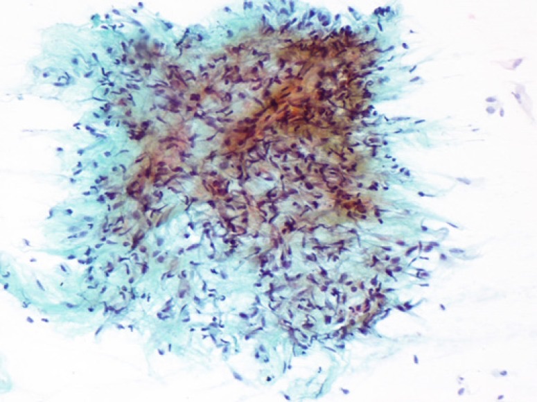
- Cytologic findings. Smears contained few spindled and histiocytoid cells in an abundant mucoid background. Spindle cells had long cytoplasmic processes that formed fibrillar tangles. Nuclei were oval to spindled with fine chromatin and inconspicuous nucleoli. Capillaries were sparse and showed simple, non-plexiform branching (Caraway et al. 1994).
- Limits of cytology. A definitive diagnosis was often difficult because of scant cellularity and a lack of distinctive features. MRI findings supported the cytologic impression (Caraway et al. 1994).
- Frequency among myxoid aspirates. Intramuscular myxoma formed 15.5% of 129 aspirates with a prominent myxoid component, second to myxoid liposarcoma (27.1%) (Saoud, Schowinsky and Ali 2023).
- GNAS mutations. GNAS mutations were found in 23 of 34 sporadic intramuscular myxomas (68%), mostly p.R201C and p.R201H (Bekers et al. 2019). With next-generation sequencing, a GNAS mutation was found in 88% of cases (Libbrecht et al. 2019).
- CD34. All ten intramuscular myxomas in one series showed diffuse, strong CD34 expression, which is sensitive but nonspecific (Libbrecht et al. 2019).
- Differential diagnosis. Intramuscular myxoma is difficult to separate from low-grade fibromyxoid sarcoma and low-grade myxofibrosarcoma on small samples. GNAS mutations are frequent in intramuscular myxoma and are not features of these sarcomas (Bekers et al. 2019). Cellular myxomas were negative for MUC4, which marks low-grade fibromyxoid sarcoma (Doyle et al. 2011).
Tenosynovial giant cell tumor on FNA
- Definition. Tenosynovial giant cell tumor, also known as giant cell tumor of tendon sheath or pigmented villonodular synovitis, is the most common benign tumor of tendon and synovium (Zhao et al. 2017).
- Localized and diffuse types. The tumor affects the synovium of joints, bursae and tendon sheaths and is divided into localized and diffuse types (Sugita et al. 2022). The intra-articular diffuse type can form a large infiltrative mass, involve adjacent soft tissue and destroy bone, which raises clinical concern for malignancy (Zhao et al. 2017).
- Genetics. Both types carry recurrent CSF1 rearrangements, most often fused to COL6A3, which cause overexpression of CSF1 mRNA and protein (Sugita et al. 2022).
- CSF1 immunostain. CSF1 was expressed in 50 of 65 tenosynovial giant cell tumors, including 35 of 44 localized and 15 of 20 diffuse tumors. Giant cell tumor of bone, aneurysmal bone cyst and other giant cell-rich mimics were negative (Sugita et al. 2022).
- "Ladybird cells." In two aspirates of diffuse-type tumors, smears contained large mononuclear cells with eccentric nuclei, finely granular cytoplasm and a sharp peripheral rim of hemosiderin. These cells help separate tenosynovial giant cell tumor from other lesions with osteoclast-type giant cells at or near joints (Zhao et al. 2017).
- WHO category. Tenosynovial giant cell tumor is an entity of the benign category (IARC 2026, contents).
Leiomyoma on FNA
- Cellularity and cohesion. In uterine smooth muscle tumors, leiomyomas yielded few scattered, poorly cellular fragments of highly cohesive tapering cells with abundant cytoplasm and without nuclear crowding. Leiomyosarcomas yielded many single cells and loose fragments with nuclear enlargement and crowding (Barbazza et al. 1997).
- Borderline tumors. A smooth muscle tumor of uncertain malignant potential could not be reliably separated from leiomyoma or leiomyosarcoma (Barbazza et al. 1997).
- Distinction from leiomyosarcoma. In 51 aspirated smooth muscle tumors (10 leiomyomas and 41 leiomyosarcomas), well-differentiated leiomyosarcoma could be separated from leiomyoma on cytologic findings, with no false-positive results (Tao and Davidson 1993).
- Pseudoanaplastic atypia. Leiomyoma may show degenerative atypia that mimics sarcoma (Dodd and Martinez 2001).
Adult rhabdomyoma on FNA
- Clinical setting. Adult rhabdomyoma is a rare benign tumor that arises most often in the head and neck. It can mimic malignancy clinically, and its treatment is complete excision without radical surgery (Domanski and Dawiskiba 2000).
- Cytologic findings. Tumor cells have eosinophilic, finely granular cytoplasm (Jin and Saleh 2009). They resemble normal striated muscle and other tumors with abundant granular cytoplasm (Domanski and Dawiskiba 2000).
- Misinterpretations. One adult rhabdomyoma near the thyroid was diagnosed as colloid goiter because its cells were interpreted as colloid fragments (Domanski and Dawiskiba 2000). Another was misinterpreted as granular cell tumor (Jin and Saleh 2009).
- Immunostains. A panel of S100 protein, desmin and myoglobin helps separate adult rhabdomyoma from granular cell tumor (Jin and Saleh 2009).
Immunostains for benign soft tissue tumors and their mimics on cell block
| Marker | Tumor supported | Limitation | Source |
|---|---|---|---|
| Nuclear STAT6 | Solitary fibrous tumor (98%) | Weak staining in rare dedifferentiated liposarcomas | Doyle et al. 2014 |
| CD34 | Solitary fibrous tumor, intramuscular myxoma, spindle cell lipoma | Nonspecific | Doyle et al. 2014; Libbrecht et al. 2019; Ohshima et al. 2023 |
| Loss of nuclear RB1 | Spindle cell/pleomorphic lipoma | Not specific to one tumor type | Ohshima et al. 2023 |
| S100 protein and SOX10 | Schwannoma | Shared by other neural and melanocytic tumors | Bui 2025 |
| CSF1 | Tenosynovial giant cell tumor (50 of 65) | Absent in about one quarter of tumors | Sugita et al. 2022 |
| MUC4 | Low-grade fibromyxoid sarcoma (the main malignant mimic of myxoma) | Positive in 30% of monophasic synovial sarcomas | Doyle et al. 2011 |
| Desmin and myoglobin | Adult rhabdomyoma | Used with S100 protein | Jin and Saleh 2009 |
WHO categories for benign soft tissue tumors on FNA
| Situation | WHO category | Source |
|---|---|---|
| Mature adipose tissue concordant with a superficial lipoma on imaging | Benign | IARC 2026, contents |
| Tenosynovial giant cell tumor with typical cells at a tendon sheath | Benign | IARC 2026, contents |
| Bland spindle cell neoplasm with STAT6-positive cells | STNUMP | IARC 2026, contents |
| Spindle cell aspirate with degenerative atypia in which schwannoma is favored but not established | Atypical | Bui 2025 |
| Schwannoma established by cytology with S100 protein and SOX10 | Benign | IARC 2026, contents |
| Intramuscular myxoma with concordant MRI and smears | Benign | IARC 2026, contents |
| Lipoblastoma, spindle cell/pleomorphic lipoma, leiomyoma or adult rhabdomyoma with a specific cytologic diagnosis | Benign | IARC 2026, contents |
| Low-grade myxoid neoplasm without a specific diagnosis | STNUMP | Bui 2025 |
Adipocytic Sarcomas, Myxoid Lesions, Extraskeletal Myxoid Chondrosarcoma and Chordoma on Fine-Needle Aspiration
Definition. On fine-needle aspiration (FNA), liposarcomas are composed in varying proportions of round cells, spindle cells, lipoblasts, myxoid stroma and arborizing vessels, and each type has a characteristic combination (Klijanienko, Caillaud and Lagacé 2004). Myxoid lesions range from ganglion cyst and intramuscular myxoma to myxoid sarcomas. They are classified by cell type, nuclear atypia, vascular fragments and immunohistochemical and molecular results (Layfield, Dodd and Klijanienko 2021). In the Institut Curie series, myxoid aspirates had a risk of malignancy (ROM) of 84.58%, and lipomatous aspirates had a ROM of 3.00% (Gajdzis, Brisse and Klijanienko 2024). In the WHO Reporting System for Soft Tissue Cytopathology, atypical lipomatous tumor/well-differentiated liposarcoma, dedifferentiated, pleomorphic and myxoid liposarcoma, myxofibrosarcoma, myxoinflammatory fibroblastic sarcoma, extraskeletal myxoid chondrosarcoma, chordoma and low-grade fibromyxoid sarcoma are entities of the malignant category. Intramuscular and juxta-articular myxoma is an entity of the benign category (IARC 2026, contents). Entity names follow the WHO 5th edition (2020); the abstract of the 6th-edition summary confirms no change to these entities (Hornick et al. 2026).
Related page: The WHO Reporting System for Soft Tissue Cytopathology and the Approach to Soft Tissue FNA (WHO categories and the pattern-based approach) Related page: Benign Soft Tissue Tumors on FNA (lipoma, spindle cell lipoma, lipoblastoma and intramuscular myxoma) Related page: Non-Neoplastic and Reactive Soft Tissue and Skin Lesions on FNA (ganglion cyst and nodular fasciitis) Related page: Round Cell, Spindle Cell and Pleomorphic Sarcomas on FNA (undifferentiated pleomorphic sarcoma and other high-grade sarcomas) Related page: Molecular and Ancillary Testing of Soft Tissue FNA Material (MDM2, DDIT3 and NR4A3 testing on FNA material) Related page: Bone FNA (chondrosarcoma and other bone tumors)
Cytologic composition of liposarcoma types on FNA
| Liposarcoma type | Cells | Lipoblasts | Myxoid stroma and arborizing vessels | Defining genetic event |
|---|---|---|---|---|
| Atypical lipomatous tumor/well-differentiated liposarcoma | Mature fat; occasional round or spindle cells; atypical stromal cells | Reported in pure tumors in one series; the diagnosis rests on atypical stromal cells and MDM2 amplification | Absent | MDM2 and CDK4 amplification |
| Dedifferentiated liposarcoma and sclerosing well-differentiated liposarcoma (5 cases) | Spindle or round cells | Occasional | Absent | MDM2 and CDK4 amplification |
| Myxoid liposarcoma | Round or spindle cells with bland ovoid nuclei | Present, usually univacuolated | Myxoid background; plexiform capillaries | FUS::DDIT3 or EWSR1::DDIT3 |
| Pleomorphic liposarcoma | Pleomorphic cells mixed with all cellular and stromal elements | Multivacuolated lipoblasts, at least focally | Variable | No MDM2 or CDK4 amplification |
Sources: Klijanienko, Caillaud and Lagacé 2004 (86 liposarcomas on FNA); Wakely and Jin 2016; Sirvent et al. 2007; Baranov et al. 2021; Hornick et al. 2004; Bui 2025; Kilpatrick 2024.
Atypical lipomatous tumor/well-differentiated liposarcoma on FNA
- Cytologic findings. In the Klijanienko series of 14 well-differentiated liposarcomas, pure tumors were frequently composed of lipoblasts, and round or spindle cells were occasionally seen. Myxoid and arborizing vascular structures were absent (Klijanienko, Caillaud and Lagacé 2004).
- Atypical cells. Two sclerosing well-differentiated liposarcomas yielded large cells with multilobulated nuclei together with mature-looking fat (Dey 2000).
- Distinction from lipoma. The distinction of atypical lipomatous tumor/well-differentiated liposarcoma from lipoma is simple on resection specimens. It becomes difficult when the characteristic atypical cells are poorly represented, as in small samples, obscured by inflammation or not recognized (Kilpatrick 2024).
- Cytologic differential. Hibernoma and spindle cell lipoma are in the cytologic differential diagnosis of well-differentiated liposarcoma (Klijanienko, Caillaud and Lagacé 2004). The atypical spindle cell/pleomorphic lipomatous tumor also mimics it and lacks MDM2 amplification (Kilpatrick 2024; Wakely 2023).
- Site and behavior. Deep-seated tumors with large, bizarre cells are excised widely because they recur more often (Dey 2000).
MDM2 testing in adipocytic tumors
| Method | Performance and role | Source |
|---|---|---|
| MDM2 and CDK4 FISH | Interpretable in 45 of 50 (90%) well-defined tumors; more specific and sensitive than quantitative PCR and immunohistochemistry; allows visualization of individual cells when tumor cells are a minor component | Sirvent et al. 2007 |
| Quantitative PCR | Concordant with FISH in most cases; fewer noninterpretable results | Sirvent et al. 2007 |
| MDM2 immunohistochemistry | Supports the diagnosis; often insufficient alone | Sirvent et al. 2007 |
| Limitations | A small subset of otherwise typical tumors lacks MDM2 amplification; testing does not separate cellular atypical lipomatous tumor from low-grade dedifferentiated liposarcoma, which share the amplification | Kilpatrick 2024 |
- Indication. MDM2 and CDK4 testing supplements immunohistochemistry when an adipose tumor cannot be diagnosed on clinical and morphologic grounds alone (Sirvent et al. 2007).
- Overuse. The author of a 2024 review described overuse of and overreliance on MDM2 FISH and proposed evidence-based criteria for cost-effective testing (Kilpatrick 2024).
Myxoid liposarcoma on FNA
- Genetics. Myxoid liposarcoma is defined by t(12;16), which produces FUS::DDIT3 in about 95% of cases, or more rarely by t(12;22), which produces EWSR1::DDIT3 (Baranov et al. 2021).
- Histologic spectrum. Low-grade tumors contain bland spindle cells in a myxoid matrix with delicate "chicken-wire" vessels. High-grade (round cell) tumors may be indistinguishable from other round cell sarcomas (Baranov et al. 2021).
- FNA series. In 24 aspirates (mean patient age 52 years), a specific diagnosis was made in 87.5%. Two cases were diagnosed as spindle or round cell neoplasm and one as suspicious for myxoid liposarcoma. There were two false-positive diagnoses and no false negatives. Nearly all tumors arose in the extremities, most often the thigh (Wakely and Jin 2016).
- Cytologic findings. The principal findings were discrete myxocellular microfragments, a plexiform capillary pattern and a variable number of univacuolated lipoblasts. Uniformly bland ovoid nuclei were scattered randomly within the microfragments (Wakely and Jin 2016).
- Frequency among myxoid aspirates. Myxoid liposarcoma was the commonest tumor among 129 aspirates with a prominent myxoid component (27.1%) (Saoud, Schowinsky and Ali 2023).
- DDIT3 FISH on cytology material. Break-apart FISH for DDIT3 on unstained smears or cell block was positive in 13 cases and unsuccessful in 3, without false-positive or false-negative results (Wakely and Jin 2016).
- DDIT3 immunostain. Diffuse, moderate-to-strong nuclear DDIT3 staining was present in all 46 myxoid liposarcomas tested, including six high-grade tumors (Scapa et al. 2021). In high-grade myxoid liposarcoma, DDIT3 immunostaining had a sensitivity of 96% and a specificity of 98%. Strong, diffuse staining was 100% specific among round cell sarcoma mimics (Baranov et al. 2021).
- DDIT3 staining in other tumors. Limited, weak staining in fewer than 10% of cells occurred in dedifferentiated liposarcoma (15%), solitary fibrous tumor (25%), pleomorphic liposarcoma (7%) and high-grade myxofibrosarcoma (12%) (Scapa et al. 2021).
- Cytologic differential. The cytologic differential diagnosis of myxoid liposarcoma includes myxoma, myxoid chondrosarcoma, chordoma, myxoid leiomyosarcoma and myxofibrosarcoma (reported as myxoid malignant fibrous histiocytoma) (Klijanienko, Caillaud and Lagacé 2004). Lipoblastoma also contains lipoblasts in myxoid matrix but arises in infants and carries PLAG1 rearrangement (Ferreira et al. 2017).
Pleomorphic liposarcoma on FNA
- Clinical features. In 57 cases, patients were aged 27 to 95 years (median 54), with a male-to-female ratio of 1.2:1. The lower limb (47%) and upper limb (18%) were the commonest sites. Median size was 8 cm, and most tumors were deep (Hornick et al. 2004).
- Defining histologic feature. All tumors showed pleomorphic sarcoma with at least focal multivacuolated lipoblasts (Hornick et al. 2004).
- Histologic patterns. Tumors were pleomorphic or spindle cell sarcomas with scattered or sheet-like lipoblasts (60%), pleomorphic sarcomas with epithelioid areas (28%) or myxofibrosarcoma-like sarcomas with lipoblasts (12%) (Hornick et al. 2004).
- Immunohistochemistry. Focal smooth muscle actin was present in 45%, S100 protein in lipoblasts in 33%, keratin in 21% (38% of epithelioid tumors) and desmin in 13% (Hornick et al. 2004).
- Cytologic findings. Aspirates of pleomorphic liposarcoma contained an admixture of all the cellular and stromal elements seen in liposarcoma (Klijanienko, Caillaud and Lagacé 2004).
- Molecular finding. Pleomorphic liposarcoma lacks MDM2 and CDK4 amplification, which separates it from dedifferentiated liposarcoma (Bui 2025).
- Outcome. Local recurrence, metastasis and tumor-related death each occurred in about one third of patients. Five-year overall survival was 63% (Hornick et al. 2004).
- Distinction from other high-grade sarcomas. On FNA, pleomorphic liposarcoma is separated from other high-grade sarcomas when lipoblasts are identified (Klijanienko, Caillaud and Lagacé 2004).
Differential diagnosis of myxoid soft tissue lesions on FNA
| Lesion | Cells | Vessels | Matrix and other features | Key ancillary finding |
|---|---|---|---|---|
| Ganglion cyst | Rare histiocytes | Not a feature | Thick gelatinous fluid, juxta-articular site | None required |
| Intramuscular myxoma | Few bland spindle and histiocytoid cells with long processes | Sparse, simple branching | Abundant mucoid matrix; clear viscous aspirate | GNAS mutation; diffuse CD34 |
| Nodular fasciitis | Single and loosely cohesive plump spindle cells | Not a defining feature | Myxoid stroma; tissue-culture appearance | USP6 rearrangement |
| Myxoid liposarcoma | Bland ovoid cells in microfragments; univacuolated lipoblasts | Plexiform capillaries | Myxoid microfragments | DDIT3 rearrangement; nuclear DDIT3 |
| Myxofibrosarcoma | Spindle cells with nuclear pleomorphism; multinucleated cells | Curvilinear | Myxoid matrix | No specific marker |
| Low-grade fibromyxoid sarcoma | Deceptively bland spindle cells | Not a defining feature | Alternating collagenous and myxoid areas | MUC4; FUS::CREB3L2 |
| Extraskeletal myxoid chondrosarcoma | Plasmacytoid to fusiform bland cells in cords, strands and lace-like patterns | Not a defining feature | Abundant chondromyxoid matrix | NR4A3 rearrangement |
| Chordoma | Large cells with abundant vacuolated and non-vacuolated cytoplasm | Not a defining feature | Myxoid or chondromyxoid stroma; axial site | Nuclear brachyury |
Sources: Dodd and Layfield 1996; Caraway et al. 1994; Bekers et al. 2019; Libbrecht et al. 2019; Allison et al. 2018; Erickson-Johnson et al. 2011; Wakely and Jin 2016; Olson and Ali 2012; Doyle et al. 2011; Santos, Martins and Lemos 2018; Huang et al. 2023; Wakely, Saoud and Ali 2023; Jo, Hornick and Qian 2014.
- Spectrum in a myxoid FNA series. Myxoid lesions formed 2.4% of all soft tissue aspirates at one center. The commonest tumors were myxoid liposarcoma (27.1%), intramuscular myxoma (15.5%) and myxofibrosarcoma (13.1%) (Saoud, Schowinsky and Ali 2023).
- Accuracy for benign versus malignant. FNA had a sensitivity of 98% and a specificity of 100% for the benign or malignant nature of myxoid lesions (Saoud, Schowinsky and Ali 2023).
- WHO categories in myxoid lesions. Categories were distributed as benign 7.8%, atypical 34.1%, STNUMP 18.6%, suspicious 3.1% and malignant 36.4%. The ROMs were 10%, 31.8%, 50%, 100% and 100% (Saoud, Schowinsky and Ali 2023).
Myxofibrosarcoma on FNA
Frequency of features. In 22 patients, myxoid matrix was the commonest finding (88%), followed by spindle cells (82%), nuclear pleomorphism (76%), multinucleated cells (71%) and curvilinear vessels (65%) (Olson and Ali 2012).
FNA diagnoses. Diagnoses included high-grade sarcoma (32%), recurrent myxofibrosarcoma (23%), spindle cell neoplasm (18%), indeterminate-grade sarcoma (14%), low-grade sarcoma (9%) and pleomorphic adenoma (4%) (Olson and Ali 2012).
Colin series. In 14 samples from 12 patients, smears contained regular small spindle and stellate cells with pale cytoplasmic processes, moderate atypia, abundant myxoid background and curvilinear vessels. Three cases were false negative, and the distinction from other low-grade myxoid lesions was difficult in most cases (Colin et al. 2010).
Clinical consequence. A cytologic finding of a myxoid tumor leads to surgical removal and histologic analysis, which limits the consequence of misclassification (Colin et al. 2010).
Low-grade fibromyxoid sarcoma. Low-grade fibromyxoid sarcoma has alternating collagenous and myxoid areas, deceptively bland spindle cells, a whorled architecture and a t(7;16) translocation involving FUS and CREB3L2 (Doyle et al. 2011).
MUC4 in low-grade fibromyxoid sarcoma. All 49 tumors (100%) showed cytoplasmic MUC4 staining. Other tumors were negative apart from 6 of 20 monophasic synovial sarcomas (30%). Myxofibrosarcomas, cellular myxomas, extraskeletal myxoid chondrosarcomas and myxoid liposarcomas were negative (Doyle et al. 2011).
Extraskeletal myxoid chondrosarcoma on FNA
- Definition and genetics. Extraskeletal myxoid chondrosarcoma is a rare sarcoma with myxoid and reticular histology and NR4A3 translocation. In 58 tumors, the fusions were EWSR1::NR4A3 in 79%, TAF15::NR4A3 in 16% and TCF12::NR4A3 in 3% (Huang et al. 2023).
- Cytologic findings. Smears were moderately hypercellular. Plasmacytoid to fusiform cells with scant pale cytoplasm, bland nuclei and inconspicuous nucleoli were arranged in cords, strands and occasionally a lace-like pattern within abundant chondromyxoid matrix (Santos, Martins and Lemos 2018).
- Accuracy of FNA. Of 10 preoperative aspirates of primary tumors, 8 were diagnosed as extraskeletal myxoid chondrosarcoma and 2 as myxoid neoplasm (Santos, Martins and Lemos 2018).
- Ancillary studies on FNA material. Immunostains showed focal S100 protein and neuron-specific enolase. FISH on three FNA specimens showed EWSR1 rearrangement in all three and NR4A3 rearrangement in one (Santos, Martins and Lemos 2018).
- Immunostains in tissue. Moderate-to-strong pan-Trk, CD117 and INSM1 staining was present in 35.4%, 52.6% and 54.6% of tumors (Huang et al. 2023).
- Morphologic range. Forms other than the classic tumor include cellular, rhabdoid or anaplastic, solid and mixed tumor-like forms (Huang et al. 2023).
- Prognosis. Size greater than 10 cm and metastasis at presentation were independent adverse factors. Most TAF15::NR4A3 tumors measured more than 10 cm (Huang et al. 2023).
- Differential diagnosis. The main differential diagnoses on FNA are myoepithelial tumors of soft tissue, myxoid liposarcoma and myxofibrosarcoma (Santos, Martins and Lemos 2018).
Chordoma on FNA
- Clinical features. In 47 specimens from 44 patients, the male-to-female ratio was 1.8:1 and the mean age was 55 years (range 5–81). Forty-two aspirates (89%) came from the axial and peri-axial skeleton and surrounding soft tissue (Wakely, Saoud and Ali 2023).
- Cytologic findings. Smears were cellular, with large cells that had abundant vacuolated and non-vacuolated cytoplasm, in an abundant myxoid or chondromyxoid stroma (Wakely, Saoud and Ali 2023).
- Accuracy. Specific diagnoses of chordoma or consistent with chordoma were made in 44 cases (94%). Diagnostic accuracy was 91%, including one benign notochordal tumor misdiagnosed as chordoma (Wakely, Saoud and Ali 2023).
- Brachyury. Brachyury was expressed in 23 of 24 chordomas (96%) in the same series (Wakely, Saoud and Ali 2023). In a second series, brachyury was positive in 17 of 20 chordomas (85%) sampled by FNA or core needle biopsy, with moderate-to-strong nuclear staining (Jo, Hornick and Qian 2014).
- Specificity of brachyury. No nuclear brachyury staining occurred in chondrosarcoma or metastatic mucinous adenocarcinoma. Two adenocarcinomas showed cytoplasmic staining only (Jo, Hornick and Qian 2014).
- Conventional markers. S100 protein with keratin or EMA was positive in 9 of 13 chordomas (69%). Brachyury alone was more sensitive than this panel, and adding it to the panel raised sensitivity to 100% without loss of specificity (Jo, Hornick and Qian 2014).
- Cell block limitation. In one of five paired samples, brachyury was positive in the core biopsy and negative in the FNA cell block (Jo, Hornick and Qian 2014).
Immunostain and molecular panel for myxoid and adipocytic tumors on FNA material
| Test | Positive result supports | Source |
|---|---|---|
| MDM2 and CDK4 amplification | Atypical lipomatous tumor/well-differentiated and dedifferentiated liposarcoma | Sirvent et al. 2007 |
| DDIT3 rearrangement or nuclear DDIT3 | Myxoid liposarcoma | Wakely and Jin 2016; Scapa et al. 2021; Baranov et al. 2021 |
| MUC4 | Low-grade fibromyxoid sarcoma | Doyle et al. 2011 |
| NR4A3 rearrangement | Extraskeletal myxoid chondrosarcoma | Huang et al. 2023 |
| Nuclear brachyury | Chordoma | Jo, Hornick and Qian 2014; Wakely, Saoud and Ali 2023 |
| GNAS mutation | Intramuscular myxoma | Bekers et al. 2019 |
| PLAG1 rearrangement | Lipoblastoma | Ferreira et al. 2017 |
| USP6 rearrangement | Nodular fasciitis | Erickson-Johnson et al. 2011 |
Round Cell, Spindle Cell and Pleomorphic Sarcomas on Fine-Needle Aspiration
Definition. The high-grade sarcomas that fine-needle aspiration (FNA) most often samples in the round cell, spindle cell and pleomorphic cytologic patterns are rhabdomyosarcoma, Ewing sarcoma and the other undifferentiated round cell sarcomas, synovial sarcoma and undifferentiated pleomorphic sarcoma (UPS). Soft tissue aspirates are grouped by the dominant cell pattern (lipomatous, myxoid, spindled, pleomorphic, round cell or epithelioid), and the pattern determines the differential diagnosis and the choice of ancillary tests (Chebib and Jo 2018). Most round cell sarcomas need molecular studies for exact classification, and cytologic samples are usually high-quality material for these tests (Gajdzis and Klijanienko 2026, pediatric review).
Related page: The WHO Reporting System for Soft Tissue Cytopathology and the Approach to Soft Tissue FNA (reporting categories and the general approach to soft tissue FNA) Related page: Molecular and Ancillary Testing of Soft Tissue FNA Material (fusion and amplification testing, immunohistochemical surrogates and cell block use) Related page: Adipocytic Sarcomas, Myxoid Lesions, Extraskeletal Myxoid Chondrosarcoma and Chordoma on FNA (myxoid and pleomorphic liposarcoma and other myxoid lesions) Related page: Bone FNA (Ewing sarcoma and osteosarcoma of bone) Related page: Lymphoma and Metastatic Carcinoma in Soft Tissue FNA (lymphoma and metastatic carcinoma presenting as a soft tissue mass)
Classification of round cell, spindle cell and pleomorphic sarcomas in the WHO 6th edition
- Current edition. The WHO Classification of Tumours of Soft Tissue and Bone, 6th edition (2026), contains 116 soft tissue tumors, 46 bone tumors, 18 vascular tumors of soft tissue and bone, 4 undifferentiated spindle and round cell sarcomas, 6 hematopoietic and dendritic cell neoplasms and 6 genetic syndromes, with 62 sarcomas in total (Hornick et al. 2026).
- New entities. Nineteen new tumor types were added (Hornick et al. 2026).
- Terminology. A tumor type differs from other tumor types in several parameters. A subtype differs from the main type in at least one dimension (clinical, histopathologic or genetic), preferably one that changes treatment or outcome. A "morphological pattern" replaces the former term "variant" and does not differ in clinical behavior (Hornick et al. 2026).
- Previous-edition rule for round cell sarcomas. The 5th edition (2020) introduced the chapter "Undifferentiated small round cell sarcomas of bone and soft tissue". It contains Ewing sarcoma and three types kept separate from it by their gene rearrangements: round cell sarcoma with EWSR1–non-ETS fusions (including NFATC2 and PATZ1), CIC-rearranged sarcoma and sarcoma with BCOR genetic alterations (Sbaraglia, Bellan and Dei Tos 2021; Dehner, Lazar and Chrisinger 2024). Desmoplastic small round cell tumor was classified with tumors of uncertain differentiation (Sbaraglia, Bellan and Dei Tos 2021). The 6th edition summary names a chapter "undifferentiated spindle and round cell sarcomas" with four tumor types and does not list them (Hornick et al. 2026).
- Previous-edition rule for spindle cell and sclerosing rhabdomyosarcoma. Spindle cell and sclerosing rhabdomyosarcoma was designated a stand-alone rhabdomyosarcoma type in the 4th edition (2013) (Agaram et al. 2019). The 5th edition (2020) kept it among malignant skeletal muscle tumors and reported no major changes to rhabdomyosarcoma (Sbaraglia, Bellan and Dei Tos 2021).
- New and provisional entities on cytology. Newly described and provisional soft tissue tumors, many defined by high-throughput molecular techniques and by immunohistochemical surrogates, have been reported on cytology. They include sarcomas with BCOR alterations, CIC-rearranged sarcoma and MYOD1-positive spindle cell and sclerosing rhabdomyosarcoma (Rekhi, Qian and Field 2026).
Reporting high-grade sarcoma aspirates in the WHO Reporting System for Soft Tissue Cytopathology
- Reporting system. The WHO Reporting System for Soft Tissue Cytopathology is one of the joint IAC-IARC-WHO systems published between 2022 and 2025 (Field et al. 2026). It has six categories: nondiagnostic, benign, atypical, soft tissue neoplasm of uncertain malignant potential, suspicious for malignancy and malignant (Gajdzis, Brisse and Klijanienko 2024).
- Risk of malignancy (ROM) of the malignant category. In 2,214 Institut Curie aspirates, the ROM of the malignant category was 97.69% (Gajdzis, Brisse and Klijanienko 2024).
- ROM by cytologic pattern. In the same series, the ROM varied by pattern: round cell 100%, pleomorphic 94.26%, myxoid 84.58%, epithelioid 78.15%, spindle cell 62.78% and lipomatous 3.00% (Gajdzis, Brisse and Klijanienko 2024).
- Overall accuracy. Overall diagnostic accuracy of the original cytologic evaluation was 94.2% in that series (Gajdzis, Brisse and Klijanienko 2024).
- Agreement between observers. In a consultation series, kappa between three cytopathologists ranged from 0.2183 to 0.3465, and agreement was only fair (Layfield, Dodd and Esebua 2024). In a second series, interobserver agreement was moderate (kappa 0.45) (Ahuja et al. 2025).
Ewing sarcoma on FNA
- Clinical features. Ewing sarcoma is the second most frequent bone tumor of childhood and adolescence and also arises in soft tissue. Survival is 70–80% for standard-risk localized disease and about 30% for metastatic disease (Grünewald et al. 2018).
- Genetics. Ewing sarcoma is defined by a balanced translocation that fuses a FET family gene to an ETS family transcription factor. EWSR1::FLI1 is the most common fusion. Additional mutations at diagnosis are rare and mainly involve STAG2, TP53 and CDKN2A deletions (Grünewald et al. 2018).
- Typical cytology. In 22 cases, 15 showed typical features: small round cells, scant cytoplasm, round nuclei, fine chromatin and inconspicuous but distinct basophilic nucleoli (Renshaw et al. 1996).
- Atypical large cell morphology. Two cases had abundant eosinophilic cytoplasm, large irregular nuclei, vesicular chromatin and prominent eosinophilic nucleoli. Five more had intermediate features, including abundant cytoplasm, intranuclear grooves and pale vesicular nuclei. All had the Ewing immunophenotype, including CD99 reactivity, or a t(11;22)(q24;q12) (Renshaw et al. 1996).
- Diagnostic performance of FNA. In 50 primary Ewing sarcomas sampled by FNA, all were reported as malignant and 46 (92%) as Ewing sarcoma. No suspicious or false-negative results were rendered. Fusion transcripts were detected on the aspirate in 44 cases (88%), and karyotyping was interpretable in 17 of 20 attempts (Klijanienko et al. 2012).
- CD99. Ewing sarcoma cells express high levels of CD99 (Grünewald et al. 2018).
- NKX2.2 on cell blocks. In 107 FNA cell blocks, NKX2.2 had a sensitivity of 100% and a specificity of 85% for Ewing sarcoma (Russell-Goldman et al. 2018).
- NKX2.2 in mimics. NKX2.2 stained 80% of small cell carcinomas, 45% of well-differentiated neuroendocrine tumors and 42% of the mesenchymal mimics. It was absent in melanoma, adenoid cystic carcinoma and lymphoproliferative neoplasms. NKX2.2 is therefore interpreted within a panel and often with molecular confirmation (Russell-Goldman et al. 2018).
- SATB2. SATB2 was expressed in 1.3% of 371 genetically confirmed Ewing sarcomas and in 87.5% of small cell osteosarcomas (Machado et al. 2016).
Undifferentiated round cell sarcomas and desmoplastic small round cell tumor on FNA
- CIC-rearranged sarcoma: cytology. In five cases with CIC::DUX4 or CIC::NUTM1 fusions, smears were cellular, with isolated and clustered round, oval and short spindle cells. Rosettes were present in three. Cytoplasm varied and sometimes showed microvacuolation. Marked pleomorphism and rhabdoid features occurred in two (Gajdzis and Klijanienko 2026, CIC).
- CIC-rearranged sarcoma: CD99. CD99 was positive in three of the five cases (Gajdzis and Klijanienko 2026, CIC). In a review of 436 cases, CD99 staining was typically non-diffuse and highly nonspecific (Cloutier et al. 2025).
- CIC-rearranged sarcoma: other markers. Sensitivities were WT1 83%, ETV4 85% and DUX4 97%. Specificities were WT1 81–90%, ETV4 95% and DUX4 100% (Cloutier et al. 2025).
- CIC-rearranged sarcoma: molecular tests. CIC break-apart fluorescence in situ hybridization (FISH) has a false-negative rate of 26–43%. RNA next-generation sequencing (NGS) also has false negatives (Cloutier et al. 2025).
- Sarcoma with BCOR genetic alterations. In five BCOR::CCNB3 sarcomas from patients aged 8.5–22 years, smears showed mainly dispersed round cells with some spindle and rhabdoid-like cells. Pseudopapillary structures with vascular cores were present in four, and scant connective tissue fragments in four. No case formed rosettes (Gajdzis et al. 2020). A later report described oval to short spindle cells with finely dispersed chromatin and inconspicuous nucleoli arranged around vessels as pseudopapillae (Wang et al. 2025).
- BCOR immunohistochemistry. Strong diffuse nuclear BCOR staining was present in all round cell sarcomas with BCOR::CCNB3 or BCOR::MAML3 fusions, in 93% of those with BCOR internal tandem duplication, in all with YWHAE::NUTM2B and in all clear cell sarcomas of kidney. SATB2 was also positive in most of these tumors, including 71% of BCOR::CCNB3 sarcomas (Kao et al. 2016).
- Stroma in the smear background. Fibromyxoid stroma occurs in CIC-rearranged sarcoma and sarcoma with BCOR alterations. Desmoplastic stromal fragments occur in desmoplastic small round cell tumor (Gajdzis and Klijanienko 2026, pediatric review).
- Desmoplastic small round cell tumor (DSRCT). Desmoplastic small round cell tumor is classified with tumors of uncertain differentiation (Sbaraglia, Bellan and Dei Tos 2021). In 10 aspirates, smears were hypercellular with round, kidney-shaped or heart-shaped nuclei and nuclear molding. Paranuclear cytoplasmic densities were present in 7 and purple-stained stroma in 8. The EWSR1::WT1 transcript was found on the aspirate in all 8 patients tested (Klijanienko et al. 2014).
- Desmoplastic small round cell tumor: immunohistochemistry. In 109 tumors, tumor cells were usually positive for keratin (86%), EMA (93%), desmin (90%) and neuron-specific enolase (81%). They were negative for myogenin (0 of 8) and variably positive for CD99 (20%) (Gerald et al. 1998). Desmin showed a dot pattern in 81% of 32 tumors (Laé et al. 2002). A WT1 antibody to the C-terminus (WT1 C-19) stained all 15 tumors and no Ewing sarcoma or neuroblastoma (Barnoud et al. 2000). Dot-like keratin, desmin and WT1 were also shown on cytology material (Chhetri Aryal et al. 2026).
| Entity | Cytologic findings | Immunohistochemistry | Defining genetics |
|---|---|---|---|
| Ewing sarcoma | Monotonous small round cells; fine chromatin; small distinct nucleoli; atypical large cell morphology in a minority | CD99 high; NKX2.2 sensitive (100%) and moderately specific (85%) | FET::ETS fusion, most often EWSR1::FLI1 |
| CIC-rearranged sarcoma | Round, oval and short spindle cells; microvacuolated cytoplasm; rosettes in some; pleomorphism or rhabdoid cells in some | CD99 non-diffuse; WT1, ETV4, DUX4 | CIC::DUX4 and other CIC fusions |
| Sarcoma with BCOR alterations | Round to short spindle cells; perivascular pseudopapillae; connective tissue fragments; no rosettes | BCOR (strong diffuse nuclear); SATB2 in most | BCOR::CCNB3 and other BCOR alterations |
| Desmoplastic small round cell tumor | Molding; paranuclear densities; purple stroma | Keratin, EMA, desmin (dot-like), nuclear WT1 (C-terminal antibody); CD99 variable | EWSR1::WT1 |
| Alveolar rhabdomyosarcoma | Cellular smears; alveolar structures; multinucleated giant cells; frequent mitoses | Desmin; myogenin and MyoD1 with extensive staining | PAX3::FOXO1 or PAX7::FOXO1 |
Sources: Grünewald et al. 2018; Renshaw et al. 1996; Russell-Goldman et al. 2018; Gajdzis and Klijanienko 2026, CIC; Cloutier et al. 2025; Gajdzis et al. 2020; Wang et al. 2025; Kao et al. 2016; Klijanienko et al. 2014; Gerald et al. 1998; Laé et al. 2002; Barnoud et al. 2000; Klijanienko et al. 2007; Morotti et al. 2006; Missiaglia et al. 2012.
Rhabdomyosarcoma on FNA
- Frequency. Rhabdomyosarcoma is the most common round cell soft tissue sarcoma in children (Gajdzis and Klijanienko 2026, pediatric review).
- Diagnostic performance. In 180 FNA samples from 109 patients, 3 (1.7%) were reported as suspicious, 1 was unsatisfactory and no false-negative reports occurred. Of the samples, 58 were primary tumors, 34 recurrences and 88 metastases (Klijanienko et al. 2007).
- Alveolar versus nonalveolar cytology. Alveolar rhabdomyosarcomas were more cellular than nonalveolar tumors (91.4% versus 64.9%). They more often showed rhabdomyoblastic cells (77.1% versus 52.7%), alveolar structures (67.6% versus 10.8%), giant multinucleated cells (22.9% versus 6.7%), mitoses (57.1% versus 18.9%) and nuclear atypia (77.1% versus 43.2%). Spindle cells were more frequent in nonalveolar tumors (37.8% versus 20.9%) (Klijanienko et al. 2007).
- Myogenin and MyoD1. In 956 rhabdomyosarcomas, myogenin and MyoD1 were each positive in 97%, with specificities of 90% and 91%. Both showed a significantly greater extent of staining in alveolar than in embryonal rhabdomyosarcoma. Neither was expressed in undifferentiated sarcomas, and reactive or regenerating myocytes were positive (Morotti et al. 2006).
- Sources of false-positive myogenin. Rare non-rhabdomyosarcomas (7 of 107) showed focal nuclear myogenin, and entrapped atrophic or regenerating skeletal muscle fibers were positive. Alveolar rhabdomyosarcoma showed strong nuclear staining, while embryonal rhabdomyosarcoma was more variable (Cessna et al. 2001).
- Fusion status and prognosis. Among patients with nonmetastatic rhabdomyosarcoma, PAX3::FOXO1-positive tumors had a significantly poorer outcome than fusion-negative and PAX7::FOXO1-positive tumors. Fusion status accounted for most of the prognostic value of gene-expression signatures (Missiaglia et al. 2012).
- Spindle cell and sclerosing rhabdomyosarcoma: genetics. Three genomic groups are recognized: an infantile group with VGLL2-related fusions, a MYOD1-mutant group often with sclerosing morphology and a group without recurrent alterations. All 30 MYOD1-mutant tumors carried MYOD1 p.L122R, and 33% also had a PIK3CA mutation (Agaram et al. 2019).
- Spindle cell and sclerosing rhabdomyosarcoma: behavior. Of 22 MYOD1-mutant patients with follow-up, 15 (68%) died of disease. Prognosis was equally poor in children and adults (Agaram et al. 2019).
- Spindle cell and sclerosing rhabdomyosarcoma: cytology. Two reported aspirates showed clusters of round, oval and spindle cells with scant to moderate cytoplasm and wisps of metachromatic stroma or interspersed stromal fragments. The tumors were positive for desmin and myogenin, and one carried MYOD1 p.L122R (Rekhi et al. 2022).
- SOX10 in rhabdomyosarcoma. SOX10 is virtually absent in mesenchymal tumors other than nerve sheath tumors and is occasionally expressed in alveolar rhabdomyosarcoma (Miettinen et al. 2015).
Synovial sarcoma on FNA
- Genetics. Synovial sarcoma carries t(X;18)(p11;q11), which produces SS18::SSX fusions. It includes monophasic, biphasic and poorly differentiated forms (Baranov et al. 2020).
- Clinical setting of aspirated cases. In 51 cytology specimens (44 FNAs, 6 imprint smears and 1 pleural fluid) from 49 patients aged 12–79 years (mean 40), the sites were the extremities (47%), trunk (24%), lung (16%), head and neck (12%) and pleural fluid (2%). Of the specimens, 71% were primary tumors, 24% metastases and 5% recurrences (Wakely, Saoud and Ali 2023).
- Cytology. Smears contained dense cell aggregates and single cells, with a monotonous population of fusiform, rounded or ovoid bland nuclei and scant cytoplasm. Poorly differentiated tumors showed large epithelioid cells and small cells (Wakely, Saoud and Ali 2023).
- Additional cytologic features. In 38 aspirates, smears were highly cellular with mixed round, oval and spindle cells. Pericapillary arrangement was frequent, and pink background stroma occurred occasionally. Glandular or epithelial structures were seen in most tumors classified as biphasic on histology. Pleomorphism and mitoses were rare (Zhang et al. 2019).
- Cytologic diagnoses rendered. The aspirates were reported as synovial sarcoma (69%), suspicious for synovial sarcoma (12%), malignancy (10%), spindle cell neoplasm (4%) and malignancy other than synovial sarcoma (6%) (Wakely, Saoud and Ali 2023).
- Immunocytochemistry. Vimentin, epithelial membrane antigen (EMA), BCL2 and, in recent cases, TLE1 were frequently positive. Pan-keratin and CK7 were occasionally positive in biphasic tumors (Zhang et al. 2019).
- TLE1. In a prospective study, TLE1 showed intense diffuse nuclear staining in 35 of 35 molecularly confirmed synovial sarcomas. It was positive in 1 of 43 malignant peripheral nerve sheath tumors, 1 fibrosarcoma and 1 pleomorphic sarcoma. Its positive predictive value was 92% and negative predictive value 100%. TLE1 was more sensitive and specific than BCL2, EMA and keratin (Jagdis et al. 2009).
- SS18-SSX fusion-specific antibody. The SS18-SSX antibody showed strong diffuse nuclear staining in 95 of 100 synovial sarcomas and in none of 300 mimics (sensitivity 95%, specificity 100%). An antibody to the SSX C-terminus had a sensitivity of 100% and a specificity of 96% (Baranov et al. 2020).
- Molecular confirmation on FNA. SS18 immunohistochemistry or genetic testing was positive in all 19 aspirates and 3 core biopsies tested (Wakely, Saoud and Ali 2023). The SS18::SSX fusion was detected by polymerase chain reaction (PCR) or FISH in all 38 aspirates of another series (Zhang et al. 2019).
Undifferentiated pleomorphic sarcoma and its mimics on FNA
- Definition. UPS of soft tissue is a sarcoma with no recognizable line of differentiation. Ancillary studies have narrowed the category by excluding more specific malignancies (Cannatella, Ganapathi and Horvai 2020).
- Hematolymphoid mimics. In 61 UPS screened with an immunostain panel, none met criteria for anaplastic large cell lymphoma, diffuse large B-cell lymphoma, myeloid sarcoma, follicular dendritic cell sarcoma or histiocytic sarcoma. Histiocytic sarcoma was the most difficult to exclude, and PU.1 was proposed as an easier nuclear marker to interpret (Cannatella, Ganapathi and Horvai 2020).
- High-grade myxofibrosarcoma. In 66 aspirates of myxofibrosarcoma, 64% were classified correctly and 20% were interpreted as UPS. All tumors reported as UPS on FNA were high-grade myxofibrosarcoma, and the cause was absent or scant myxoid stroma. Arborizing capillaries were common and pseudolipoblasts uncommon (Wakely 2021).
- Dedifferentiated liposarcoma. Dedifferentiated liposarcoma is a nonlipogenic sarcoma with MDM2 and CDK4 amplification. On FNA, MDM2 was expressed in 96% and CDK4 in 80% of 25 tumors (Mariño-Enríquez et al. 2014).
- Pleomorphic liposarcoma. Marked pleomorphism, abundant lipoblasts and microvacuolated cytoplasm favor pleomorphic liposarcoma. None of 13 pleomorphic liposarcomas coexpressed MDM2 and CDK4 (Mariño-Enríquez et al. 2014).
- Dedifferentiated and undifferentiated melanoma. By definition, the undifferentiated component lacks S100, SOX10, Melan-A, HMB45 and pan-melanoma staining. Of 66 cases with a known initial diagnosis, 30 were first diagnosed as undifferentiated pleomorphic sarcoma, and melanoma was considered in only 11 (16.6%) (Agaimy et al. 2021).
- Molecular support for melanoma. In patients with a prior melanoma, a melanoma-compatible mutation was found in 35 of 48 evaluable tumors (73%): BRAF in 40.8% and NRAS in 30.6%. Features that support melanoma include a history of melanoma, a minimal differentiated clone, sites unusual for UPS such as the axilla, groin and neck, and multifocal spread (Agaimy et al. 2021).
- MiTF in UPS. Nuclear MiTF was present in 17 of 19 UPS (89%), whereas SOX10 and Melan-A were negative in all. MiTF supports melanoma only when several melanocytic markers are positive (Choy et al. 2017).
| Pleomorphic or spindle cell sarcoma | Findings that favor it on FNA | Key ancillary test |
|---|---|---|
| Undifferentiated pleomorphic sarcoma | Pleomorphic cells without a line of differentiation | Exclusion panel; diagnosis of exclusion |
| High-grade myxofibrosarcoma | Myxoid stroma and arborizing capillaries in an older adult | Morphology; immunohistochemistry has a minor role |
| Dedifferentiated liposarcoma | Spindled or epithelioid cells; curvilinear vessels in some | MDM2 and CDK4 coexpression or MDM2 amplification |
| Pleomorphic liposarcoma | Marked pleomorphism; abundant lipoblasts; microvacuolated cytoplasm | Absence of MDM2 and CDK4 coexpression |
| Dedifferentiated or undifferentiated melanoma | Prior melanoma; axillary, groin or neck site; multifocal disease | BRAF or NRAS mutation analysis |
| Poorly differentiated synovial sarcoma | Large epithelioid or small cells | SS18-SSX antibody or SS18 testing |
Sources: Cannatella, Ganapathi and Horvai 2020; Wakely 2021; Mariño-Enríquez et al. 2014; Agaimy et al. 2021; Wakely, Saoud and Ali 2023; Baranov et al. 2020.
Sarcoma grading on FNA material
- FNCLCC and NCI grading systems. The French Federation of Cancer Centers Sarcoma Group (FNCLCC) system and the National Cancer Institute (NCI) system are both three-grade histologic systems. They are based mainly on histologic type and subtype, tumor necrosis and mitotic activity (Coindre 2006).
- Prognostic value of FNCLCC grade. In 410 adults with nonmetastatic soft tissue sarcoma, high grade in either system, size of 10 cm or more and deep location were independent predictors of metastasis. Grade had higher predictive value than size or depth. The FNCLCC system had slightly greater ability than the NCI system to predict metastasis and tumor mortality. The two systems assigned discordant grades in 34.6% of cases (Guillou et al. 1997).
- Limits of FNCLCC grading. Histologic type is defined before grading. Grading is not used for tumors of intermediate malignancy, such as atypical fibroxanthoma, or for some sarcomas, such as dedifferentiated liposarcoma. Current grading is not suitable for core needle biopsies (Coindre 2006).
- Two-tier grading of sarcomas on FNA. In a blind review of 84 soft tissue FNAs, the cytologic grade matched the histologic grade in 90% of sarcomas when tumors were separated into low and high grade. The pleomorphic, small round cell and epithelioid or polygonal cytologic subgroups corresponded to high-grade sarcomas in all cases. Major grading discrepancies occurred in 50% of myxoid and 9% of spindle cell sarcomas (Palmer et al. 2001).
- Three-tier grading of spindle cell sarcomas on FNA. In 54 spindle cell sarcomas graded on smears with a three-tier cytologic system and on histology with the FNCLCC system, grades agreed in 74% overall. Agreement was 69% for grade I, 76% for grade II and 75% for grade III. Major disagreement occurred in 18.5% and minor disagreement in 7.4%. Sampling error due to morphologic heterogeneity accounted for some discordant cases (Mathur, Kapila and Verma 2003).
- FNCLCC grading on needle core biopsy with surrogate markers. A modified FNCLCC system that replaced the mitotic count with Ki-67 and histologic necrosis with radiologic necrosis separated high-grade (grades II and III) from low-grade sarcomas in 70% of 53 core biopsies. It matched the resection grade in 49%. Grades of lipomatous tumors were often underestimated because of sampling error. Five tumors graded as low grade on core biopsy had Ki-67 scores of 2 or 3 and were high-grade dedifferentiated liposarcoma at resection (Lin et al. 2016).
- Limits of grading on small samples. Specimen adequacy requires attention, and grading of myxoid and spindle cell sarcomas on FNA requires caution (Palmer et al. 2001). On needle core biopsy, small sample size limits assessment of necrosis and mitotic activity (Lin et al. 2016).
Specimen triage for suspected sarcoma on FNA
- Material for molecular tests. In the Ewing series, fusion transcripts and karyotypes were obtained directly from aspirated material (Klijanienko et al. 2012).
- Core biopsy alongside FNA. In the synovial sarcoma series, 11 aspirates had a concurrent core needle biopsy (Wakely, Saoud and Ali 2023).
- Selection of tests. Immunohistochemical surrogates, FISH and fusion sequencing are chosen from the pattern-based differential diagnosis (Chebib and Jo 2018).
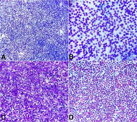
Molecular and Ancillary Testing of Soft Tissue Fine-Needle Aspiration Material
Definition. Ancillary testing of soft tissue fine-needle aspiration (FNA) material comprises immunohistochemistry (IHC), including IHC surrogates for specific genetic alterations, in situ hybridization, polymerase chain reaction (PCR) and next-generation sequencing (NGS). These tests are applied to cell blocks, smears and liquid-based or rinse material to reach a specific diagnosis on a limited sample (Rottmann, Abdulfatah and Pantanowitz 2023). More than 100 distinct soft tissue tumor types exist, including more than 80 benign and intermediate tumors and about 40 sarcomas, and their architectural features are often absent from FNA specimens (Hornick 2019). A morphology-based framework guides the differential diagnosis and the selection of ancillary tests (Lajara and Jo 2024).
Related page: The WHO Reporting System for Soft Tissue Cytopathology and the Approach to Soft Tissue FNA (reporting categories and the general approach to soft tissue FNA) Related page: Round Cell, Spindle Cell and Pleomorphic Sarcomas on FNA (Ewing sarcoma, round cell sarcomas, rhabdomyosarcoma, synovial sarcoma and pleomorphic sarcomas) Related page: Adipocytic Sarcomas, Myxoid Lesions, Extraskeletal Myxoid Chondrosarcoma and Chordoma on FNA (atypical lipomatous tumor, myxoid liposarcoma and extraskeletal myxoid chondrosarcoma) Related page: Bone FNA (H3 mutation-specific immunohistochemistry in giant cell-rich bone tumors)
Molecular classification of soft tissue tumors in the WHO 6th edition
- Current edition. The WHO Classification of Tumours of Soft Tissue and Bone, 6th edition (2026), integrates recent molecular discoveries with morphology. Its essential and desirable diagnostic criteria were refined with different resource settings in mind (Hornick et al. 2026).
- NTRK-rearranged tumors and round cell sarcomas in the 5th edition. The 5th edition (2020) listed NTRK-rearranged soft tissue tumors as an emerging entity and subclassified undifferentiated round cell sarcomas by their gene rearrangements (Sbaraglia, Bellan and Dei Tos 2021).
- Tumors defined by molecular testing. The diagnosis of newer entities such as CIC-rearranged sarcoma explicitly requires molecular testing (Rottmann, Abdulfatah and Pantanowitz 2023).
- IHC surrogates in cytopathology. Recently described and provisional soft tissue tumors are increasingly recognized on cytology through IHC surrogates for their molecular signatures (Rekhi, Qian and Field 2026).
- Link to the reporting system. The WHO Reporting Systems provide standardized information on ancillary testing, including preanalytic handling and specimen preparation (Field et al. 2026).
Preanalytic handling of soft tissue FNA material
- Formalin-fixed cell blocks. Paraffin-embedded cell blocks from samples collected in and fixed with formalin are the simplest material for ancillary tests. Most tests were developed for formalin-fixed paraffin-embedded (FFPE) tissue, and these cell blocks are stained with the same IHC protocols and on-slide controls without additional validation (Suikkanen, Remes and Tuominen 2026).
- Formalin fixation time. Nucleic acid quality and quantity decrease with time in formalin, so fixation time of cell block material is controlled (Suikkanen, Remes and Tuominen 2026).
- Alcohol fixation. Ethanol and methanol alter the tertiary structure of proteins. Depending on the antibody, staining of alcohol-fixed cells is weaker or absent (Suikkanen, Remes and Tuominen 2026).
- Other preparations. Air-dried or methanol-fixed cytospins, liquid-based samples, cell-free supernatants and stained or unstained smears can be used for ancillary tests after protocol optimization and validation. Appropriate on-slide controls for immunocytochemistry can be prepared from cell lines or residual patient samples (Suikkanen, Remes and Tuominen 2026).
| Preparation | Use in ancillary testing | Validation requirement |
|---|---|---|
| Formalin-fixed paraffin cell block | IHC, FISH, DNA and RNA tests designed for FFPE tissue | Same protocols and on-slide controls as histology |
| Alcohol-fixed Papanicolaou smear | FISH; RNA fusion testing after scraping | Protocol optimization and validation |
| Air-dried Romanowsky smear | RNA fusion testing after scraping | Protocol optimization and validation |
| Cytospin or liquid-based preparation | Immunocytochemistry and molecular tests | Protocol optimization and validation |
| Cell-free supernatant | Molecular tests | Validation for each sample type |
Sources: Suikkanen, Remes and Tuominen 2026; Wang et al. 2026; Zreik et al. 2015.
Fluorescence in situ hybridization on soft tissue FNA material
- Break-apart probes for gene rearrangements. In 81 small round cell tumors tested on formalin-fixed paraffin-embedded tissue with an EWSR1 break-apart probe, the result was positive in 59, negative in 13 and failed in 9 (Murthy et al. 2021). The same probes are applied to formalin-fixed cell blocks (Suikkanen, Remes and Tuominen 2026).
- EWSR1 rearrangement in tumors other than Ewing sarcoma. Among EWSR1-rearranged tumors in that series, 89.83% were Ewing sarcoma family tumors. Final diagnoses in the series also included angiomatoid fibrous histiocytoma, desmoplastic small round cell tumor and myxoid chondrosarcoma (Murthy et al. 2021).
- Extraskeletal myxoid chondrosarcoma. FISH on three aspirates showed EWSR1 rearrangement in all, with concomitant NR4A3 rearrangement in one (Santos, Martins and Lemos 2018).
- Desmoplastic small round cell tumor. The EWSR1::WT1 transcript was identified on aspirated material in all 8 patients tested (Klijanienko et al. 2014).
- CIC break-apart FISH. CIC FISH is helpful and inexpensive, with a false-negative rate of 26–43% (Cloutier et al. 2025).
- Amplification FISH. MDM2 amplification has been shown by FISH on a Papanicolaou-stained smear of a dedifferentiated liposarcoma (Zreik et al. 2015). Ring or giant marker chromosomes or MDM2 amplification by FISH were present in all six dedifferentiated liposarcomas tested in an FNA series (Mariño-Enríquez et al. 2014).
PCR and next-generation sequencing on soft tissue FNA material
- Reverse transcription PCR for fusion transcripts. Ewing sarcoma fusion transcripts were detected on FNA material in 44 of 50 primary tumors (88%) (Klijanienko et al. 2012). The SS18::SSX fusion was detected by PCR or FISH in all 38 synovial sarcoma aspirates of a second series (Zhang et al. 2019).
- Anchored multiplex PCR on cytology specimens. Of 1,609 cytology specimens tested with an anchored multiplex PCR fusion assay, 48 (3%) were mesenchymal tumors. Material was insufficient in 2 (4%). The fusion partners detected were ALK (4), STAT6 (4), EWSR1 (3) and one each of SS18, YAP1 and PHF1 (Helland, Fisch and Chebib 2024).
- Diagnostic contribution. Of the 14 mesenchymal cases with a detected fusion, 8 were disease-defining on the cytology preparation and 6 confirmed a metastasis of a known tumor (Helland, Fisch and Chebib 2024).
- RNA fusion testing on sacrificed smears. Of 8,706 fusion assays in one laboratory, 807 (9.3%) were performed on cytology specimens, and 14 used scraped direct smears. Alcohol-fixed Papanicolaou-stained and air-dried modified Giemsa-stained smears that met predefined cellularity thresholds were digitally archived before scraping. Adequate nucleic acid was obtained in all 14 (Wang et al. 2026).
- Fusions found on sacrificed smears. Fusions were detected in 9 of 14 smears (64%), including COL1A2::USP6 and COL1A1::PDGFB. The results established diagnoses that included nodular fasciitis and dermatofibrosarcoma protuberans (Wang et al. 2026).
- DNA- versus RNA-based detection of NTRK fusions. Compared with RNA-based sequencing, DNA-based NGS had a sensitivity of 81.1% and a specificity of 99.9% for NTRK fusions. False negatives occurred when breakpoints fell outside the assay coverage (Solomon et al. 2020).
- RNA NGS for CIC-rearranged sarcoma. RNA fusion analysis also has false-negative results, and manual data review reduces them in part (Cloutier et al. 2025).
Immunohistochemical surrogates for molecular alterations in soft tissue tumors
IHC surrogates detect the protein product or the downstream effect of a defining genetic alteration. They are efficient in small biopsies (Lajara and Jo 2024). Examples include STAT6 in solitary fibrous tumor, H3K27me3 in malignant peripheral nerve sheath tumor, CAMTA1 in epithelioid hemangioendothelioma, MDM2 in dedifferentiated liposarcoma, and WT1 and ETV4 in CIC::DUX4 sarcoma (Hornick 2019).
| Marker | Tumor | Genetic basis | Performance and limitations | Source |
|---|---|---|---|---|
| STAT6 (nuclear) | Solitary fibrous tumor | NAB2::STAT6 fusion | Nuclear staining in solitary fibrous tumor; cytoplasmic staining in cytologic mimics | Tani et al. 2018 |
| DDIT3 (nuclear) | Myxoid liposarcoma | FUS::DDIT3 or EWSR1::DDIT3 | Diffuse in all 46 cases; weak staining in <10% of cells in some dedifferentiated liposarcomas (15%), solitary fibrous tumors (25%), pleomorphic liposarcomas (7%) and high-grade myxofibrosarcomas (12%) | Scapa et al. 2021 |
| MDM2 and CDK4 | Dedifferentiated liposarcoma | MDM2 and CDK4 amplification | MDM2 96% and CDK4 80% on FNA; no pleomorphic liposarcoma coexpressed both | Mariño-Enríquez et al. 2014 |
| SS18-SSX | Synovial sarcoma | SS18::SSX fusion | Sensitivity 95%, specificity 100% | Baranov et al. 2020 |
| SSX C-terminus | Synovial sarcoma | SS18::SSX fusion | Sensitivity 100%, specificity 96% | Baranov et al. 2020 |
| NKX2.2 | Ewing sarcoma | FET::ETS fusion | Sensitivity 100%, specificity 85% on cell blocks; positive in 80% of small cell carcinomas | Russell-Goldman et al. 2018 |
| DUX4 | CIC-rearranged sarcoma | CIC::DUX4 fusion | Sensitivity 97%, specificity 100% | Cloutier et al. 2025 |
| ETV4 and WT1 | CIC-rearranged sarcoma | CIC fusion | ETV4 85% and 95%; WT1 83% and 81–90% (sensitivity and specificity) | Cloutier et al. 2025 |
| BCOR (nuclear) | Sarcoma with BCOR alterations; clear cell sarcoma of kidney | BCOR fusion or internal tandem duplication; YWHAE::NUTM2B | Strong diffuse in all BCOR::CCNB3 and BCOR::MAML3 sarcomas and 93% with BCOR internal tandem duplication; SATB2 also positive in most | Kao et al. 2016 |
| H3K27me3 (loss) | Malignant peripheral nerve sheath tumor | Loss of H3K27 trimethylation | Complete loss in 44% of MPNST and 9% of synovial sarcomas; partial loss of limited use | Pekmezci et al. 2017 |
| Pan-TRK | NTRK-rearranged tumors | NTRK1–3 fusions | Overall sensitivity 87.9% and specificity 81.1%; NTRK3 sensitivity 79%; poor in sarcomas | Solomon et al. 2020 |
| H3G34W | Giant cell tumor of bone | H3-3A G34 mutation | Positive in 85% of FNA and core biopsies; negative in mimics | Schaefer et al. 2018 |
| H3K36M | Chondroblastoma | H3-3B K36M mutation | Positive in all 8 chondroblastomas; negative in mimics | Schaefer et al. 2018 |
| IDH1 R132H | Central cartilaginous tumors | IDH1 R132H mutation | Immunoreactivity confirmed R132H mutations | Amary et al. 2011 |
- STAT6 on cytology. In 28 solitary fibrous tumors sampled by FNA, benign and malignant tumors had similar cytology, with nuclear pleomorphism the most notable difference. Nuclear STAT6 immunoreactivity was a valuable cytologic marker (Tani et al. 2018).
- DDIT3 and radiation. DDIT3 highlighted a vasculocentric growth pattern in 7 myxoid liposarcomas treated with neoadjuvant radiation. Minimal DDIT3 expression in other sarcomas was related to prior radiation or cytotoxic chemotherapy (Scapa et al. 2021).
- SS18-SSX in practice. IHC with the SS18-SSX antibody could replace molecular genetic or cytogenetic testing in most cases of synovial sarcoma (Baranov et al. 2020).
- H3K27me3 in practice. Complete loss of H3K27me3 is a moderately sensitive and relatively specific marker of malignant peripheral nerve sheath tumor when the differential includes synovial sarcoma and fibrosarcomatous dermatofibrosarcoma protuberans (Pekmezci et al. 2017).
- Pan-TRK in practice. Pan-TRK IHC had a specificity of 100% in carcinomas of the colon, lung, thyroid, pancreas and biliary tract. Staining was frequent in tumors with neural differentiation. Assay selection depends on tumor type, the genes involved, available material and the need for concurrent comprehensive testing (Solomon et al. 2020).
Selection of ancillary tests by cytologic pattern in soft tissue FNA
Soft tissue tumors are grouped on cytology as lipomatous, myxoid, spindled, pleomorphic, round cell and epithelioid. Entities that share a pattern can differ greatly in behavior, and defining molecular alterations are detected by FISH or IHC surrogates (Chebib and Jo 2018).
| Cytologic pattern | Common differential diagnosis | First-line ancillary tests | Source |
|---|---|---|---|
| Round cell | Ewing sarcoma, CIC-rearranged sarcoma, sarcoma with BCOR alterations, rhabdomyosarcoma, desmoplastic small round cell tumor | CD99, NKX2.2, desmin, myogenin, WT1 (C-terminal antibody), ETV4, DUX4, BCOR, keratin; EWSR1 or CIC FISH; RNA fusion testing | Russell-Goldman et al. 2018; Cloutier et al. 2025; Morotti et al. 2006; Kao et al. 2016; Barnoud et al. 2000 |
| Spindle cell | Synovial sarcoma, solitary fibrous tumor, malignant peripheral nerve sheath tumor, nodular fasciitis | SS18-SSX, STAT6, H3K27me3, SOX10; USP6 fusion testing | Baranov et al. 2020; Tani et al. 2018; Pekmezci et al. 2017; Wang et al. 2026 |
| Myxoid | Myxoid liposarcoma, extraskeletal myxoid chondrosarcoma, myxofibrosarcoma | DDIT3; EWSR1 and NR4A3 FISH | Scapa et al. 2021; Santos, Martins and Lemos 2018 |
| Pleomorphic | Dedifferentiated liposarcoma, pleomorphic liposarcoma, UPS, dedifferentiated melanoma | MDM2, CDK4, MDM2 FISH; melanocytic markers; BRAF and NRAS mutation analysis | Mariño-Enríquez et al. 2014; Agaimy et al. 2021 |
| Giant cell-rich (bone) | Giant cell tumor of bone, chondroblastoma, giant cell-rich osteosarcoma | H3G34W, H3K36M | Schaefer et al. 2018 |
- SOX10 in spindle cell tumors. SOX10 is consistently expressed in benign Schwann cell tumors and metastatic melanoma and variably in malignant peripheral nerve sheath tumor. It is absent in many mimics of nerve sheath tumors, including cellular neurothekeoma, meningioma, gastrointestinal stromal tumor, perivascular epithelioid cell tumor and fibroblastic-myofibroblastic tumors (Miettinen et al. 2015).
- Entrapped normal cells. SOX10 expression in entrapped non-neoplastic Schwann cells or melanocytes is a source of false-positive interpretation (Miettinen et al. 2015). Entrapped regenerating skeletal muscle fibers stain for myogenin (Cessna et al. 2001).
Integration of ancillary results in soft tissue FNA reports
- Integrated diagnosis. An accurate diagnosis of CIC-rearranged sarcoma requires morphology combined with IHC and cytogenetic or molecular assays (Cloutier et al. 2025). The same principle applies to NKX2.2, which is read within a panel and often with confirmatory molecular testing (Russell-Goldman et al. 2018).
- Negative results from assays with known false negatives. A negative break-apart FISH result does not exclude a rearrangement when the assay has a known false-negative rate, as with CIC FISH (Cloutier et al. 2025). DNA-based panels miss fusions whose breakpoints fall outside the covered regions (Solomon et al. 2020).
Melanoma, Merkel Cell Carcinoma and Fine-Needle Aspiration of the Skin
Definition. Fine-needle aspiration (FNA) of the skin and subcutis samples dermal and subcutaneous nodules, most often to confirm metastatic or recurrent malignancy. Melanoma and Merkel cell carcinoma (MCC) are the two primary cutaneous malignancies most often diagnosed by FNA, usually in regional lymph nodes or subcutaneous sites (Lindsey et al. 2016; Shield and Crous 2014). The Tzanck smear is a cytologic preparation from erosive, vesicular, bullous or pustular skin lesions. It is used mainly for mucocutaneous herpesvirus infections and the pemphigus group (Durdu, Baba and Seçkin 2008).
Related page: Metastatic Melanoma in Lymph Nodes (Cytology) (metastatic melanoma in lymph nodes and the melanocytic immunostain panel) Related page: Small Cell Lung Carcinoma (Cytology) (small cell carcinoma of the lung) Related page: Round Cell, Spindle Cell and Pleomorphic Sarcomas on FNA (undifferentiated pleomorphic sarcoma and dedifferentiated melanoma) Related page: Non-Neoplastic and Reactive Soft Tissue and Skin Lesions on FNA (epidermal inclusion cyst, pilomatrixoma and other benign skin and subcutaneous lesions) Related page: Lymphoma and Metastatic Carcinoma in Soft Tissue FNA (metastatic carcinoma and lymphoma presenting as a soft tissue mass) Related page: The WHO Reporting System for Soft Tissue Cytopathology and the Approach to Soft Tissue FNA (reporting categories for soft tissue and subcutaneous aspirates)
Skin tumors in the WHO Classification of Skin Tumours, 5th edition
- Current edition. The WHO Classification of Skin Tumours, 5th edition (Skin5), is the current skin volume. It follows the standardized structure of the 5th-edition series and gives essential and desirable diagnostic criteria for each entity (Goldman-Lévy et al. 2026).
- New chapters. Skin5 added two chapters: "Tumours of the nail unit" and "Metastases to skin" (Goldman-Lévy et al. 2026).
- Terminology. In the 5th-edition series, "variant" refers only to a genetic variant. A "subtype" differs from the main type in at least one clinical, histologic or genetic characteristic (Goldman-Lévy et al. 2026).
- Metastases to skin. These involve mainly the dermis and subcutis and rarely the epidermis. They are usually epithelial, melanocytic or mesenchymal, and they can be the first sign of the malignancy. The most frequent primary cancers are breast carcinoma in women and melanoma in men (Goldman-Lévy et al. 2026).
- Melanocytic tumors. Skin5 classifies melanocytic tumors along progression pathways to melanoma and applies the term "melanocytoma" to lesions such as atypical Spitz tumor, cellular blue/atypical cellular blue melanocytoma and nodule in congenital nevus (Goldman-Lévy et al. 2026).
Diagnostic performance of FNA for primary cutaneous tumors
- Primary skin tumors. In 28 primary skin tumors sampled by FNA, a specific diagnosis was accurate in 42.8% and the benign or malignant nature was correct in 82.1% (Bharani et al. 2017).
- Causes of error. Sampling error was the most common cause of an incorrect diagnosis. A definitive diagnosis was not reached in adnexal tumors because of overlapping cytologic features and the lack of architecture (Bharani et al. 2017).
- Acral and cutaneous melanoma. In 128 patients with stage 0–III acral and cutaneous melanoma, 5.5% had a negative cytologic diagnosis. The negative rate was 12.2% for primary lesions and 2.3% for lymph nodes (Yang et al. 2020).
- Prognostic association. In the same matched cohort, patients who underwent FNA had worse overall survival than matched patients who did not (Yang et al. 2020).
Cytomorphology of melanoma on FNA of skin and subcutaneous nodules
- Clinical setting. FNA of metastatic melanoma can establish the diagnosis before the primary lesion is identified in up to 20% of cases. In a patient with a known melanoma, a negative result from a subcutaneous or pulmonary nodule is regarded as a possible false-negative result (Lindsey et al. 2016).
- Smear pattern. Smears usually show poorly cohesive cells. Frankly malignant spindled and epithelioid cells are the most common shapes (Lindsey et al. 2016).
- Melanin. Melanin is diagnostic when present and is seen in about 50% of cases (Lindsey et al. 2016).
- Accessory nuclear and cytoplasmic features. Eccentric nuclei, nucleoli and nuclear pseudoinclusions support the diagnosis. Cytoplasmic vacuoles can usually be found on Romanowsky-stained smears (Lindsey et al. 2016).
- Accuracy in lymph nodes. In a meta-analysis of 10 studies with 5,518 cases, FNA had a pooled sensitivity of 0.97 and specificity of 0.98 for metastatic melanoma (Hall et al. 2013).
| Cytologic feature | BRAF V600-mutated (n = 18) | BRAF wild-type (n = 21) |
|---|---|---|
| Binucleation or multinucleation | 78% | 57% |
| Nuclear pleomorphism | 72% | 67% |
| Spindle cell morphology | 61% | 29% |
| Macronucleoli | 56% | 52% |
| Intranuclear inclusions | 50% | 33% |
| Pigment | 44% | 24% |
| Cytoplasmic vacuolization | 22% | 29% |
| Necrosis | 11% | 10% |
Source: Lee et al. 2019. The mean age was 52.2 years in the BRAF-mutated group and 65.2 years in the wild-type group. Spindle cell morphology correlated most with BRAF V600 mutation (Lee et al. 2019).
Immunocytochemistry and molecular testing of melanoma on FNA
- SOX10 and S100 on cell blocks. In 38 nodal melanoma metastases, S100 and SOX10 each had a sensitivity of 100%, higher than Melan-A and HMB45. SOX10 had the best staining performance (Ronchi et al. 2022).
- Marker sensitivity in a larger cytology series. In 93 melanomas, sensitivity was SOX10 100%, PRAME 86%, HMB45 84%, Melan-A 82% and S100 71%. Four of 10 spindle cell melanomas were PRAME negative (Saad et al. 2024).
- PRAME specificity. PRAME stained 23% of carcinomas, 33% of sarcomas and 11% of hematologic malignancies. Its specificity for melanoma was 82% (Saad et al. 2024).
- SOX10 in nonmelanocytic tumors. SOX10 is consistently expressed in benign Schwann cell tumors and variably in malignant peripheral nerve sheath tumors. It is expressed in myoepitheliomas, basal-type breast carcinomas, 6% of head and neck squamous cell carcinomas and 7% of pulmonary small cell carcinomas (Miettinen et al. 2015).
- Direct smears for immunocytochemistry. In 17 metastatic melanomas, S100 was positive in all on direct smears. HMB45 and Melan-A were positive in 81% and 88% (Hookim et al. 2012).
- BRAF testing on smears. BRAF-activating mutations are present in at least 40% of melanomas. BRAF mutations were found in 8 of 15 archived Diff-Quik-stained smears (53%; 5 V600E and 3 V600K), and corresponding cell blocks were concordant in 14 of 15. One cell block result was falsely negative (Hookim et al. 2012).
- Clinical use of BRAF status. Clinicians used the BRAF result on FNA material to select BRAF inhibitor treatment (Lee et al. 2019).
- Dedifferentiated and undifferentiated melanoma. The undifferentiated component lacks S100, SOX10, Melan-A, HMB45 and pan-melanoma staining. In patients with a prior melanoma, 73% of evaluable tumors carried a melanoma-compatible mutation (Agaimy et al. 2021).
| Marker | Sensitivity for metastatic melanoma on cytology | Specificity issues |
|---|---|---|
| SOX10 | 100% (Ronchi 2022; Saad 2024) | Schwann cell tumors, myoepitheliomas, basal-type breast carcinoma, some small cell and squamous carcinomas (Miettinen 2015) |
| S100 | 100% (Ronchi 2022); 71% (Saad 2024) | Not assessed in these series |
| PRAME | 86% overall; 60% in spindle cell melanoma (4 of 10 negative) (Saad 2024) | 23% of carcinomas, 33% of sarcomas, 11% of hematologic malignancies; specificity 82% (Saad 2024) |
| HMB45 | 84% (Saad 2024); 81% on smears (Hookim 2012) | Less sensitive than SOX10 (Ronchi 2022) |
| Melan-A | 82% (Saad 2024); 88% on smears (Hookim 2012) | Less sensitive than SOX10 (Ronchi 2022) |
| MiTF | Not assessed in these series | Positive in 89% of undifferentiated pleomorphic sarcomas (Choy 2017) |
Merkel cell carcinoma on FNA
- Definition and incidence. MCC is an aggressive, poorly differentiated neuroendocrine carcinoma of the skin whose incidence has increased over recent decades (Harms 2026). It occurs mainly in elderly or immunocompromised patients (Bechert, Schnadig and Nawgiri 2013).
- Etiology. Clonal integration of Merkel cell polyomavirus (MCPyV) DNA, with persistent expression of viral T antigens, causes at least 60% of MCC. Ultraviolet damage with highly mutated genomes causes the nonviral form. Both forms have similar presentation, prognosis and response to therapy (DeCaprio 2021).
- Clinical setting of aspirated cases. In 69 MCC sampled by FNA, the median age was 78 years. Sites included lymph nodes of the neck, axilla and groin and the parotid gland. Most patients had a history of MCC (68%) or of another malignancy (70%) (Shield and Crous 2014).
- Smear pattern. Smears were cellular, with malignant cells mostly dispersed and in variable numbers of disorganized groups (Shield and Crous 2014).
- Cell features. Cytoplasm was scant or absent. Nuclei showed mild to moderate anisokaryosis, stippled chromatin, inconspicuous nucleoli and molding. Apoptotic bodies were often numerous (Shield and Crous 2014).
- Features that separate MCC from lymphoma. In a nodal FNA first reported as suspicious for lymphoma, review showed nuclear molding, Indian filing and rare cytoplasmic pale bodies and no lymphoglandular bodies (Liu et al. 2022). Lymphoglandular bodies also occur in some nonlymphoid malignancies, including small cell carcinoma (Flanders et al. 1993).
- Concurrent lymphoma. MCC can coexist with a low-grade lymphoma in the same patient (Bechert, Schnadig and Nawgiri 2013).
Immunohistochemistry of Merkel cell carcinoma on cell blocks
- Keratins. Cell blocks usually showed cytokeratin staining in a perinuclear dot pattern. CK20 was positive in 88% of cases (Shield and Crous 2014).
- Neuroendocrine markers. CD56 was the most sensitive neuroendocrine marker on cell blocks (95%) and was positive by flow cytometry in all 9 cases tested (Shield and Crous 2014).
- Distinction from small cell lung carcinoma. All 13 MCC were CK20 positive and TTF-1 negative, and 12 of 13 were neurofilament positive. Of 13 small cell lung carcinomas, 11 were TTF-1 positive, all were neurofilament negative and all but one were CK20 negative. The other markers tested, including chromogranin, synaptophysin, neuron-specific enolase and CD56, did not separate the two tumors (Bobos et al. 2006).
- MCPyV immunohistochemistry. MCPyV was detected in 24 of 53 MCC cell blocks (45%). It was 97% specific; one follicular lymphoma among the mimics stained strongly (Mubeen and Mito 2024).
- Newer markers. SATB2, H3K27me3 and POU4F3 have been proposed as MCC markers. The interpretation of CK20, TTF-1, neurofilament and MCPyV has limitations (Harms 2026).
- Differential diagnosis of MCC. The differential includes extracutaneous small cell carcinoma. Vulvar small cell carcinoma is at substantial risk of misdiagnosis because of immunohistochemical overlap with MCC. WNT/β-catenin-activated nonpilomatrical carcinoma can express CK20 and neuroendocrine markers. Basal cell carcinoma and squamous cell carcinoma are included in the differential in some settings (Harms 2026).
| Tumor | Cytology | CK20 | TTF-1 | Neurofilament | Other |
|---|---|---|---|---|---|
| Merkel cell carcinoma | Dispersed small cells, molding, stippled chromatin, apoptotic bodies | Positive, perinuclear dot (88–100%) | Negative | Positive (12 of 13) | MCPyV in 45%; CD56 95% |
| Small cell lung carcinoma | Small cells with molding | Negative (12 of 13) | Positive (11 of 13) | Negative | Shares neuroendocrine markers with MCC |
Lymphoma shows discohesive lymphoid cells and lymphoglandular bodies and is confirmed by flow cytometry. Metastatic melanoma shows discohesive epithelioid or spindle cells with pseudoinclusions and is SOX10 positive.
Sources: Shield and Crous 2014; Bobos et al. 2006; Mubeen and Mito 2024; Liu et al. 2022; Flanders et al. 1993; Ronchi et al. 2022.
Tzanck smear in vesiculobullous and pustular skin lesions
- Method. In a series of 400 patients with erosive, vesicular, bullous or pustular lesions, Tzanck smears were stained with May-Grünwald-Giemsa. Methylene blue, Gram stain or direct immunofluorescence was added in some patients (Durdu, Baba and Seçkin 2008).
- Pemphigus. Acantholytic cells had a sensitivity of 100% for pemphigus (Durdu, Baba and Seçkin 2008). In a later series, sensitivity and specificity against histopathology were 80.70% and 68.18%, and against anti-desmoglein antibodies 82.75% and 100% (Chaudhari et al. 2025).
- Acantholytic cell morphology. Incomplete acantholytic cells predominated in 60.32% of pemphigus cases and complete acantholytic cells in 39.18%. Rounding was the most consistent feature, followed by nuclear enlargement (Chaudhari et al. 2025).
- Herpesvirus cytopathic effect. The typical Tzanck changes of HSV and VZV infection are multinucleation, margination of chromatin and nuclear molding. Their absence was a useful negative finding in a smear from an mpox lesion (Derrick et al. 2023). Herpesvirus cytopathic effect was recorded in 25 cases of a recent Tzanck series (Chaudhari et al. 2025).
- Herpesvirus infection. Multinucleated giant cells and acantholytic cells had a sensitivity of 84.7% for herpetic infections (Durdu, Baba and Seçkin 2008).
- Comparison with culture and PCR. In clinically suspected cases, the Tzanck smear was positive in 60% of herpes simplex virus (HSV) and 75% of varicella-zoster virus (VZV) infections. Viral culture was positive in 83% and 44%. PCR detected viral DNA on the stained Tzanck smears in 73% of HSV and 88% of VZV cases, and on unstained smears in 83% and 97%. No viral DNA was found in 15 controls (Nahass et al. 1992).
- PCR sources. PCR detected HSV and VZV DNA in Tzanck smear debris, vesicle fluid swabs, crusts and formalin-fixed tissue. Detection was slightly more frequent from smear debris, crusts and swabs than from fixed tissue (Nahass et al. 1995).
- Other diagnoses. Dyskeratotic acantholytic cells with cocci had a sensitivity of 92% for bullous impetigo, pseudohyphae 100% for candidiasis and more than 10 tadpole cells per low-power field 81.5% for spongiotic dermatitis (Durdu, Baba and Seçkin 2008). Molluscum contagiosum is also recognized on Tzanck smears (Chaudhari et al. 2025).
| Condition | Tzanck finding | Sensitivity | Confirmatory test | Source |
|---|---|---|---|---|
| Herpes simplex or varicella-zoster infection | Multinucleated giant cells with nuclear molding and chromatin margination; acantholytic cells | 84.7% | PCR on the smear or vesicle swab | Durdu 2008; Derrick 2023; Nahass 1992 |
| Pemphigus | Acantholytic cells (complete or incomplete) | 80.70–100% | Histopathology, direct immunofluorescence, anti-desmoglein antibodies | Durdu 2008; Chaudhari 2025 |
| Bullous impetigo | Dyskeratotic acantholytic cells and cocci | 92% | Bacterial culture | Durdu 2008 |
| Candidiasis | Pseudohyphae | 100% | Potassium hydroxide examination or culture | Durdu 2008 |
| Spongiotic dermatitis | More than 10 tadpole cells | 81.5% | Clinical and histologic correlation | Durdu 2008 |
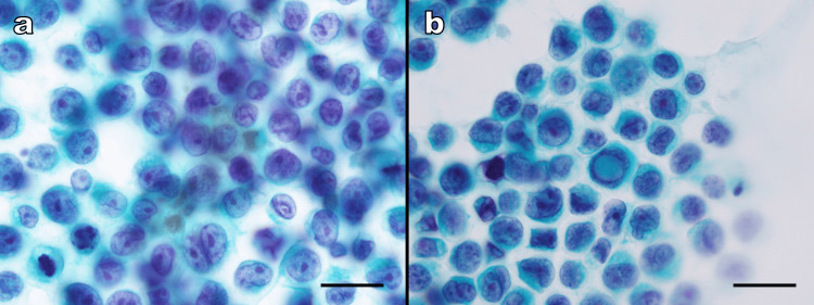
Fine-Needle Aspiration of Bone
Definition. Fine-needle aspiration (FNA) of bone samples lytic, blastic or mixed skeletal lesions, usually under imaging guidance, to separate neoplastic from nonneoplastic disease and metastatic from primary tumors. FNA of bone is reliable for metastatic and primary bone tumors, and its main limitations are inadequate sampling and misclassification of the tumor type (Bommer, Ramzy and Mody 1997). Final interpretation integrates the cytologic findings with the clinical and radiologic findings (Kabukçuoğlu et al. 1998).
Related page: Round Cell, Spindle Cell and Pleomorphic Sarcomas on FNA (Ewing sarcoma and other round cell sarcomas) Related page: Molecular and Ancillary Testing of Soft Tissue FNA Material (fusion testing and immunohistochemical surrogates on FNA material) Related page: Adipocytic Sarcomas, Myxoid Lesions, Extraskeletal Myxoid Chondrosarcoma and Chordoma on FNA (chordoma and extraskeletal myxoid chondrosarcoma) Related page: Lymphoma and Metastatic Carcinoma in Soft Tissue FNA (lymphoma and metastatic carcinoma presenting as a soft tissue mass) Related page: The WHO Reporting System for Soft Tissue Cytopathology and the Approach to Soft Tissue FNA (reporting categories and the general approach to soft tissue and bone FNA)
Bone tumors in the WHO Classification of Tumours of Soft Tissue and Bone, 6th edition
- Current edition. The 6th edition (2026) contains 46 bone tumors and 18 vascular tumors of soft tissue and bone (Hornick et al. 2026).
- Gene nomenclature. The gene mutated in giant cell tumor of bone is H3-3A, previously designated H3F3A. The gene mutated in chondroblastoma is H3-3B, previously H3F3B (Wągrodzki et al. 2022; Schaefer et al. 2018).
- Previous-edition rule for low-grade central cartilaginous tumors. In the 5th edition (2020), a low-grade central cartilaginous tumor of the appendicular skeleton is an atypical cartilaginous tumor (intermediate, locally aggressive). A histologically identical tumor of the axial skeleton, including the pelvic bones and skull base, is chondrosarcoma grade 1 (malignant). The term atypical cartilaginous tumor was first introduced in the 2013 classification (Kim and Lee 2023).
Indications and diagnostic performance of bone FNA
- Case mix. In 450 bone FNAs from 427 patients aged 5–94 years, the spine was the most frequent site (49%), followed by the ilium, sacrum, mandible, ribs and femur (Bommer, Ramzy and Mody 1997).
- Adequacy and results. Of the 450 aspirates, 385 (86%) were adequate. Of these, 215 were positive for malignancy, 11 suspicious, 2 inconclusive and 157 negative (Bommer, Ramzy and Mody 1997).
- Metastatic carcinoma as the most frequent malignancy. Metastatic carcinoma accounted for 175 of the 215 malignant aspirates, and 67% of these were adenocarcinomas. The 40 primary malignant bone neoplasms included myeloma, lymphoma, Ewing sarcoma, chondrosarcoma, ameloblastoma, chordoma, malignant peripheral nerve sheath tumor and unclassified high-grade sarcoma (Bommer, Ramzy and Mody 1997).
- False-negative and false-positive results. Ten aspirates were false negative, and in 6 of these the material did not represent the lesion. Five malignancies were misclassified by type. The false-positive rate was 0.2% (Bommer, Ramzy and Mody 1997).
- Accuracy in bone and soft tissue together. In 1,114 bone and soft tissue FNAs, sensitivity was 96%, specificity 98%, positive predictive value 99% and negative predictive value 92%. The 18 errors were caused by sampling (9), interpretation (7) and screening (2) (Khalbuss, Teot and Monaco 2010).
- Comparison with core and open biopsy. In one institution, diagnostic accuracy was 89% for open biopsy, 82% for FNA and 78% for core biopsy. Clinically significant errors occurred with all three methods (Layfield et al. 2014).
- Lesions that cause difficulty. In 38 clinically malignant bone lesions, FNA accuracy was 89.5%. Chondrosarcoma caused the greatest difficulty in separation from other chondroid tumors. Giant cell tumor was difficult when the aspirate was hemorrhagic and hypocellular. Metastatic carcinoma was diagnosed with high accuracy (Kabukçuoğlu et al. 1998).
- On-site adequacy assessment. In 1,995 bone and soft tissue FNAs for suspected metastatic carcinoma, melanoma or lymphoma, on-site adequacy downgrade rates were equivalent for cytotechnologists and cytopathologists (4.1% versus 3.6%) (Olson et al. 2013).
Giant cell tumor of bone and chondroblastoma on FNA
- Biology. Giant cell tumor of bone is an intermediate-grade, locally destructive primary bone neoplasm that recurs and rarely metastasizes or transforms to malignancy. The neoplastic cell is the mononuclear stromal cell, which carries an H3-3A G34 mutation and induces osteoclastogenesis through dysregulated RANKL signaling. The tumor contains osteoclast-type giant cells, mononuclear histiocytic cells and neoplastic stromal cells (Carneiro et al. 2026).
- H3G34W immunohistochemistry on FNA and core biopsy. H3G34W was positive in 22 of 26 giant cell tumors (85%) and negative in all histologic mimics, including aneurysmal bone cyst and osteosarcoma. Two of the four negative tumors carried alternate G34L or G34V mutations (Schaefer et al. 2018).
- Concordance. IHC results were concordant between biopsy and surgical specimens in 152 of 158 samples (96%) (Schaefer et al. 2018).
- Sequencing. In 214 patients, H3-3A Sanger sequencing had a sensitivity and specificity of 100%. IHC with G34W and G34V antibodies had a sensitivity of 94.32% and a specificity of 100%. Tissue was decalcified in EDTA before testing. Proper processing and decalcification are required (Wągrodzki et al. 2022).
- Chondroblastoma. H3K36M IHC was positive in all 8 chondroblastomas and negative in all mimics. H3-3B K36M mutations are present in nearly all chondroblastomas (Schaefer et al. 2018).
- Denosumab. Denosumab is a monoclonal antibody against RANK ligand used to treat giant cell tumor of bone. Denosumab-treated tumors show bone formation and morphologic overlap with malignancy, and malignant transformation during treatment has been reported (Li et al. 2020).
- Mutation after denosumab. The H3-3A mutation was detected in 9 of 13 tumors after denosumab and in 2 of 9 secondary malignant giant cell tumors. Denosumab did not eradicate the neoplastic cell population, and the presence of the mutation did not exclude malignancy (Wągrodzki et al. 2022).
| Giant cell-rich lesion | Ancillary finding | Source |
|---|---|---|
| Giant cell tumor of bone | H3G34W positive (85% on FNA and core biopsy); H3-3A G34 mutation | Schaefer et al. 2018; Wągrodzki et al. 2022 |
| Chondroblastoma | H3K36M positive; H3-3B K36M mutation | Schaefer et al. 2018 |
| Aneurysmal bone cyst | H3G34W and H3K36M negative | Schaefer et al. 2018 |
| Giant cell-rich osteosarcoma | H3G34W and H3K36M negative; SATB2 often positive | Schaefer et al. 2018; Machado et al. 2016 |
| Langerhans cell histiocytosis | Multinucleated giant cells in a mixed infiltrate; CD1a positive | Pohar-Marinsek and Us-Krasovec 1996; Hang, Siddiqui and Ali 2017 |
Cartilaginous tumors and chondrosarcoma on FNA
- Accuracy for primary chondrosarcoma. In 34 aspirates from 32 patients, 18 of 27 primary chondrosarcomas were diagnosed definitively as chondrosarcoma or malignant chondroid neoplasm (accuracy 67%). Eight were equivocal and required another procedure, and two false negatives were caused by inadequate sampling (Dodd 2006).
- Recurrent and metastatic chondrosarcoma. Accuracy was higher for recurrent or metastatic lesions (86%, 6 of 7). FNA is reliable in patients with a documented history of chondrosarcoma (Dodd 2006).
- Chondroblastic osteosarcoma. Two tumors diagnosed as chondrosarcoma on FNA were chondroblastic osteosarcomas (Dodd 2006).
- Dedifferentiated chondrosarcoma. The diagnosis requires recognition of a high-grade non-cartilage-producing sarcoma juxtaposed against a low-grade chondrosarcoma. In 4 aspirates from 3 patients, no FNA or core biopsy showed both components: two primary aspirates showed only pleomorphic sarcoma and one showed only low-grade chondrosarcoma. Clinical and radiographic correlation suggested the diagnosis (Rinas, Ward and Kilpatrick 2005).
- IDH1 and IDH2 mutations. Heterozygous somatic IDH1 or IDH2 mutations were found only in central and periosteal cartilaginous tumors, in at least 56% of them, among about 1,200 mesenchymal tumors screened. None occurred in osteosarcomas, peripheral chondrosarcomas or osteochondromas (Amary et al. 2011).
- Spectrum of IDH-mutant tumors and mutation types. The mutations were present from enchondroma through conventional central and dedifferentiated chondrosarcoma. The ratio of IDH1 to IDH2 mutations was 10.6:1, and about 40% were IDH1 R132C. IDH1 R132H was confirmed by immunoreactivity (Amary et al. 2011).
- SATB2 and SOX9 in cartilaginous tumors. SATB2 was expressed in 46.6% of chondrosarcomas, more often in grade III tumors, and could not separate chondroblastic osteosarcoma from high-grade chondrosarcoma (Machado et al. 2016). SOX9 was expressed in 94% of chondrosarcomas and 98% of osteosarcomas on biopsy material (Sharma, Pytel and Cipriani 2022).
Osteosarcoma on FNA
- Skeletal osteosarcoma. Of 12 adequate primary skeletal osteosarcomas sampled by FNA, 10 (83%) were diagnosed correctly. One was diagnosed as giant cell tumor and proved to be a high-grade fibroblastic osteosarcoma arising in a giant cell tumor; another was reported as a spindle cell neoplasm (Nicol et al. 1998).
- Extraskeletal osteosarcoma. None of the primary extraskeletal osteosarcomas sampled by FNA was recognized as osteosarcoma, whereas both metastatic extraskeletal osteosarcomas were recognized. Extraskeletal osteosarcoma most likely requires incisional biopsy (Nicol et al. 1998).
- Osteoid. The identification of malignant osteoid matrix, integrated with clinical and radiologic data, is the main criterion for the diagnosis of osteosarcoma (Machado et al. 2016).
- SATB2. SATB2 was positive in 90.4% of osteosarcomas, 87.5% of small cell osteosarcomas, 46.6% of chondrosarcomas and 1.3% of Ewing sarcomas. Sensitivity was 90.4% and specificity 95.3%, with a positive predictive value of 66.6% and a negative predictive value of 98.9% (Machado et al. 2016).
- Limits of SATB2 and SOX9. On biopsy material, SATB2 was expressed in 86% of osteosarcomas and 50% of chondrosarcomas. The two markers were coexpressed in 89% of osteosarcomas and 44% of chondrosarcomas, and they did not reliably separate the two tumors (Sharma, Pytel and Cipriani 2022).
| Marker | Osteosarcoma | Chondrosarcoma | Source |
|---|---|---|---|
| SATB2 | 86–90.4% | 46.6–50% | Machado et al. 2016; Sharma, Pytel and Cipriani 2022 |
| SOX9 | 98% | 94% | Sharma, Pytel and Cipriani 2022 |
| IDH1 or IDH2 mutation | None | ≥56% of central and periosteal tumors | Amary et al. 2011 |
Plasma cell neoplasms and lymphoma of bone on FNA
- Frequency among primary bone malignancies. Myeloma and lymphoma were among the primary malignant bone neoplasms diagnosed by FNA in the 450-case series (Bommer, Ramzy and Mody 1997). Multiple myeloma was identified on FNA when interpreted with the coexisting clinical findings (Kabukçuoğlu et al. 1998).
- Plasma cell grade on FNA of extraosseous masses. In 30 soft tissue masses aspirated from 27 patients with multiple myeloma, 27 were positive for myeloma. Graded by the Bartl classification, 18.5% were low grade, 55.6% intermediate grade and 25.9% high grade (blastic) (Mukunyadzi et al. 2001).
- Prognostic value of grade. Of patients with high-grade myeloma in a soft tissue mass, 43% died by a median of 2 months, compared with 12% of those with low- or intermediate-grade disease. Concurrent bone marrow involvement was present in 71% of the high-grade cases (Mukunyadzi et al. 2001).
- Anaplastic plasma cells in extramedullary plasmacytoma. In 18 extramedullary plasmacytomas of the skin, lymph nodes, liver and spleen sampled by FNA, smears showed anaplastic plasma cells in 13. Some cases had been thought clinically to be infections, and two had a prior histologic diagnosis of non-Hodgkin lymphoma (Bangerter et al. 2000).
- Ancillary studies. FNA material provides samples for flow cytometry and cytogenetic studies in plasma cell neoplasms (Mukunyadzi et al. 2001). Lymphoma in bone and soft tissue is covered in Lymphoma and Metastatic Carcinoma in Soft Tissue FNA.
- CD138 and clonality. CD138 (syndecan-1) identifies plasma cells but is not specific for them. Prostatic, colonic, renal cell and hepatocellular carcinomas can express it. A metastatic hepatocellular carcinoma in a soft tissue mass was CD138 positive by immunohistochemistry and flow cytometry and clinically mimicked myeloma (Ramalingam et al. 2008). Clonality is shown by restriction of cytoplasmic kappa or lambda light chain. In bone marrow, the cytoplasmic kappa/lambda ratio of CD138-positive plasma cells separated plasma cell myeloma from reactive plasmacytosis with a sensitivity of 100% and a specificity of 98.0% (Nakayama-Ichiyama et al. 2012).
Langerhans cell histiocytosis of bone on FNA
- Clinical setting. Langerhans cell histiocytosis (LCH) predominantly occurs in children and young adults. Of 37 patients diagnosed by FNA, 28 had bone lesions, 6 head and neck soft tissue lesions and 3 lymph node lesions (Hang, Siddiqui and Ali 2017).
- Diagnostic performance. Thirty-one patients (84%) were diagnosed as LCH. Four (11%) received a descriptive diagnosis of scant histiocyte-like cells with mixed inflammation, and 2 (5%) were nondiagnostic (Hang, Siddiqui and Ali 2017).
- Langerhans cells. Langerhans cells have oval to reniform nuclei with nuclear grooving and abundant pale cytoplasm (Elsheikh et al. 1991).
- Background. Smears contain eosinophils, neutrophils, lymphocytes, macrophages, multinucleated giant cells and capillary fragments, in varying proportions (Pohar-Marinsek and Us-Krasovec 1996). Eosinophils can be sparse (Elsheikh et al. 1991).
- Sources of error. In 37 smears, LCH was recognized in 22 and suspected in 5, and 6 were nondiagnostic. Three reactive lesions were misinterpreted as LCH (Pohar-Marinsek and Us-Krasovec 1996).
- Immunohistochemistry on cell blocks. CD1a was positive in 24 of 24 cases (100%), S100 in 22 of 23 (96%) and CD68 in 3 of 3 (Hang, Siddiqui and Ali 2017). Birbeck granules were shown by electron microscopy in one case (Elsheikh et al. 1991).
- BRAF V600E. The BRAF V600E mutation was found in 35 of 61 LCH specimens (57%). It was more frequent in younger patients and was not associated with disease site or stage. Phospho-MEK and phospho-ERK staining was present regardless of mutation status (Badalian-Very et al. 2010).
- Other MAPK pathway alterations. MAP2K1 mutations also occur in LCH, and both BRAF V600E and MAP2K1 alterations are shared with Erdheim-Chester disease (McKinney and Wang 2025).
Metastatic carcinoma to bone on FNA
- Frequency. Metastatic carcinoma is the most frequent malignancy in bone FNA (Bommer, Ramzy and Mody 1997).
- Prostatic origin. NKX3.1 had a sensitivity of 98.6% for metastatic prostatic adenocarcinoma in lymph node, bone and soft tissue metastases and a specificity of 99.7% in 349 nonprostatic tumors. (Gurel et al. 2010).
- Other primary sites. Immunohistochemistry for the primary site of a metastasis is covered in Lymphoma and Metastatic Carcinoma in Soft Tissue FNA.
| Bone lesion on FNA | Key cytologic finding | Key ancillary test | Source |
|---|---|---|---|
| Metastatic carcinoma | Malignant epithelial cells; adenocarcinoma in 67% | Site-specific panels such as NKX3.1 and PAX8 | Bommer, Ramzy and Mody 1997; Gurel et al. 2010; Barr et al. 2015 |
| Plasma cell neoplasm | Plasma cells, sometimes anaplastic | CD138 with kappa/lambda light-chain restriction; flow cytometry; cytogenetics | Mukunyadzi et al. 2001; Bangerter et al. 2000; Nakayama-Ichiyama et al. 2012 |
| Giant cell tumor of bone | Osteoclast-type giant cells and mononuclear cells | H3G34W | Schaefer et al. 2018 |
| Chondrosarcoma | Chondroid matrix with atypical chondrocytes | IDH1 or IDH2 mutation | Dodd 2006; Amary et al. 2011 |
| Osteosarcoma | Malignant cells with osteoid | SATB2 | Machado et al. 2016 |
| Langerhans cell histiocytosis | Grooved Langerhans cells, eosinophils, giant cells | CD1a, S100; BRAF V600E | Hang, Siddiqui and Ali 2017; Badalian-Very et al. 2010 |
| Ewing sarcoma | Small round cells | CD99, NKX2.2; FET::ETS fusion | Russell-Goldman et al. 2018; Grünewald et al. 2018 |
Lymphoma and Metastatic Carcinoma in Soft Tissue Fine-Needle Aspiration
Definition. A soft tissue mass sampled by fine-needle aspiration (FNA) can be a hematolymphoid neoplasm or a metastasis. Both can mimic a primary sarcoma clinically and pathologically (Plaza et al. 2008; Wakely, Frable and Kneisl 2001). Malignant lymphoma and leukemia involve soft tissue infrequently (Wakely, Frable and Kneisl 2001). Metastases presenting as soft tissue masses are relatively rare and are a source of diagnostic confusion (Plaza et al. 2008).
Related page: The WHO Reporting System for Soft Tissue Cytopathology and the Approach to Soft Tissue FNA (reporting categories and the general approach to soft tissue FNA) Related page: Round Cell, Spindle Cell and Pleomorphic Sarcomas on FNA (undifferentiated pleomorphic sarcoma and its mimics) Related page: Melanoma, Merkel Cell Carcinoma and Skin FNA (melanoma and Merkel cell carcinoma in skin and subcutis) Related page: Bone FNA (plasma cell neoplasms and metastatic carcinoma in bone) Related page: Diffuse Large B-Cell Lymphoma (Cytology) (diffuse large B-cell lymphoma in lymph nodes) Related page: Peripheral T-Cell Lymphomas and Anaplastic Large Cell Lymphoma in Cytology (T-cell lymphomas including anaplastic large cell lymphoma in lymph nodes) Related page: Metastatic Carcinoma in Lymph Nodes (Cytology) (metastatic carcinoma in lymph nodes)
Classification and reporting frameworks for hematolymphoid tumors in soft tissue
- Soft tissue and bone classification. The WHO Classification of Tumours of Soft Tissue and Bone, 6th edition (2026), includes 6 hematopoietic and dendritic cell neoplasms (Hornick et al. 2026).
- Reporting system for hematolymphoid aspirates. The WHO Reporting System for Lymph Node, Spleen, and Thymus Cytopathology uses five categories: inadequate/insufficient/nondiagnostic, benign, atypical, suspicious for malignancy and malignant. Final and specific diagnoses often require flow cytometry, immunocytochemistry, in situ hybridization and molecular tests (Ehinger et al. 2026).
- Pattern-based approach to hematolymphoid aspirates. The lymph node system defines four cytologic patterns: mixed lymphoid cells; predominantly small or intermediate cells; predominantly large, pleomorphic or blastic cells; and single very large atypical cells (Ehinger et al. 2026).
Lymphoma and leukemia in soft tissue on FNA
- Case mix. In 21 patients with soft tissue lymphoma or leukemia, after exclusion of lymph node-rich sites and masses near enlarged nodes or skin lesions, ages ranged from 10 months to 87 years (mean 51). Seven masses were superficial and 14 deep. Sites were the extremities (10), trunk (8) and head (3) (Wakely, Frable and Kneisl 2001).
- Diagnoses. Cytologic diagnoses were large cell lymphoma (11), Hodgkin lymphoma (1), lymphoblastic leukemia (3), myelogenous leukemia (2) and atypical lymphoid cells (4). All non-Hodgkin lymphomas were of large B-cell type (Wakely, Frable and Kneisl 2001).
- Outcome of atypical aspirates. Of the four aspirates reported as atypical lymphoid cells, three proved to be lymphoma and one acute leukemia. One was a mantle cell lymphoma (Wakely, Frable and Kneisl 2001).
- Immunophenotyping. Cytology was confirmed by flow cytometry (5 cases), cytospin (3) and cell block (4). In one patient, the FNA diagnosis of lymphoma reversed a prior core biopsy diagnosis of liposarcoma (Wakely, Frable and Kneisl 2001).
- Diagnostic performance. Sensitivity was 82% and specificity 100% for a definitive diagnosis of lymphoma or leukemia (Wakely, Frable and Kneisl 2001).
- FNA with flow cytometry. In 81 patients with suspected nodal or extranodal lymphoma, FNA with flow cytometry achieved a diagnosis in 79%. Thirteen (16.1%) were not malignant, 2 (2.5%) were false negative and 2 (2.5%) needed histology (Swart et al. 2007).
- Chest wall lymphomas. In 11 chest wall lymphoid neoplasms, the reviewed FNA diagnoses were anaplastic large cell lymphoma (5), centroblastic lymphoma (2) and one each of small lymphocytic lymphoma, post-transplant peripheral T-cell lymphoma, Hodgkin lymphoma and plasmacytoma (Das et al. 2017).
Lymphoglandular bodies in lymphoma and other malignancies on soft tissue FNA
- Definition. Lymphoglandular bodies are cytoplasmic fragments of lymphocytes, usually less than half the size of a red blood cell, and a feature of lymphoid tissue (Murakami et al. 2008).
- Frequency in lymphoma. In Diff-Quik-stained smears, 24 of 28 lymphomas had easily identifiable (more than 2 per high-power field) or numerous (more than 10 per high-power field) lymphoglandular bodies (Flanders et al. 1993).
- Lymphoglandular bodies in nonlymphoid malignancies. Nine of 104 nonlymphoid malignancies also had easily identifiable or numerous lymphoglandular bodies: two small cell carcinomas, four non-small cell carcinomas, one ganglioneuroblastoma, one melanoma and one seminoma (Flanders et al. 1993).
- In histologic sections. Lymphoglandular bodies were found in about 40% of lymphomas and in 3.8% of nonlymphoma malignancies in hematoxylin and eosin sections (Murakami et al. 2008).
Anaplastic large cell lymphoma in soft tissue on FNA
- Mimicry of sarcoma. Two anaplastic large cell lymphomas (ALCL) in patients aged 16 and 9 years showed pleomorphic cells in loosely cohesive groups around blood vessels and singly, with prominent nucleoli, finely vacuolated cytoplasm and a few cells with embryoid nuclei. Rhabdomyosarcoma was included in the differential diagnosis (Rekhi et al. 2010).
- Immunophenotype. The tumor cells were positive for vimentin, CD45, EMA, CD30 and ALK (Rekhi et al. 2010).
- Multifocal presentation. One patient had multiple nodular soft tissue and bone lesions. ALCL is included in the differential diagnosis of pediatric soft tissue tumors, especially with multifocal involvement (Rekhi et al. 2010).
- ALK-negative ALCL. In 37 ALCL, ALK-negative smears had more anaplastic cells than ALK-positive smears, and their hallmark cells were not classic. Severe pleomorphism and anaplasia correlated with ALK-negative status (Rapkiewicz et al. 2007).
- Triage. ALK-positive ALCL can be diagnosed accurately by FNA alone with immunocytochemistry. Material for cell block and molecular studies is collected at the time of FNA (Rapkiewicz et al. 2007).
Plasma cell neoplasms in soft tissue on FNA
- Soft tissue myeloma. In 30 soft tissue masses from 27 patients with multiple myeloma, 27 aspirates were positive for myeloma. The others were a lipoma, a keratinous cyst and aspergillosis (Mukunyadzi et al. 2001).
- Grading. Graded by the Bartl classification, 18.5% were low grade, 55.6% intermediate grade and 25.9% high grade (blastic). Of the patients with high-grade disease, 43% died by a median of 2 months, compared with 12% of those with low- or intermediate-grade disease (Mukunyadzi et al. 2001).
- Extramedullary plasmacytoma. In 18 patients with extramedullary plasmacytoma diagnosed by FNA, lesions were in the skin (11), lymph nodes (7), liver (1) and spleen (1). Smears showed anaplastic plasma cells in 13 (Bangerter et al. 2000).
- Clinical mimics. Two patients with known plasma cell disease were thought clinically to have an infection, and two had a prior histologic diagnosis of non-Hodgkin lymphoma (Bangerter et al. 2000).
- CD138 and clonality. CD138 identifies plasma cells but is not specific for them. Prostatic, colonic, renal cell and hepatocellular carcinomas can express it. A metastatic hepatocellular carcinoma in a soft tissue mass of the back was CD138 positive by immunohistochemistry and flow cytometry and mimicked myeloma (Ramalingam et al. 2008). Clonality is shown by kappa or lambda light-chain restriction. The cytoplasmic kappa/lambda ratio of CD138-positive plasma cells separated plasma cell myeloma from reactive plasmacytosis with a sensitivity of 100% and a specificity of 98.0% in bone marrow (Nakayama-Ichiyama et al. 2012).
Metastatic carcinoma and melanoma in soft tissue on FNA
- Primary sites. In 118 metastases to soft tissue, mainly skeletal muscle and subcutis of the limbs, trunk, shoulders and buttocks, the primary sites were skin (19), lung (13), breast (13), kidney (12), colon and rectum (12), uterus (8), ovary (5), head and neck (5) and others (Plaza et al. 2008).
- Histologic types. The metastases were carcinoma (83), melanoma (20), sarcoma and carcinosarcoma (9), malignant mixed Müllerian tumor (2) and one each of seminoma, malignant teratoma, gastrointestinal stromal tumor and neuroblastoma (Plaza et al. 2008).
- First manifestation of cancer. The soft tissue metastasis was the first manifestation of disease in 27% (32 of 118), and the primary site was never identified in 13.5% (16 of 118) (Plaza et al. 2008).
- Sites of metastasis. The most frequent sites were the abdominal wall (25), back including the scapular region (20), thigh (17), chest wall (15) and arm (15) (Plaza et al. 2008).
- Sarcoma-like histology. Many metastases resembled primary soft tissue sarcomas, and a basic immunohistochemical panel was needed to define the cell type (Plaza et al. 2008).
| Primary site of soft tissue metastasis | Number of patients (n = 118) |
|---|---|
| Skin | 19 |
| Lung | 13 |
| Breast | 13 |
| Kidney | 12 |
| Colon and rectum | 12 |
| Uterus | 8 |
| Ovary | 5 |
| Head and neck | 5 |
| Other sites (esophagus, stomach, cervix, small bowel, bone, adrenal, eye, testis, bladder, salivary gland) | 1–2 each |
| Unknown primary | 16 (13.5%) |
Source: Plaza et al. 2008.
Immunohistochemistry for the primary site of a soft tissue metastasis
- Need for a panel. No single marker is both site-specific and site-sensitive. Combined profiles of CDX2, CK7, CK20, TTF-1, CEA, MUC2, MUC5AC, estrogen receptor and GCDFP-15 correctly predicted the primary site in 75% of metastatic adenocarcinomas with known primaries (Park et al. 2007).
- Colorectal profile. The colorectal profile was TTF-1 negative, CDX2 positive, CK7 negative and CK20 positive (Park et al. 2007).
- Breast profile. The breast profile was GCDFP-15 positive, TTF-1 negative, CDX2 negative, CK7 positive and CK20 negative, or estrogen receptor positive with negative TTF-1, CDX2, CK20, CEA and MUC5AC (Park et al. 2007).
- Lung profile. The lung profile was TTF-1 positive, or a TTF-1-negative CK7-positive profile negative for CDX2, CK20, GCDFP-15, estrogen receptor, CEA and MUC5AC (Park et al. 2007).
- Prostate. NKX3.1 had a sensitivity of 98.6% for metastatic prostatic adenocarcinoma in lymph node, bone and soft tissue metastases. Its specificity was 99.7% in 349 nonprostatic tumors (Gurel et al. 2010).
- Kidney. PAX8 was positive in 80% of primary clear cell, 95% of papillary and 100% of chromophobe renal cell carcinomas, and its expression was found in metastatic sites. Clear cell carcinoma is less often positive, so a negative PAX8 does not exclude renal origin (Barr et al. 2015).
- Metastatic clear cell renal cell carcinoma on FNA. In 43 FNAs of metastatic clear cell renal cell carcinoma, 91% were reported as malignant and 9% as suspicious or atypical. FNA established the first diagnosis of renal cell carcinoma in 42%. PAX8, CD10 and RCC marker confirmed renal origin, and cell blocks were used for immunocytochemistry in 60.4% (Sorice et al. 2026).
- Liver. Arginase-1 was the most specific marker for hepatocellular carcinoma: no nonhepatic tumor was positive, compared with 2.7% for HepPar-1 and 3.1% for glypican-3. A panel of the three markers is recommended (Timek et al. 2012).
| Marker | Primary site supported | Performance | Source |
|---|---|---|---|
| NKX3.1 | Prostate | Sensitivity 98.6% in metastases; specificity 99.7% | Gurel et al. 2010 |
| PAX8 | Kidney (and other PAX8-positive sites) | Positive in 80% of clear cell, 95% of papillary, 100% of chromophobe RCC | Barr et al. 2015 |
| Arginase-1 | Liver (hepatocellular carcinoma) | No nonhepatic tumor positive | Timek et al. 2012 |
| CDX2 with CK20 and negative CK7 | Colon and rectum | Part of the colorectal profile | Park et al. 2007 |
| GCDFP-15 or estrogen receptor | Breast | Part of the breast profile | Park et al. 2007 |
| TTF-1 | Lung | Part of the lung profile | Park et al. 2007 |
| SOX10 | Melanoma | Sensitivity 100% on cell blocks | Ronchi et al. 2022 |
Differential diagnosis of lymphoma and metastasis versus primary sarcoma in soft tissue FNA
| Finding on FNA | Favors | Confirmation | Source |
|---|---|---|---|
| Discohesive atypical cells with numerous lymphoglandular bodies | Lymphoma | Flow cytometry; immunocytochemistry | Flanders et al. 1993; Wakely, Frable and Kneisl 2001 |
| Pleomorphic cells in perivascular groups with embryoid or hallmark nuclei in a child or young adult | Anaplastic large cell lymphoma | CD30, ALK, EMA | Rekhi et al. 2010 |
| Plasma cells, sometimes anaplastic, in a patient with myeloma | Plasma cell neoplasm | CD138 with kappa/lambda light-chain restriction; flow cytometry; clinical history | Mukunyadzi et al. 2001; Bangerter et al. 2000 |
| Poorly cohesive epithelioid and spindled cells, melanin or pseudoinclusions | Metastatic melanoma | SOX10 | Lindsey et al. 2016; Ronchi et al. 2022 |
| Malignant epithelial cells, usually in an adult (ages 20–87 years, median 53.5, in one series) | Metastatic carcinoma | Site-specific panel | Plaza et al. 2008; Park et al. 2007 |
| Sarcoma-like spindle or pleomorphic cells | Primary sarcoma or sarcomatoid metastasis | Immunohistochemical panel | Plaza et al. 2008 |
References
- Agaimy A, Stoehr R, Hornung A, et al. Dedifferentiated and undifferentiated melanomas: report of 35 new cases with literature review and proposal of diagnostic criteria. Am J Surg Pathol. 2021;45(2):240–254. doi:10.1097/PAS.0000000000001645. PMID 33428337
- Agaram NP, LaQuaglia MP, Alaggio R, et al. MYOD1-mutant spindle cell and sclerosing rhabdomyosarcoma: an aggressive subtype irrespective of age. A reappraisal for molecular classification and risk stratification. Mod Pathol. 2019;32(1):27–36. doi:10.1038/s41379-018-0120-9. PMID 30181563
- Ahn D, Lee GJ, Sohn JH, Jeong JY. Fine-needle aspiration cytology versus core-needle biopsy for the diagnosis of extracranial head and neck schwannoma. Head Neck. 2018;40(12):2695–2700. doi:10.1002/hed.25520. PMID 30457183
- Ahuja S, Fattahi-Darghlou M, Ahuja P, Ahuja R, Zaheer S. Systematic review and meta-analysis of the diagnostic accuracy of the World Health Organization reporting system for soft tissue cytopathology. J Am Soc Cytopathol. 2026;15(1):29–38. doi:10.1016/j.jasc.2025.08.003. PMID 40967999
- Ahuja S, Khan AA, Ahluwalia C, Ranga S. Application of the WHO reporting system for soft tissue cytopathology with assessment of risk of malignancy: a retrospective study. J Am Soc Cytopathol. 2025;14(1):44–54. doi:10.1016/j.jasc.2024.09.004. PMID 39424465
- Ahuja S, Zaheer S. Cytomorphological spectrum of pilomatricoma: a series of 28 cases. Cytopathology. 2023;34(3):232–238. doi:10.1111/cyt.13219. PMID 36790316
- Allison DB, VandenBussche CJ, Rooper LM, et al. Nodular fasciitis of the parotid gland: a challenging diagnosis on FNA. Cancer Cytopathol. 2018;126(10):872–880. doi:10.1002/cncy.22049. PMID 30311731
- Amary MF, Bacsi K, Maggiani F, et al. IDH1 and IDH2 mutations are frequent events in central chondrosarcoma and central and periosteal chondromas but not in other mesenchymal tumours. J Pathol. 2011;224(3):334–343. doi:10.1002/path.2913. PMID 21598255
- Badalian-Very G, Vergilio JA, Degar BA, et al. Recurrent BRAF mutations in Langerhans cell histiocytosis. Blood. 2010;116(11):1919–1923. doi:10.1182/blood-2010-04-279083. PMID 20519626
- Bangerter M, Hildebrand A, Waidmann O, Griesshammer M. Fine needle aspiration cytology in extramedullary plasmacytoma. Acta Cytol. 2000;44(3):287–291. doi:10.1159/000328466. PMID 10833979
- Baranov E, Black MA, Fletcher CDM, Charville GW, Hornick JL. Nuclear expression of DDIT3 distinguishes high-grade myxoid liposarcoma from other round cell sarcomas. Mod Pathol. 2021;34(7):1367–1372. doi:10.1038/s41379-021-00782-1. PMID 33731886
- Baranov E, McBride MJ, Bellizzi AM, et al. A novel SS18-SSX fusion-specific antibody for the diagnosis of synovial sarcoma. Am J Surg Pathol. 2020;44(7):922–933. doi:10.1097/PAS.0000000000001447. PMID 32141887
- Barbazza R, Chiarelli S, Quintarelli GF, Manconi R. Role of fine-needle aspiration cytology in the preoperative evaluation of smooth muscle tumors. Diagn Cytopathol. 1997;16(4):326–330. doi:10.1002/(sici)1097-0339(199704)16:4<326::aid-dc4>3.0.co;2-b. PMID 9143825
- Barnoud R, Sabourin JC, Pasquier D, et al. Immunohistochemical expression of WT1 by desmoplastic small round cell tumor: a comparative study with other small round cell tumors. Am J Surg Pathol. 2000;24(6):830–836. doi:10.1097/00000478-200006000-00008. PMID 10843285
- Barr ML, Jilaveanu LB, Camp RL, Adeniran AJ, Kluger HM, Shuch B. PAX-8 expression in renal tumours and distant sites: a useful marker of primary and metastatic renal cell carcinoma? J Clin Pathol. 2015;68(1):12–17. doi:10.1136/jclinpath-2014-202259. PMID 25315900
- Barwad A, Banik T, Gorsi U, et al. Fine needle aspiration cytology of myositis ossificans. Diagn Cytopathol. 2011;39(6):432–434. doi:10.1002/dc.21448. PMID 20954272
- Bechert CJ, Schnadig V, Nawgiri R. The Merkel cell carcinoma challenge: a review from the fine needle aspiration service. Cancer Cytopathol. 2013;121(4):179–188. doi:10.1002/cncy.21237. PMID 23225406
- Bekers EM, Eijkelenboom A, Grünberg K, et al. Myositis ossificans: another condition with USP6 rearrangement, providing evidence of a relationship with nodular fasciitis and aneurysmal bone cyst. Ann Diagn Pathol. 2018;34:56–59. doi:10.1016/j.anndiagpath.2018.01.006. PMID 29661729
- Bekers EM, Eijkelenboom A, Rombout P, et al. Identification of novel GNAS mutations in intramuscular myxoma using next-generation sequencing with single-molecule tagged molecular inversion probes. Diagn Pathol. 2019;14(1):15. doi:10.1186/s13000-019-0787-3. PMID 30736805
- Bharani V, Kumar R, Gupta N, et al. Fine-needle aspiration cytology in primary cutaneous tumors. Diagn Cytopathol. 2017;45(8):681–688. doi:10.1002/dc.23741. PMID 28474476
- Blumenfeld W, Hildebrandt RH. Fine needle aspiration of abdominal fat for the diagnosis of amyloidosis. Acta Cytol. 1993;37(2):170–174. PMID 8465636
- Bobos M, Hytiroglou P, Kostopoulos I, Karkavelas G, Papadimitriou CS. Immunohistochemical distinction between Merkel cell carcinoma and small cell carcinoma of the lung. Am J Dermatopathol. 2006;28(2):99–104. doi:10.1097/01.dad.0000183701.67366.c7. PMID 16625069
- Bommer KK, Ramzy I, Mody D. Fine-needle aspiration biopsy in the diagnosis and management of bone lesions: a study of 450 cases. Cancer. 1997;81(3):148–156. doi:10.1002/(sici)1097-0142(19970625)81:3<148::aid-cncr4>3.0.co;2-n. PMID 9196013
- Bui MM. World Health Organization Reporting System for Soft Tissue Cytopathology: a concise review with a practical diagnostic approach. J Clin Transl Pathol. 2025;5(2):72–78. doi:10.14218/JCTP.2025.00016
- Cannatella J, Ganapathi K, Horvai A. Hematolymphoid neoplasms rarely mimic undifferentiated pleomorphic sarcoma of soft tissue. Arch Pathol Lab Med. 2020;144(12):1547–1552. doi:10.5858/arpa.2019-0580-OA. PMID 32167380
- Caraway NP, Staerkel GA, Fanning CV, Varma DG, Pollock RE. Diagnosing intramuscular myxoma by fine-needle aspiration: a multidisciplinary approach. Diagn Cytopathol. 1994;11(3):255–261. doi:10.1002/dc.2840110312. PMID 7867468
- Carneiro BD, Brilhante S, Faria CS, Fonseca S, Pozza DH. Giant cell tumor of bone: biology, pathophysiology, and histopathology in the era of H3F3A. Biomedicines. 2026;14(2):449. doi:10.3390/biomedicines14020449. PMID 41751348
- Cessna MH, Zhou H, Perkins SL, et al. Are myogenin and myoD1 expression specific for rhabdomyosarcoma? A study of 150 cases, with emphasis on spindle cell mimics. Am J Surg Pathol. 2001;25(9):1150–1157. doi:10.1097/00000478-200109000-00005. PMID 11688574
- Chan EF, Gat U, McNiff JM, Fuchs E. A common human skin tumour is caused by activating mutations in beta-catenin. Nat Genet. 1999;21(4):410–413. doi:10.1038/7747. PMID 10192393
- Chatterjee D, Agnihotri M, Kothari K. Fine-needle aspiration cytology of cutaneous endometriosis: a series of 17 cases with literature review. J Midlife Health. 2025;16(3):344–346. doi:10.4103/jmh.jmh_97_25. PMID 40951852
- Chaudhari S, Meshram N, Bhatkule M, Srivastava A, Gadkari R. Tzanck smear: old handy tool in modern dermatology. Diagn Cytopathol. 2025;53(7):333–343. doi:10.1002/dc.25470. PMID 40129299
- Chebib I, Jo VY. Application of ancillary studies in soft tissue cytology using a pattern-based approach. Cancer Cytopathol. 2018;126(Suppl 8):691–710. doi:10.1002/cncy.22030. PMID 30156774
- Chhetri Aryal S, Shittu K, Mustafa M, et al. Desmoplastic small round cell tumor: study of cytomorphologic and immunophenotypical features in seven cases, one with unusual rhabdoid morphology. Diagn Cytopathol. 2026;54(4):E100–E107. doi:10.1002/dc.70079. PMID 41520348
- Choy B, Hyjek E, Montag AG, et al. High prevalence of MiTF staining in undifferentiated pleomorphic sarcoma: caution in the use of melanocytic markers in sarcoma. Histopathology. 2017;70(5):734–745. doi:10.1111/his.13139. PMID 27926791
- Cloutier JM, Al-Rohil RN, Patel RM, Ko JS, Linos K. Ancillary tools for the diagnosis of CIC-rearranged sarcoma: a comprehensive review. J Cutan Pathol. 2025;52(4):324–331. doi:10.1111/cup.14792. PMID 39921488
- Coindre JM. Grading of soft tissue sarcomas: review and update. Arch Pathol Lab Med. 2006;130(10):1448–1453. doi:10.5858/2006-130-1448-GOSTSR. PMID 17090186
- Colin P, Lagacé R, Caillaud JM, Sastre-Garau X, Klijanienko J. Fine-needle aspiration in myxofibrosarcoma: experience of Institut Curie. Diagn Cytopathol. 2010;38(5):343–346. doi:10.1002/dc.21206. PMID 19899126
- College of American Pathologists. Cancer protocol templates: Bone and Soft Tissue (Bone, Biopsy and Resection, v4.2.0.0, June 2024; Soft Tissue, Biopsy and Resection, v4.2.0.0, June 2024) and Gastrointestinal (GIST, Biopsy and Resection, v4.4.0.0, September 2026). https://www.cap.org/protocols-and-guidelines/cancer-reporting-tools/cancer-protocol-templates. Accessed 2026-11-04.
- Das DK, Pathan SK, Al-Waheeb SKM, et al. Chest wall lymphomas: fine needle aspiration cytodiagnosis and review of the literature. Cytopathology. 2017;28(5):364–370. doi:10.1111/cyt.12433. PMID 28730684
- DeCaprio JA. Molecular pathogenesis of Merkel cell carcinoma. Annu Rev Pathol. 2021;16:69–91. doi:10.1146/annurev-pathmechdis-012419-032817. PMID 33228463
- Dehner CA, Lazar AJ, Chrisinger JSA. Updates on WHO classification for small round cell tumors: Ewing sarcoma vs. everything else. Hum Pathol. 2024;147:101–113. doi:10.1016/j.humpath.2024.01.007. PMID 38280658
- Derrick KM, Marson JW, Chakka S, Heilman ER. Mpox virus and coinfections: an approach to rapid diagnosis. J Cutan Pathol. 2023;50(10):878–883. doi:10.1111/cup.14490. PMID 37423617
- Dey P. Fine needle aspiration cytology of well-differentiated liposarcoma: a report of two cases. Acta Cytol. 2000;44(3):459–462. doi:10.1159/000328499. PMID 10834012
- Dhingra S, Krishnani N, Kumari N, Pandey R. Evaluation of abdominal fat pad aspiration cytology and grading for detection in systemic amyloidosis. Acta Cytol. 2007;51(6):860–864. doi:10.1159/000325861. PMID 18077977
- Dodd LG, Layfield LJ. Fine-needle aspiration cytology of ganglion cysts. Diagn Cytopathol. 1996;15(5):377–381. doi:10.1002/(SICI)1097-0339(199612)15:5<377::AID-DC4>3.0.CO;2-9. PMID 8989538
- Dodd LG, Martinez S. Fine-needle aspiration cytology of pseudosarcomatous lesions of soft tissue. Diagn Cytopathol. 2001;24(1):28–35. doi:10.1002/1097-0339(200101)24:1<28::aid-dc1004>3.0.co;2-g. PMID 11135465
- Dodd LG. Fine-needle aspiration of chondrosarcoma. Diagn Cytopathol. 2006;34(6):413–418. doi:10.1002/dc.20470. PMID 16680768
- Domanski HA, Dawiskiba S. Adult rhabdomyoma in fine needle aspirates: a report of two cases. Acta Cytol. 2000;44(2):223–226. doi:10.1159/000326364. PMID 10740610
- Domanski HA. Role of fine needle aspiration cytology in the diagnosis of soft tissue tumours. Cytopathology. 2020;31(4):271–279. doi:10.1111/cyt.12836. PMID 32298511
- Doyle LA, Möller E, Dal Cin P, Fletcher CD, Mertens F, Hornick JL. MUC4 is a highly sensitive and specific marker for low-grade fibromyxoid sarcoma. Am J Surg Pathol. 2011;35(5):733–741. doi:10.1097/PAS.0b013e318210c268. PMID 21415703
- Doyle LA, Vivero M, Fletcher CD, Mertens F, Hornick JL. Nuclear expression of STAT6 distinguishes solitary fibrous tumor from histologic mimics. Mod Pathol. 2014;27(3):390–395. doi:10.1038/modpathol.2013.164. PMID 24030747
- Durdu M, Baba M, Seçkin D. The value of Tzanck smear test in diagnosis of erosive, vesicular, bullous, and pustular skin lesions. J Am Acad Dermatol. 2008;59(6):958–964. doi:10.1016/j.jaad.2008.07.059. PMID 18929431
- Ehinger M, Calaminici M, Cozzolino I, Zeppa P, Field AS. Practical implications of the World Health Organization Reporting System for Lymph Node, Spleen, and Thymus Cytopathology: categories and ancillary testing for subtyping of hematolymphoid tumors on FNA biopsy cytopathology using a pattern-based approach. Cancer Cytopathol. 2026;134(1):e70066. doi:10.1002/cncy.70066. PMID 41432088
- Elsheikh T, Silverman JF, Wakely PE Jr, Holbrook CT, Joshi VV. Fine-needle aspiration cytology of Langerhans' cell histiocytosis (eosinophilic granuloma) of bone in children. Diagn Cytopathol. 1991;7(3):261–266. doi:10.1002/dc.2840070310. PMID 1879261
- Erdogan-Durmus S, Aktas-Senkaya SO, Tanritanir R, Arslan MC, Comunoglu C. Application of the WHO Reporting System for Soft Tissue Cytopathology in bone and soft tissue FNAs: diagnostic accuracy and risk of malignancy analysis. Diagn Cytopathol. 2026;54(11):798–804. doi:10.1002/dc.70181. PMID 42444089
- Erickson-Johnson MR, Chou MM, Evers BR, et al. Nodular fasciitis: a novel model of transient neoplasia induced by MYH9-USP6 gene fusion. Lab Invest. 2011;91(10):1427–1433. doi:10.1038/labinvest.2011.118. PMID 21826056
- Ferreira J, Esteves G, Fonseca R, et al. Fine-needle aspiration of lipoblastoma: cytological, molecular, and clinical features. Cancer Cytopathol. 2017;125(12):934–939. doi:10.1002/cncy.21916. PMID 28902468
- Field AS, Pitman MB, Rekhi B, Cree IA, Lokuhetty D, Schmitt F. WHO Reporting Systems in cytopathology: a review of current status and the future. Acta Cytol. 2026 (online ahead of print). doi:10.1159/acy/abjag010. PMID 42814652
- Flanders E, Kornstein MJ, Wakely PE Jr, Kardos TF, Frable WJ. Lymphoglandular bodies in fine-needle aspiration cytology smears. Am J Clin Pathol. 1993;99(5):566–569. doi:10.1093/ajcp/99.5.566. PMID 8388161
- Flucke U, Bekers EM, Creytens D, van Gorp JM. COL1A1 is a fusion partner of USP6 in myositis ossificans: FISH analysis of six cases. Ann Diagn Pathol. 2018;36:61–62. doi:10.1016/j.anndiagpath.2018.06.009. PMID 29980413
- Gajdzis P, Brisse HJ, Klijanienko J. Diagnostic performance of fine-needle aspiration in soft tissue tumors: application of the World Health Organization System for Reporting Soft Tissue Cytopathology and risk of malignancy assessment. Cancer Cytopathol. 2024;132(12):768–778. doi:10.1002/cncy.22897. PMID 39212584
- Gajdzis P, Klijanienko J. Cytological features of CIC-rearranged sarcoma: a report of 5 new cases and review of the literature. Acta Cytol. 2026 (online ahead of print). doi:10.1159/000553217. PMID 42378138
- Gajdzis P, Klijanienko J. Cytological, immunocytochemical, and molecular findings of extraskeletal round cell sarcomas in paediatric patients. Acta Cytol. 2026;70(3):346–355. doi:10.1159/000550765. PMID 41615867
- Gajdzis P, Laé M, Pierron G, et al. Fine-needle aspiration features of BCOR-CCNB3 sarcoma. Am J Clin Pathol. 2020;153(3):315–321. doi:10.1093/ajcp/aqz159. PMID 31679010
- Gerald WL, Ladanyi M, de Alava E, et al. Clinical, pathologic, and molecular spectrum of tumors associated with t(11;22)(p13;q12): desmoplastic small round-cell tumor and its variants. J Clin Oncol. 1998;16(9):3028–3036. doi:10.1200/JCO.1998.16.9.3028. PMID 9738572
- Giorgadze TA, Shiina N, Baloch ZW, Tomaszewski JE, Gupta PK. Improved detection of amyloid in fat pad aspiration: an evaluation of Congo red stain by fluorescent microscopy. Diagn Cytopathol. 2004;31(5):300–306. doi:10.1002/dc.20131. PMID 15468138
- Goldman-Lévy G, Barnhill R, Bastian BC, et al. WHO classification of skin tumours: key updates in the fifth edition. Histopathology. 2026;88(3):555–568. doi:10.1111/his.15562. PMID 41263762
- Grünewald TGP, Cidre-Aranaz F, Surdez D, et al. Ewing sarcoma. Nat Rev Dis Primers. 2018;4(1):5. doi:10.1038/s41572-018-0003-x. PMID 29977059
- Guillou L, Coindre JM, Bonichon F, et al. Comparative study of the National Cancer Institute and French Federation of Cancer Centers Sarcoma Group grading systems in a population of 410 adult patients with soft tissue sarcoma. J Clin Oncol. 1997;15(1):350–362. doi:10.1200/JCO.1997.15.1.350. PMID 8996162
- Guo Z, Voytovich M, Kurtycz DF, et al. Fine-needle aspiration diagnosis of spindle-cell lipoma: a case report and review of the literature. Diagn Cytopathol. 2000;23(5):362–365. doi:10.1002/1097-0339(200011)23:5<362::aid-dc17>3.0.co;2-6. PMID 11074636
- Gurel B, Ali TZ, Montgomery EA, et al. NKX3.1 as a marker of prostatic origin in metastatic tumors. Am J Surg Pathol. 2010;34(8):1097–1105. doi:10.1097/PAS.0b013e3181e6cbf3. PMID 20588175
- Hall BJ, Schmidt RL, Sharma RR, Layfield LJ. Fine-needle aspiration cytology for the diagnosis of metastatic melanoma: systematic review and meta-analysis. Am J Clin Pathol. 2013;140(5):635–642. doi:10.1309/AJCPWSDDHLLW40WI. PMID 24124141
- Han K, Ha HJ, Kong JS, et al. Importance of individual ghost cells in fine-needle aspiration cytology diagnosis of pilomatricoma. J Pathol Transl Med. 2018;52(1):45–50. doi:10.4132/jptm.2017.10.18. PMID 29370510
- Handa U, Chhabra S, Mohan H. Epidermal inclusion cyst: cytomorphological features and differential diagnosis. Diagn Cytopathol. 2008;36(12):861–863. doi:10.1002/dc.20923. PMID 18855902
- Hang JF, Siddiqui MT, Ali SZ. Fine needle aspiration of Langerhans cell histiocytosis: a cytopathologic study of 37 cases. Acta Cytol. 2017;61(2):96–102. doi:10.1159/000468505. PMID 28407623
- Harms PW. Merkel cell carcinoma: an updated guide for pathologists. Histopathology. 2026 (online ahead of print). doi:10.1111/his.70244. PMID 42543834
- Helland TL, Fisch AS, Chebib I. Utility of an anchored multiplex polymerase chain reaction-based fusion assay for diagnosis of soft tissue tumors in cytology. Cancer Cytopathol. 2024;132(9):580–587. doi:10.1002/cncy.22881. PMID 38944697
- Hookim K, Roh MH, Willman J, et al. Application of immunocytochemistry and BRAF mutational analysis to direct smears of metastatic melanoma. Cancer Cytopathol. 2012;120(1):52–61. doi:10.1002/cncy.20180. PMID 21793228
- Hornick JL, Bosenberg MW, Mentzel T, McMenamin ME, Oliveira AM, Fletcher CD. Pleomorphic liposarcoma: clinicopathologic analysis of 57 cases. Am J Surg Pathol. 2004;28(10):1257–1267. doi:10.1097/01.pas.0000135524.73447.4a. PMID 15371941
- Hornick JL, Bovee JVMG, Antonescu CR, et al. The 2026 World Health Organization Classification of Tumors of Soft Tissue and Bone: 6th edition. Am J Surg Pathol. 2026 (online ahead of print). doi:10.1097/PAS.0000000000002624. PMID 42839882
- Hornick JL. Limited biopsies of soft tissue tumors: the contemporary role of immunohistochemistry and molecular diagnostics. Mod Pathol. 2019;32(Suppl 1):27–37. doi:10.1038/s41379-018-0139-y. PMID 30600320
- Huang SC, Lee JC, Hsu YC, et al. Extraskeletal myxoid chondrosarcomas: the uncommon clinicopathologic manifestations and significance of TAF15::NR4A3 fusion. Mod Pathol. 2023;36(7):100161. doi:10.1016/j.modpat.2023.100161. PMID 36948401
- IAC-IARC-WHO Joint Editorial Board. WHO Reporting System for Soft Tissue Cytopathology. IAC-IARC-WHO Cytopathology Reporting Systems, 1st ed., vol. 4. Lyon: International Agency for Research on Cancer; 2026. ISBN 978-92-832-4536-0. https://publications.iarc.who.int/651 (cited as IARC 2026; "contents" refers to the volume's table of contents)
- Jagdis A, Rubin BP, Tubbs RR, Pacheco M, Nielsen TO. Prospective evaluation of TLE1 as a diagnostic immunohistochemical marker in synovial sarcoma. Am J Surg Pathol. 2009;33(12):1743–1751. doi:10.1097/PAS.0b013e3181b7ed36. PMID 19809272
- Jaiprakash P, Radhakrishnan B, Kudva R, Valiathan M, Prasad S. Nodular fasciitis: fine needle aspiration cytology diagnosis and its pitfalls, with review of literature. Iran J Pathol. 2019;14(1):76–82. doi:10.30699/IJP.14.1.76. PMID 31531104
- Jin B, Saleh H. Pitfalls in the diagnosis of adult rhabdomyoma by fine needle aspiration: report of a case and a brief literature review. Diagn Cytopathol. 2009;37(7):483–486. doi:10.1002/dc.21033. PMID 19217050
- Jo VY, Hornick JL, Qian X. Utility of brachyury in distinction of chordoma from cytomorphologic mimics in fine-needle aspiration and core needle biopsy. Diagn Cytopathol. 2014;42(8):647–652. doi:10.1002/dc.23100. PMID 24554551
- Kabukçuoğlu F, Kabukçuoğlu Y, Kuzgun U, Evren I. Fine needle aspiration of malignant bone lesions. Acta Cytol. 1998;42(4):875–882. doi:10.1159/000331962. PMID 9684571
- Kao YC, Sung YS, Zhang L, et al. BCOR overexpression is a highly sensitive marker in round cell sarcomas with BCOR genetic abnormalities. Am J Surg Pathol. 2016;40(12):1670–1678. doi:10.1097/PAS.0000000000000697. PMID 27428733
- Khalbuss WE, Teot LA, Monaco SE. Diagnostic accuracy and limitations of fine-needle aspiration cytology of bone and soft tissue lesions: a review of 1114 cases with cytological-histological correlation. Cancer Cytopathol. 2010;118(1):24–32. doi:10.1002/cncy.20058. PMID 20091838
- Kilpatrick SE. Atypical lipomatous tumor/well differentiated liposarcoma and related mimics with updates: when is molecular testing most cost-effective, necessary, and indicated? Hum Pathol. 2024;147:82–91. doi:10.1016/j.humpath.2023.12.005. PMID 38135062
- Kim JH, Lee SK. Classification of chondrosarcoma: from characteristic to challenging imaging findings. Cancers (Basel). 2023;15(6):1703. doi:10.3390/cancers15061703. PMID 36980590
- Klapsinou E, Despoina P, Dimitra D. Cytologic findings and potential pitfalls in proliferative myositis and myositis ossificans diagnosed by fine needle aspiration cytology: report of four cases and review of the literature. Diagn Cytopathol. 2012;40(3):239–244. doi:10.1002/dc.21549. PMID 20890998
- Klijanienko J, Caillaud JM, Lagacé R. Fine-needle aspiration in liposarcoma: cytohistologic correlative study including well-differentiated, myxoid, and pleomorphic variants. Diagn Cytopathol. 2004;30(5):307–312. doi:10.1002/dc.10424. PMID 15108227
- Klijanienko J, Caillaud JM, Orbach D, et al. Cyto-histological correlations in primary, recurrent and metastatic rhabdomyosarcoma: the Institut Curie's experience. Diagn Cytopathol. 2007;35(8):482–487. doi:10.1002/dc.20662. PMID 17636492
- Klijanienko J, Colin P, Couturier J, et al. Fine-needle aspiration in desmoplastic small round cell tumor: a report of 10 new tumors in 8 patients with clinicopathological and molecular correlations with review of the literature. Cancer Cytopathol. 2014;122(5):386–393. doi:10.1002/cncy.21415. PMID 24639098
- Klijanienko J, Couturier J, Bourdeaut F, et al. Fine-needle aspiration as a diagnostic technique in 50 cases of primary Ewing sarcoma/peripheral neuroectodermal tumor. Institut Curie's experience. Diagn Cytopathol. 2012;40(1):19–25. doi:10.1002/dc.21491. PMID 22180234
- Köster J, Ghanei I, Domanski HA. Comparative cytological and histological assessment of 828 primary soft tissue and bone lesions, and proposal for a system for reporting soft tissue cytopathology. Cytopathology. 2021;32(1):7–19. doi:10.1111/cyt.12914. PMID 32926591
- Laé ME, Roche PC, Jin L, Lloyd RV, Nascimento AG. Desmoplastic small round cell tumor: a clinicopathologic, immunohistochemical, and molecular study of 32 tumors. Am J Surg Pathol. 2002;26(7):823–835. doi:10.1097/00000478-200207000-00001. PMID 12131150
- Lajara S, Jo VY. Soft tissue fine-needle aspiration: current and future impact on patient care. Surg Pathol Clin. 2024;17(3):483–507. doi:10.1016/j.path.2024.04.009. PMID 39129144
- Layfield LJ, Dodd L, Esebua M. World Health Organization Reporting System for Soft Tissue Cytopathology: risk of malignancy and reproducibility of categories among observers. Diagn Cytopathol. 2024;52(9):480–484. doi:10.1002/dc.25340. PMID 38733149
- Layfield LJ, Dodd L, Klijanienko J. Myxoid neoplasms of bone and soft tissue: a pattern-based approach. J Am Soc Cytopathol. 2021;10(3):278–292. doi:10.1016/j.jasc.2020.09.009. PMID 33168472
- Layfield LJ, Schmidt RL, Sangle N, Crim JR. Diagnostic accuracy and clinical utility of biopsy in musculoskeletal lesions: a comparison of fine-needle aspiration, core, and open biopsy techniques. Diagn Cytopathol. 2014;42(6):476–486. doi:10.1002/dc.23005. PMID 24644128
- Lee PJ, Dennis K, Madan R, Fan F. BRAF V600 mutational status in melanoma correlates with cytologic features in fine-needle aspirate specimens. Acta Cytol. 2019;63(1):10–16. doi:10.1159/000491814. PMID 30396186
- Lemos MM, Kindblom LG, Meis-Kindblom JM, et al. Fine-needle aspiration features of pilomatrixoma. Cancer. 2001;93(4):252–256. doi:10.1002/cncr.9038. PMID 11507698
- Li H, Gao J, Gao Y, Lin N, Zheng M, Ye Z. Denosumab in giant cell tumor of bone: current status and pitfalls. Front Oncol. 2020;10:580605. doi:10.3389/fonc.2020.580605. PMID 33123484
- Libbrecht L, Vanden Bempt I, Schubert T, Sciot R, Galant C. Next generation sequencing for GNAS uncovers CD34 as a sensitive marker for intramuscular myxoma. Ann Diagn Pathol. 2019;43:151409. doi:10.1016/j.anndiagpath.2019.151409. PMID 31726379
- Lin X, Davion S, Bertsch EC, Omar I, Nayar R, Laskin WB. Federation Nationale des Centers de Lutte Contre le Cancer grading of soft tissue sarcomas on needle core biopsies using surrogate markers. Hum Pathol. 2016;56:147–154. doi:10.1016/j.humpath.2016.06.008. PMID 27346575
- Lindsey KG, Ingram C, Bergeron J, Yang J. Cytological diagnosis of metastatic malignant melanoma by fine-needle aspiration biopsy. Semin Diagn Pathol. 2016;33(4):198–203. doi:10.1053/j.semdp.2016.04.004. PMID 27199077
- Liu CY, Lai FJ, Chang ST, Chuang SS. Diagnostic clues for differentiating Merkel cell carcinoma from lymphoma in fine-needle aspiration cytology. Diagn Cytopathol. 2022;50(1):E23–E27. doi:10.1002/dc.24872. PMID 34491618
- Machado I, Navarro S, Picci P, Llombart-Bosch A. The utility of SATB2 immunohistochemical expression in distinguishing between osteosarcomas and their malignant bone tumor mimickers, such as Ewing sarcomas and chondrosarcomas. Pathol Res Pract. 2016;212(9):811–816. doi:10.1016/j.prp.2016.06.012. PMID 27465835
- Makise N, Mori T, Motoi T, et al. Recurrent FOS rearrangement in proliferative fasciitis/proliferative myositis. Mod Pathol. 2021;34(5):942–950. doi:10.1038/s41379-020-00725-2. PMID 33318581
- Mariño-Enríquez A, Hornick JL, Dal Cin P, Cibas ES, Qian X. Dedifferentiated liposarcoma and pleomorphic liposarcoma: a comparative study of cytomorphology and MDM2/CDK4 expression on fine-needle aspiration. Cancer Cytopathol. 2014;122(2):128–137. doi:10.1002/cncy.21362. PMID 24227706
- Mathur S, Kapila K, Verma K. Accuracy of cytological grading of spindle-cell sarcomas. Diagn Cytopathol. 2003;29(2):79–83. doi:10.1002/dc.10328. PMID 12889044
- McKinney RA, Wang G. Langerhans cell histiocytosis and other histiocytic lesions. Head Neck Pathol. 2025;19(1):26. doi:10.1007/s12105-025-01766-2. PMID 39998733
- Miettinen M, McCue PA, Sarlomo-Rikala M, et al. Sox10—a marker for not only schwannian and melanocytic neoplasms but also myoepithelial cell tumors of soft tissue: a systematic analysis of 5134 tumors. Am J Surg Pathol. 2015;39(6):826–835. doi:10.1097/PAS.0000000000000398. PMID 25724000
- Missiaglia E, Williamson D, Chisholm J, et al. PAX3/FOXO1 fusion gene status is the key prognostic molecular marker in rhabdomyosarcoma and significantly improves current risk stratification. J Clin Oncol. 2012;30(14):1670–1677. doi:10.1200/JCO.2011.38.5591. PMID 22454413
- Mooney EE, Layfield LJ, Dodd LG. Fine-needle aspiration of neural lesions. Diagn Cytopathol. 1999;20(1):1–5. doi:10.1002/(sici)1097-0339(199901)20:1<1::aid-dc1>3.0.co;2-j. PMID 9884818
- Morotti RA, Nicol KK, Parham DM, et al. An immunohistochemical algorithm to facilitate diagnosis and subtyping of rhabdomyosarcoma: the Children's Oncology Group experience. Am J Surg Pathol. 2006;30(8):962–968. doi:10.1097/00000478-200608000-00005. PMID 16861966
- Mubeen A, Mito JK. The diagnostic utility of Merkel cell polyoma virus immunohistochemistry in cytology specimens. Cytopathology. 2024;35(3):390–397. doi:10.1111/cyt.13366. PMID 38353321
- Mukunyadzi P, Bardales RH, Wilson CS, Sawyer JR, Stanley MW. Soft tissue masses in patients with multiple myeloma: a fine-needle aspiration study of 30 cases with flow cytometry and clinical correlation. Cancer. 2001;93(4):257–262. doi:10.1002/cncr.9039. PMID 11507699
- Murakami T, Kayano H, Itoh T, et al. Lymphoglandular bodies in malignant tumors: with special reference to histologic specimens. Ann Diagn Pathol. 2008;12(4):249–251. doi:10.1016/j.anndiagpath.2007.10.001. PMID 18620990
- Murthy SS, Gundimeda SD, Challa S, et al. FISH for EWSR1 in Ewing's sarcoma family of tumors: experience from a tertiary care cancer center. Indian J Pathol Microbiol. 2021;64(1):96–101. doi:10.4103/IJPM.IJPM_267_20. PMID 33433416
- Nahass GT, Goldstein BA, Zhu WY, Serfling U, Penneys NS, Leonardi CL. Comparison of Tzanck smear, viral culture, and DNA diagnostic methods in detection of herpes simplex and varicella-zoster infection. JAMA. 1992;268(18):2541–2544. PMID 1328700
- Nahass GT, Mandel MJ, Cook S, Fan W, Leonardi CL. Detection of herpes simplex and varicella-zoster infection from cutaneous lesions in different clinical stages with the polymerase chain reaction. J Am Acad Dermatol. 1995;32(5 Pt 1):730–733. doi:10.1016/0190-9622(95)91450-1. PMID 7722016
- Nakayama-Ichiyama S, Yokote T, Hirata Y, et al. Immunohistological diagnosis of plasma cell myeloma based on cytoplasmic kappa/lambda ratio of CD138-positive plasma cells. Leuk Lymphoma. 2012;53(11):2205–2209. doi:10.3109/10428194.2012.685734. PMID 22506669
- Nicol KK, Ward WG, Savage PD, Kilpatrick SE. Fine-needle aspiration biopsy of skeletal versus extraskeletal osteosarcoma. Cancer. 1998;84(3):176–185. doi:10.1002/(sici)1097-0142(19980625)84:3<176::aid-cncr10>3.0.co;2-r. PMID 9678733
- Nigam JS, Rath A, Bharti JN, Pradeep I. Diagnostic accuracy of soft tissue cytology and risk of malignancy across the IAC-IARC-WHO six-tier reporting system: a systematic review and meta-analysis with retrospective category mapping. Diagn Cytopathol. 2026;54(11):849–860. doi:10.1002/dc.70199. PMID 42578281
- Ohshima Y, Nishio J, Nakayama S, et al. Spindle cell lipoma and pleomorphic lipoma: an update and review. Cancer Diagn Progn. 2023;3(3):282–290. doi:10.21873/cdp.10213. PMID 37168965
- Olson MT, Ali SZ. Myxofibrosarcoma: cytomorphologic findings and differential diagnosis on fine needle aspiration. Acta Cytol. 2012;56(1):15–24. doi:10.1159/000333134. PMID 22236741
- Olson MT, Novak A, Kirby J, Shahid H, Boonyaarunnate T, Ali SZ. Cytotechnologist-attended on-site evaluation of adequacy for metastatic disease involving bone and soft tissue. Acta Cytol. 2013;57(6):550–556. doi:10.1159/000354079. PMID 24107299
- Pachori G, Sharma R, Sunaria RK, Bayla T. Scar endometriosis: diagnosis by fine needle aspiration. J Cytol. 2015;32(1):65–67. doi:10.4103/0970-9371.155243. PMID 25948953
- Palmer HE, Mukunyadzi P, Culbreth W, Thomas JR. Subgrouping and grading of soft-tissue sarcomas by fine-needle aspiration cytology: a histopathologic correlation study. Diagn Cytopathol. 2001;24(5):307–316. doi:10.1002/dc.1067. PMID 11335959
- Park SY, Kim BH, Kim JH, Lee S, Kang GH. Panels of immunohistochemical markers help determine primary sites of metastatic adenocarcinoma. Arch Pathol Lab Med. 2007;131(10):1561–1567. doi:10.5858/2007-131-1561-POIMHD. PMID 17922593
- Pekmezci M, Cuevas-Ocampo AK, Perry A, Horvai AE. Significance of H3K27me3 loss in the diagnosis of malignant peripheral nerve sheath tumors. Mod Pathol. 2017;30(12):1710–1719. doi:10.1038/modpathol.2017.97. PMID 28776579
- Plaza JA, Mayerson J, Wakely PE Jr. Nodular fasciitis of the hand: a potential diagnostic pitfall in fine-needle aspiration cytopathology. Am J Clin Pathol. 2005;123(3):388–393. doi:10.1309/PWD0-HB51-1L3V-R56W. PMID 15716235
- Plaza JA, Perez-Montiel D, Mayerson J, Morrison C, Suster S. Metastases to soft tissue: a review of 118 cases over a 30-year period. Cancer. 2008;112(1):193–203. doi:10.1002/cncr.23151. PMID 18040999
- Pohar-Marinsek Z, Us-Krasovec M. Cytomorphology of Langerhans cell histiocytosis. Acta Cytol. 1996;40(6):1257–1264. doi:10.1159/000333990. PMID 8960038
- Ramalingam P, Adeagbo B, Bollag R, Lee J, Reid-Nicholson M. Metastatic hepatocellular carcinoma with CD138 positivity: an unusual mimic of multiple myeloma? Diagn Cytopathol. 2008;36(10):742–748. doi:10.1002/dc.20888. PMID 18773447
- Rapkiewicz A, Wen H, Sen F, Das K. Cytomorphologic examination of anaplastic large cell lymphoma by fine-needle aspiration cytology. Cancer. 2007;111(6):499–507. doi:10.1002/cncr.23120. PMID 17941004
- Rekhi B, Dodd L, Dharavath B, Dutt A. Cytomorphology of spindle cell/sclerosing rhabdomyosarcoma, including MYOD1 (L122R) mutation result. Diagn Cytopathol. 2022;50(12):E367–E372. doi:10.1002/dc.25032. PMID 35929754
- Rekhi B, Qian X, Field AS. Updates on soft tissue cytopathology: focusing on new and provisional entities, distinct types, new diagnostic tools, and evolving concepts. Acta Cytol. 2026 (online ahead of print). doi:10.1159/acy/abjag008. PMID 42704753
- Rekhi B, Sridhar E, Viswanathan S, Shet TM, Jambhekar NA. ALK+ anaplastic large cell lymphoma with cohesive, perivascular arrangements on cytology, mimicking a soft tissue sarcoma: a report of 2 cases. Acta Cytol. 2010;54(1):75–78. doi:10.1159/000324972. PMID 20306994
- Renshaw AA, Perez-Atayde AR, Fletcher JA, Granter SR. Cytology of typical and atypical Ewing's sarcoma/PNET. Am J Clin Pathol. 1996;106(5):620–624. doi:10.1093/ajcp/106.5.620. PMID 8929472
- Rinas AC, Ward WG, Kilpatrick SE. Potential sampling error in fine needle aspiration biopsy of dedifferentiated chondrosarcoma: a report of 4 cases. Acta Cytol. 2005;49(5):554–559. doi:10.1159/000326205. PMID 16334036
- Robinson DR, Wu YM, Kalyana-Sundaram S, et al. Identification of recurrent NAB2-STAT6 gene fusions in solitary fibrous tumor by integrative sequencing. Nat Genet. 2013;45(2):180–185. doi:10.1038/ng.2509. PMID 23313952
- Ronchi A, Zito Marino F, Toni G, et al. Diagnostic performance of melanocytic markers for immunocytochemical evaluation of lymph-node melanoma metastases on cytological samples. J Clin Pathol. 2022;75(1):45–49. doi:10.1136/jclinpath-2020-206962. PMID 33509946
- Rottmann D, Abdulfatah E, Pantanowitz L. Molecular testing of soft tissue tumors. Diagn Cytopathol. 2023;51(1):12–25. doi:10.1002/dc.25013. PMID 35808975
- Russell-Goldman E, Hornick JL, Qian X, Jo VY. NKX2.2 immunohistochemistry in the distinction of Ewing sarcoma from cytomorphologic mimics: diagnostic utility and pitfalls. Cancer Cytopathol. 2018;126(11):942–949. doi:10.1002/cncy.22056. PMID 30376220
- Saad M, Cantley R, Hao W, et al. Performance of preferentially expressed antigen in melanoma (PRAME) immunohistochemistry for metastatic melanoma in cytology specimens. Diagn Cytopathol. 2024;52(7):362–368. doi:10.1002/dc.25313. PMID 38558495
- Sable M, Shameema Farween M, Mishra P, et al. Assessment of risk of malignancy by application of the proposed IAC-IARC-WHO cytopathology system for classification and reporting of soft tissue lesions: an experience from tertiary care center from India. Diagn Cytopathol. 2025;53(6):290–303. doi:10.1002/dc.25464. PMID 40079122
- Santos F, Martins C, Lemos MM. Fine-needle aspiration features of extraskeletal myxoid chondrosarcoma: a study of cytological and molecular features. Diagn Cytopathol. 2018;46(11):950–957. doi:10.1002/dc.24028. PMID 30353688
- Saoud C, Schowinsky J, Ali SZ. Myxoid soft tissue tumors: a 20-year experience on fine needle aspiration with application of the proposed WHO Reporting System for Soft Tissue Cytopathology. Acta Cytol. 2023;67(5):468–481. doi:10.1159/000531051. PMID 37231794
- Sbaraglia M, Bellan E, Dei Tos AP. The 2020 WHO classification of soft tissue tumours: news and perspectives. Pathologica. 2021;113(2):70–84. doi:10.32074/1591-951X-213. PMID 33179614
- Scapa JV, Cloutier JM, Raghavan SS, et al. DDIT3 immunohistochemistry is a useful tool for the diagnosis of myxoid liposarcoma. Am J Surg Pathol. 2021;45(2):230–239. doi:10.1097/PAS.0000000000001564. PMID 32815829
- Schaefer IM, Fletcher JA, Nielsen GP, et al. Immunohistochemistry for histone H3G34W and H3K36M is highly specific for giant cell tumor of bone and chondroblastoma, respectively, in FNA and core needle biopsy. Cancer Cytopathol. 2018;126(8):552–566. doi:10.1002/cncy.22000. PMID 29757500
- Sharma AE, Pytel P, Cipriani NA. SOX9 and SATB2 immunohistochemistry cannot reliably distinguish between osteosarcoma and chondrosarcoma on biopsy material. Hum Pathol. 2022;121:56–64. doi:10.1016/j.humpath.2021.12.011. PMID 35016891
- Shield PW, Crous H. Fine-needle aspiration cytology of Merkel cell carcinoma: a review of 69 cases. Diagn Cytopathol. 2014;42(11):924–928. doi:10.1002/dc.23151. PMID 24678011
- Sirvent N, Coindre JM, Maire G, et al. Detection of MDM2-CDK4 amplification by fluorescence in situ hybridization in 200 paraffin-embedded tumor samples: utility in diagnosing adipocytic lesions and comparison with immunohistochemistry and real-time PCR. Am J Surg Pathol. 2007;31(10):1476–1489. doi:10.1097/PAS.0b013e3180581fff. PMID 17895748
- Solomon JP, Linkov I, Rosado A, et al. NTRK fusion detection across multiple assays and 33,997 cases: diagnostic implications and pitfalls. Mod Pathol. 2020;33(1):38–46. doi:10.1038/s41379-019-0324-7. PMID 31375766
- Sorice I, Carillo AM, Salatiello M, et al. Cytological features of metastatic clear cell renal cell carcinoma diagnosed on cytology: a single-center experience with literature review. Acta Cytol. 2026;70(2):175–184. doi:10.1159/000549506. PMID 41296663
- Sugita S, Takenami T, Kido T, et al. Diagnostic utility of CSF1 immunohistochemistry in tenosynovial giant cell tumor for differentiating from giant cell-rich tumors and tumor-like lesions of bone and soft tissue. Diagn Pathol. 2022;17(1):88. doi:10.1186/s13000-022-01266-9. PMID 36320082
- Suikkanen S, Remes SM, Tuominen I. Role of preanalytical phase and laboratory process for optimal ancillary testing in cytopathology. Acta Cytol. 2026;70(1):113–125. doi:10.1159/000547240. PMID 40774154
- Swart GJ, Wright C, Brundyn K, et al. Fine needle aspiration biopsy and flow cytometry in the diagnosis of lymphoma. Transfus Apher Sci. 2007;37(1):71–79. doi:10.1016/j.transci.2007.06.001. PMID 17920333
- Tani E, Wejde J, Åström K, et al. FNA cytology of solitary fibrous tumors and the diagnostic value of STAT6 immunocytochemistry. Cancer Cytopathol. 2018;126(1):36–43. doi:10.1002/cncy.21923. PMID 28914981
- Tao LC, Davidson DD. Aspiration biopsy cytology of smooth muscle tumors: a cytologic approach to the differentiation between leiomyosarcoma and leiomyoma. Acta Cytol. 1993;37(3):300–308. PMID 8498132
- Timek DT, Shi J, Liu H, Lin F. Arginase-1, HepPar-1, and glypican-3 are the most effective panel of markers in distinguishing hepatocellular carcinoma from metastatic tumor on fine-needle aspiration specimens. Am J Clin Pathol. 2012;138(2):203–210. doi:10.1309/AJCPK1ZC9WNHCCMU. PMID 22904131
- Torous VF, Chebib I. Evaluation of abdominal fat pad aspiration highlighting challenges to interpretation. J Am Soc Cytopathol. 2024;13(3):219–226. doi:10.1016/j.jasc.2024.01.003. PMID 38493005
- Wągrodzki M, Tysarowski A, Seliga K, et al. Diagnostic utility of genetic and immunohistochemical H3-3A mutation analysis in giant cell tumour of bone. Int J Mol Sci. 2022;23(2):969. doi:10.3390/ijms23020969. PMID 35055156
- Wakely PE Jr, Almeida M, Frable WJ. Fine-needle aspiration biopsy cytology of myositis ossificans. Mod Pathol. 1994;7(1):23–25. PMID 8159649
- Wakely PE Jr, Frable WJ, Kneisl JS. Soft tissue aspiration cytopathology of malignant lymphoma and leukemia. Cancer. 2001;93(1):35–39. PMID 11241264
- Wakely PE Jr, Jin M. Myxoid liposarcoma: fine-needle aspiration cytopathology in the molecular era. A report of 24 cases. J Am Soc Cytopathol. 2016;5(3):162–169. doi:10.1016/j.jasc.2015.09.211. PMID 31042519
- Wakely PE Jr, Rekhi B. Cytopathology of solitary fibrous tumor: a series of 34 cases. J Am Soc Cytopathol. 2021;10(4):382–390. doi:10.1016/j.jasc.2021.03.005. PMID 34049841
- Wakely PE Jr, Saoud C, Ali SZ. Chordoma: analysis of 47 fine-needle aspiration biopsy, cytologic imprint, and small biopsy specimens. J Am Soc Cytopathol. 2023;12(1):30–40. doi:10.1016/j.jasc.2022.09.005. PMID 36270910
- Wakely PE Jr, Saoud C, Ali SZ. Synovial sarcoma: cytopathology of 51 cases highlighting the application of ancillary molecular testing. J Am Soc Cytopathol. 2023;12(4):284–295. doi:10.1016/j.jasc.2023.04.003. PMID 37127512
- Wakely PE Jr. Atypical spindle cell/pleomorphic lipomatous tumour (ASPLT): a report of three FNA cases and comparison with spindle cell/pleomorphic lipoma cytopathology. Cytopathology. 2023;34(4):346–352. doi:10.1111/cyt.13227. PMID 36856555
- Wakely PE Jr. Cytopathology of myxofibrosarcoma: a study of 66 cases and literature review. J Am Soc Cytopathol. 2021;10(3):300–309. doi:10.1016/j.jasc.2020.09.004. PMID 33041221
- Wang CI, Zhang ML, Madrigal E, Faquin WC, Chebib I. Diagnostic utility of anchored multiplex polymerase chain reaction-based fusion assay on sacrificed cytology slides. Cancer Cytopathol. 2026;134(6):e70105. doi:10.1002/cncy.70105. PMID 42119039
- Wang Q, Zhou D, Zhong J, Yan Z, Niu Z. Unveiling diagnostic clues of BCOR-CCNB3 sarcoma in fine-needle aspiration specimens. Cytopathology. 2025;36(6):610–613. doi:10.1111/cyt.70000. PMID 40500922
- Wong NL. Fine needle aspiration cytology of pseudosarcomatous reactive proliferative lesions of soft tissue. Acta Cytol. 2002;46(6):1049–1055. doi:10.1159/000327106. PMID 12462081
- Yang L, Sun W, Xu Y, et al. Fine needle aspiration cytology (FNAC) for Chinese patients with acral and cutaneous melanoma: accuracy and safety analysis from a single institution. Front Oncol. 2020;10:1724. doi:10.3389/fonc.2020.01724. PMID 33194572
- Zhang Y, Wessman S, Wejde J, Tani E, Haglund F. Diagnosing synovial sarcoma by fine-needle aspiration cytology and molecular techniques. Cytopathology. 2019;30(5):504–509. doi:10.1111/cyt.12736. PMID 31161693
- Zhao Z, Paquette C, Shah AA, Atkins KA, Frierson HF. Fine needle aspiration cytology of diffuse-type tenosynovial giant cell tumors. Acta Cytol. 2017;61(2):160–164. doi:10.1159/000457828. PMID 28324880
- Zreik R, Soyalp K, Ruiz S, et al. Ultrasound-guided fine-needle aspiration of a posterior neck dedifferentiated liposarcoma with MDM2 fluorescence in situ hybridization performed on a Pap-stained smear. Diagn Cytopathol. 2015;43(4):320–324. doi:10.1002/dc.23200. PMID 25132684
Image credits
- Nodular fasciitis on FNA. Image: Jaiprakash Padmapriya et al., PMC open access (Iran J Pathol. 2018 Dec 27;14(1):76–82. doi: 10.30699/IJP.14.1.76) Figure 1, CC BY.
- Ewing sarcoma recurring in a submandibular lymph node, FNA. A, a hypercellular smear of dispersed and loosely cohesive small round cells (100×). B, tumor cells in a tigroid background (400×). C, dark small round cells mixed with larger pale cells with finely vacuolated cytoplasm (400×). D, round nuclei with fine to coarse chromatin and small nucleoli (Papanicolaou stain, 400×). May-Grünwald-Giemsa stain (A–C). Image: Malik Shaivy et al., PMC open access (Autops Case Rep. 2024 Jul 12;14:e2024499. doi: 10.4322/acr.2024.499) Figure 2, CC BY.
- Melanoma on FNA (a primary melanoma of the maxillary sinus). A, discohesive tumor cells with a small area of melanin pigment. B, a tumor cell with an intranuclear cytoplasmic pseudoinclusion (center). Papanicolaou stain, 1000×; scale bars 20 µm. Image: Kobayashi Akemi et al., PMC open access (Cureus. 2025 Sep 12;17(9):e92111. doi: 10.7759/cureus.92111) Figure 4, CC BY.