Library › Cytology specimen preparation, fixation and stains
Cytology specimen preparation, fixation and stains
Last reviewed 2026-11-10 by a board-certified pathologist. 81 references.
Cytology Specimen Types and Triage
Definition. A cytology specimen consists of cells, and sometimes small tissue fragments, collected by fine-needle aspiration (FNA), touch preparation, brushing, lavage, voiding or drainage of a body fluid. Triage is the allocation of that material, at or soon after collection, to the fixatives, transport media and preparations that the diagnostic question requires. The 2020 College of American Pathologists (CAP) guideline on thoracic small biopsy and cytology specimens covers core needle biopsy (CNB), touch preparation, FNA and effusion specimens, and it addresses immunocytochemistry, fluorescence in situ hybridization, mutational analysis, flow cytometry, cytogenetics and microbiologic studies (Roy-Chowdhuri et al. 2020). The guideline contains 16 statements.
Needle passes, including EBUS-TBNA and transthoracic image-guided procedures
Direct smears, needle rinse for liquid-based preparation or cell block, dedicated passes for flow cytometry or culture
Roy-Chowdhuri et al. 2020; Sigstad et al. 2004
Touch preparation
Imprints of CNB or transbronchial forceps biopsy cores
Core touched to a slide before fixation of the core
Air-dried or alcohol-fixed imprint for adequacy assessment
Roy-Chowdhuri et al. 2020
Serous effusion
Pleural, peritoneal and pericardial fluid
Thoracentesis, paracentesis or pericardiocentesis
Direct and cytocentrifuge smears, liquid-based slides, cell block
Shidham 2022; Rooper, Ali and Olson 2014
Urine
Voided and instrumented urine
Voiding, catheterization, bladder washing
Liquid-based slides or cytocentrifuge slides
Saharti et al. 2022
Cerebrospinal fluid (CSF)
Lumbar and ventricular CSF
Lumbar puncture or ventricular drain
Cytocentrifuge slides
Nay et al. 2026
Bronchoalveolar lavage (BAL) and bronchial washing
Lavage for interstitial lung disease, tuberculosis, hemorrhage or aspiration
Bronchoscopic lavage
Nucleated cell differential, stains for organisms, lipid and iron
Meyer et al. 2012; Roy-Chowdhuri et al. 2020; Corwin and Irwin 1985; Golde et al. 1975
Cervical sample
Cervical and vaginal cells
Brush or broom device rinsed or placed in a preservative vial
Liquid-based slide
Makde and Sathawane 2022
Principles of cytology specimen triage
Material is allocated according to the diagnostic question. An initial evaluation of an effusion assists triage for microbiology, immunocytochemistry, flow cytometry and cytogenetics (Shidham 2022).
Fresh material is needed for some studies. Flow cytometric immunophenotyping of FNA material was reliable when aspirates were collected in Roswell Park Memorial Institute (RPMI) cell culture medium supplemented with 50% fetal calf serum (Sigstad et al. 2004). Mycobacterial culture of pleural fluid uses a liquid media protocol (Roy-Chowdhuri et al. 2020).
Fixed material is needed for others. Papanicolaou-stained smears require immediate wet fixation, and ethanol is the fixative specifically recommended for cytological preparations (Sathawane et al. 2022). Hematoxylin and eosin (H&E) and Papanicolaou stains both require immediately wet-fixed smears (Chan and Kung 1988).
One specimen can be used for several tests. Smears, cell blocks and liquid-based preparations may all be used for ancillary studies when the laboratory has validated each preparation type for each test (Roy-Chowdhuri et al. 2020).
Residual material is kept. A residual liquid-based specimen can be used for immunochemistry, molecular tests or a cell block (Makde and Sathawane 2022). PreservCyt specimens intended for the ThinPrep Pap test are stored at 15–30 °C and tested within 6 weeks of collection (Hologic 2022).
Collection media and handling by intended study
Intended study
Collection and handling
Limitation
Source
Papanicolaou stain
Immediate immersion of the smear in 95% ethanol for at least 20–30 minutes
Even slight air-drying alters cytomorphologic features
Sathawane et al. 2022
Romanowsky stain (Diff-Quik, May-Grünwald-Giemsa)
Air-dried smear fixed in absolute methanol at the time of staining
Heparin and potassium oxalate interfere with Romanowsky stains
Shidham 2022
Immunocytochemistry on a cell block
Sediment fixed in 10% formalin and processed to paraffin
Unfixed cells in RPMI medium with 50% fetal calf serum
Sparse or necrotic material prevented analysis in 16 of 102 referred aspirates
Sigstad et al. 2004
Mycobacterial studies
Separate material for mycobacterial smear and culture and Mycobacterium tuberculosis polymerase chain reaction (TB-PCR); liquid media protocol for pleural fluid culture
TB-PCR if available (CAP 2020, statement 15)
Roy-Chowdhuri et al. 2020
Molecular testing
Cell blocks or other cytologic preparations
Each preparation type requires validation for the assay
Lindeman et al. 2018; Roy-Chowdhuri et al. 2020
Liquid-based cytology
Methanol-based CytoLyt or PreservCyt for ThinPrep; ethanol-based preservative for SurePath
CytoLyt is hemolytic and mucolytic
Makde and Sathawane 2022
ER, PR and HER2 testing of breast carcinoma
Formalin-fixed cell block; the specimen is placed in 10% neutral buffered formalin within 1 hour and fixed for 6–72 hours
Air-dried smears are unsuitable for HER2; alcohol-fixed material requires validation
Wolff et al. 2014; Allison et al. 2020; Hammond et al. 2010; Fischer et al. 2014
CAP 2020 recommendations for thoracic small biopsy and cytology specimens
The CAP developed the guideline with eight collaborating societies (Roy-Chowdhuri et al. 2020).
Statement
Content
Strength
1
EBUS-TBNA may be used, if available, for initial evaluation (diagnosis, staging, recurrence or metastasis) of mediastinal and hilar lymph nodes and of central parenchymal lesions visible by endobronchial ultrasound
Strong recommendation
2
Endobronchial ultrasound-guided transbronchial needle aspiration (EBUS-TBNA) may use 19-, 21- or 22-gauge needles
Recommendation
3
Rapid on-site evaluation (ROSE) should be used with EBUS-TBNA, if available
Recommendation
4
Without ROSE, at minimum three and up to five EBUS-TBNA passes should be performed, if feasible; with ROSE, clinical judgment decides the number of passes; additional passes may be required for ancillary studies
Recommendation
5
ROSE should be used for adequacy assessment in transthoracic needle procedures; touch preparations may be used for CNB without concurrent FNA
Strong recommendation for ROSE; recommendation for touch preparations
6
Transthoracic FNA needles as small as 25 gauge and CNB needles as small as 20 gauge may be used
Recommendation
7
Transthoracic FNA without CNB should include multiple passes and an attempt to collect material for a tissue block (cell block or tissue clot)
Recommendation
8
Transthoracic CNB should aim for a minimum of three cores, if feasible
Recommendation
9
Image-guidance adjuncts may be used for peripheral lesions difficult to reach by conventional bronchoscopy
Recommendation
10
ROSE should be used for transbronchial needle aspirates; touch preparations may be used for forceps biopsies without concurrent aspirates
Recommendation for ROSE; expert consensus opinion for touch preparations
11
Pleural fluid for suspected malignancy: as much volume as reasonably attainable
Expert consensus opinion
12
Smears, cell blocks and liquid-based cytology may be used for ancillary studies if supported by adequate validation
Strong recommendation
13
CNB collected for ancillary studies should be fixed in 10% neutral buffered formalin
Recommendation
14
Endobronchial ultrasonography may be used to increase the yield of BAL and transbronchial biopsy for tuberculosis
Recommendation
15
EBUS-TBNA of granulomatous lymphadenopathy with suspected tuberculosis: collect for cytology, mycobacterial smear and culture, and TB-PCR, if available
Recommendation
16
Pleural fluid for extrapulmonary tuberculosis: mycobacterial culture using a liquid media protocol
Recommendation
Source: Roy-Chowdhuri et al. 2020 (statement summary published by the CAP with the guideline).
Specimen volume and adequacy of fluid specimens
Specimen
Volume finding or recommendation
Source
Pleural fluid
≥75 mL eliminated the influence of specimen volume on diagnostic adequacy in 2,540 specimens
Rooper, Ali and Olson 2014
Pleural fluid, suspected malignancy
As much fluid as reasonably attainable
Roy-Chowdhuri et al. 2020
Voided urine
Adequacy increased linearly with volume up to 30 mL in 15,731 specimens
VandenBussche, Rosenthal and Olson 2016
CSF, initial work-up
Minimum 5 mL, preferably 10–15 mL; at least 2–3 mL for cytology alone
Pleural fluid volume and malignancy fraction. The median volume in the Rooper series was 200 mL, and the overall malignancy fraction was 20.1%. The malignancy fraction rose from 10.1% for volumes below 5 mL to 23.3% for volumes of 50–75 mL. Above 75 mL it was independent of volume. Larger volumes correlated with fewer nondiagnostic and atypical results (Rooper, Ali and Olson 2014).
Low-volume urine. Voided urine specimens below 30 mL had lower fractions of malignant or suspicious diagnoses (VandenBussche, Rosenthal and Olson 2016).
CSF volume and sensitivity. Higher CSF volumes are recommended when malignancy is suspected because sensitivity increases with sample size (Nay et al. 2026).
Cell blocks from fluids. Moderate to large fluid volumes (over 5 mL) are also processed for a cell block (Shidham 2022). Cell blocks were difficult to prepare from urine because of low cellularity (Qamar et al. 2018).
Storage, anticoagulation and transport of fluid specimens
Fresh collection is preferred. Effusions are collected fresh, either without anticoagulant or with heparin, acid citrate dextrose or disodium EDTA (Shidham 2022).
Refrigeration. Effusion fluid is stored at 2–8 °C without freezing. It may be stored for 72 hours (Shidham 2022).
Fixative collection is reserved for long delays. When delayed transport or storage at high room temperature cannot be avoided, effusion fluid may be collected in 50% ethanol in a 1:1 proportion, or in a liquid-based fixative-transport medium such as CytoRich Red or CytoLyt (Shidham 2022).
Anticoagulant choice and Romanowsky stains. Heparin may cause background staining on Romanowsky-stained smears. EDTA or sodium citrate is used to prevent clotting of fluids processed by cytocentrifugation (Shidham 2022).
CSF is processed within 1–2 hours. After collection, CSF pH rises toward alkaline values within a few hours, which promotes autolysis. Lymphocyte counts fell by 10–53% within 5 hours and by up to 60% within 24 hours in published series. Data on routine refrigeration of CSF are heterogeneous, and rapid processing is the primary preanalytic priority. When full cytologic work-up is delayed, a cell count is performed within 2 hours (Nay et al. 2026).
Specimen receipt, acceptance and division for ancillary tests
Rejection or unsatisfactory classification. An effusion specimen may be rejected or classified as unsatisfactory when the container is not labeled with the patient's name and a unique identifier. It may also be rejected when its integrity has been compromised by delayed arrival or by improper preservation or collection (Shidham 2022).
Frequency of labeling errors. A systematic review of 44 articles on lost, mislabeled and mishandled pathology specimens found that 61.3% of the articles addressed mislabeling and 18.2% missing or lost specimens. Interventions reported 70–100% reductions in errors (Carmack et al. 2024).
Fixation status on arrival. In a 2024 American Society of Cytopathology survey of 294 respondents, a majority used no collection medium for FNA material. For fluids, 61% used no collection medium. For other exfoliative specimens, 64% predominantly used liquid-based preservatives (Heymann et al. 2025).
Consequences of fixation before receipt. Unfixed specimens in isotonic media allow lysis of erythrocytes and removal of blood. Specimens collected directly in formalin cannot be used for smears, and their blood cannot be removed. Ammonium chloride-based lysis does not affect immunohistochemistry. Acetic acid-based and alcohol-based lysing reagents may compromise ancillary tests (Shidham 2019).
Division of an effusion at receipt. A clot in the container is removed and fixed in 10% buffered formalin for 30 minutes or longer for a cell block. The remaining fluid is agitated to disperse the cells and processed. A 5-mL aliquot can be examined at once as a toluidine blue-stained wet film (Shidham 2022).
Cell block at receipt. When the requisition mentions a diagnosis or history of cancer, a cell block is prepared routinely (Shidham 2022). Cell blocks made from refrigerated residual specimen are described in Fixation and Preparation of Cytology Specimens.
Refrigerated effusions. Ten effusions refrigerated at 4 °C remained interpretable after 14 days. Immunostaining for AE1/AE3, EMA, calretinin and LCA was unchanged, and amplifiable DNA was present on day 14 (Manosca et al. 2007).
Cell blocks in ancillary testing. In the 2024 survey, FFPE cell blocks were used for immunohistochemistry by 89% of respondents, molecular testing by 84% and in situ hybridization by 71%. Low cellularity (49%) and lack of validation (23%) were the main factors limiting ancillary testing (Heymann et al. 2025).
Triage of fine-needle aspirates for suspected lymphoma
Flow cytometry on referred aspirates. In 102 FNA specimens sent by mail from 11 hospitals for flow cytometry, B-cell clonality could be evaluated in 86. The other 16 were sparse or necrotic (Sigstad et al. 2004).
Clonal B-cell populations. A monoclonal B-cell population was found in 17 of 86 evaluable cases, and 16 were confirmed histologically (Sigstad et al. 2004).
Transport time. An interval of 12–84 hours between sampling and analysis did not affect the results. All aspirates had been collected in RPMI medium with 50% fetal calf serum (Sigstad et al. 2004).
Limits of flow cytometry. Specimens submitted with a clinical question of Hodgkin lymphoma or T-cell lymphoma did not yield definitive flow cytometric data (Sigstad et al. 2004).
Carcinoma detected by flow cytometry. Eight aspirates contained Ber-EP4-positive epithelial cells and were diagnosed as carcinoma (Sigstad et al. 2004).
Triage of specimens for suspected mycobacterial and fungal infection
CAP recommendations. Statements 14–16 of the CAP 2020 guideline, tabulated above, cover bronchoscopic sampling, EBUS-TBNA and pleural fluid in suspected tuberculosis (Roy-Chowdhuri et al. 2020).
Special stains on granulomatous aspirates. Ziehl-Neelsen, Gomori methenamine silver and periodic acid-Schiff stains are described in Stains in Cytology.
Rapid on-site evaluation in specimen triage
ROSE assists triage, handling and processing of thoracic specimens (Roy-Chowdhuri et al. 2020). Fixation effects on ancillary tests are described in Fixation and Preparation of Cytology Specimens.
Molecular testing on cytology specimens
Cytology samples are acceptable. The 2018 CAP, International Association for the Study of Lung Cancer and Association for Molecular Pathology guideline updated its recommendations to allow testing of cytology samples for lung cancer biomarkers (Lindeman et al. 2018).
Cell-free DNA. The 2018 guideline supports cell-free DNA testing to identify targetable mutations when tissue is limited or hard to obtain (Lindeman et al. 2018).
Triage of cytology specimens in common clinical scenarios
Scenario
Handling
Reason
Source
Lymph node FNA with a monotonous small lymphoid population at ROSE
EBUS-TBNA with granulomas and suspected tuberculosis
Material for mycobacterial smear and culture and TB-PCR, in addition to cytology
Statement 15 of the CAP 2020 guideline
Roy-Chowdhuri et al. 2020
Pleural fluid with suspected malignancy
Submission of as much fluid as reasonably attainable; ≥75 mL removes the effect of volume on adequacy
Low volumes yield more nondiagnostic and atypical results
Roy-Chowdhuri et al. 2020; Rooper, Ali and Olson 2014
Lung adenocarcinoma FNA for PD-L1
Formalin-fixed cell block
Alcohol fixation can produce false-negative PD-L1 staining
Koomen et al. 2021
Breast carcinoma metastasis for ER, PR or HER2 on cytology
Formalin-fixed cell block, 10% neutral buffered formalin for 6–72 hours
ASCO/CAP fixation requirements apply to cytologic specimens; air-dried smears are unsuitable for HER2
Wolff et al. 2014; Allison et al. 2020; Fischer et al. 2014
CSF from a patient with suspected leptomeningeal disease
Processing within 1–2 hours of collection
Autolysis and loss of lymphocytes after a delay
Nay et al. 2026
Effusion awaiting processing over a weekend
Refrigeration at 2–8 °C for up to 72 hours; collection in 50% ethanol when a delay or high room temperature cannot be avoided
Preservation of morphology during storage
Shidham 2022
Fixation and Preparation of Cytology Specimens
Definition. Cytologic fixation preserves the cytomorphologic characteristics and diagnostically essential elements of the cell. Ethanol is the fixative specifically recommended for cytologic preparations. Fixation sharpens the nuclear chromatin pattern (Sathawane et al. 2022). Preparation converts the collected material into slides or a paraffin block. The most commonly used procedures are direct smears, cytocentrifuge preparations, simple centrifugation and cell blocks (Qamar et al. 2018). Liquid-based cytology (LBC) adds automated preparation of a thin cell layer from a preservative vial (Makde and Sathawane 2022). Cytology specimens prepared by any of these methods may be used for ancillary studies when the laboratory has validated them (Roy-Chowdhuri et al. 2020).
Wet fixation. Wet fixatives include 95% ethanol, 100% methanol, 95% denatured alcohol, 80% isopropanol and 80% propanol. An equal mixture of 95% ethanol and ether was used historically and has been abandoned because it is flammable and has a pungent odor (Sathawane et al. 2022).
Timing. Smears are placed in the fixative immediately, because even slight air-drying alters cytomorphologic features (Sathawane et al. 2022).
Duration. Fixation in 95% ethanol lasts at least 20–30 minutes. Fixation for several days or weeks does not alter the cells. During prolonged fixation, the jar may be refrigerated to reduce evaporation (Sathawane et al. 2022).
Air-dried preparations. Smears for Romanowsky stains are air-dried rapidly and then fixed in absolute methanol for about 1 minute at the start of staining. Methanol is preferred over ethanol for this step (Shidham 2022).
Formalin for paraffin blocks. Cell block sediments are fixed in 10% formalin for at least 2 hours before paraffin processing. Formalin-fixed paraffin-embedded (FFPE) material is the basis on which most immunohistochemistry and molecular methods are standardized (Shidham 2019).
Formalin fixation for breast receptor testing. Any specimen used for HER2 testing, cytologic specimens included, is placed in 10% neutral buffered formalin within 1 hour and fixed for 6–72 hours (Wolff et al. 2014). ER and PR testing follows the same 6–72-hour window (Allison et al. 2020). Alcohol-fixed cytology material may be uninterpretable for ER and PR unless the method is validated (Hammond et al. 2010). The 2-hour minimum used for routine cell blocks does not meet this requirement (Shidham 2019).
Fixatives used in cytology
Fixative
Class
Main use
Notes
Source
95% ethanol
Alcohol, wet
Papanicolaou and H&E smears
At least 20–30 minutes; prolonged storage does not alter cells
Sathawane et al. 2022
Absolute methanol
Alcohol
Fixation of air-dried smears for Diff-Quik and other Romanowsky stains
About 1 minute
Shidham 2022
Coating (spray or drop) fixative
Alcohol base with a wax-like coating
Smears that must be transported
Removed in 80% ethanol for 1–2 hours before staining
Sathawane et al. 2022
Carnoy solution
Mixed nonformalin fixative
Bloody cervical smears
Reduced blood contamination from 49.51% to 13.85% of slides
Shamsi et al. 2008
50% ethanol, 1:1 with the specimen
Alcohol
Effusions when delayed transport cannot be avoided
Reserved for unavoidable delays; fresh fluid is preferred
Shidham 2022
CytoLyt and PreservCyt
Methanol-based LBC preservatives
ThinPrep preparations
CytoLyt is hemolytic and mucolytic
Makde and Sathawane 2022
SurePath preservative
Ethanol-based LBC preservative
SurePath preparations
Fixes and preserves at collection
Makde and Sathawane 2022
10% formalin
Aldehyde
Cell blocks and cores for ancillary testing
At least 2 hours before processing
Shidham 2019; Roy-Chowdhuri et al. 2020
B-5, Zenker and Bouin fixatives
Heavy metal or acidic
Alternative cell block fixatives
Immunostaining may not match FFPE controls; picric acid damages DNA
Shidham 2019
Coating fixatives. Coating fixatives combine an alcohol base, which fixes the cells, with a wax-like substance that forms a protective film. The spray is held at least 10–12 inches from the slide to prevent layering and holes.
Carnoy solution in bloody smears. In 410 conventional cervical smear slides from women with contact bleeding, prepared in pairs and fixed in Carnoy solution or 96% ethanol, Carnoy-fixed slides showed better cell adequacy and clearance and more detection of inflammatory cells and organisms. Detection of epithelial abnormalities did not differ (Shamsi et al. 2008).
Effects of alcohol and formalin fixation on ancillary tests
Fixative group
Histomorphology
Immunostaining
Molecular testing
Source
Formalin
Comparable to surgical FFPE material
Standard reference condition for most published protocols
Most methods are standardized on FFPE
Shidham 2019
Alcohols (methanol in PreservCyt and CytoLyt; ethanol in SurePath; Cellient)
Comparable, with shrinkage artifacts
Immunoreactivity may be affected, especially for nuclear markers such as ER, PR, Ki-67, p53 and S-100 protein
Specific validation required
Shidham 2019
Heavy metal and acidic fixatives
Comparable
Results may differ from FFPE controls
Little data; picric acid damages DNA
Shidham 2019
PD-L1 and alcohol. In 67 paired lung carcinoma samples, formalin-fixed agar cell blocks showed substantial agreement with histology for PD-L1 (κ 0.70 and 0.67). Cellient cell blocks fixed in CytoLyt and PreservCyt showed fair to moderate agreement (κ 0.28 and 0.49) (Koomen et al. 2021).
Cell line data. Cell lines fixed in alcohol-based fixatives showed less PD-L1 immunoreactivity than cell lines fixed in formalin. Postfixation in formalin after alcohol preserved staining intensity with the 22C3 assays. It did not preserve staining intensity with SP263 (Koomen et al. 2021).
HER2. A CAP survey answered by 818 of 1,899 cytology laboratories identified air-dried smears as unsuitable for HER2 testing. The survey found no other systematic problem with any antibody or platform (Fischer et al. 2014).
Mismatched controls. Of laboratories that performed immunohistochemistry on cytology, 59.2% reported a platform for cytology that differed from that of the positive controls. Most (84%) did not alter antibody dilutions or antigen retrieval for cytology (Fischer et al. 2014).
Air-drying and rehydration of smears
Rehydration in normal saline. Air-dried smears placed in normal saline for 30 seconds before fixation in 95% alcohol were superior or identical in quality to immediately wet-fixed smears. This held when drying did not exceed 30 minutes (Chan and Kung 1988).
Rehydration and postfixation. Air-dried FNA and intraoperative smears rehydrated in saline and postfixed in 95% ethanol with 5% acetic acid showed no air-drying artifact on H&E and Papanicolaou stains. They were more cellular than wet-fixed smears. Smears stored up to 72 hours at room temperature before this processing gave comparable morphology. Diff-Quik staining was better when air-dried smears were processed within 24 hours (Shidham, Kampalath and England 2001).
Rehydration in glycerol. Air-dried smears can be rehydrated in 50% aqueous glycerol for 3 minutes and rinsed in 95% alcohol before Papanicolaou staining. Squamous cells are restored to a considerable extent, and secretory cells often suffer irreparable damage (Sathawane et al. 2022).
Rapid Papanicolaou methods. Saline rehydration of air-dried smears is the first step of the ultrafast Papanicolaou stain (Yang and Alvarez 1995). The stain is described under Stains in Cytology.
Conventional direct smears
Paired preparations. FNA material is routinely examined on alcohol-fixed wet smears and air-dried smears. The fixed smears are stained by H&E or Papanicolaou stains for nuclear detail. The air-dried smears are stained by Romanowsky stains for cytoplasm, cell size and metachromatic extracellular material (Deepthi, Prayaga and Rukmangadha 2022).
Smears from fluid sediments. Wet-fixed direct smears of effusions are made by spreading a drop of cellular sediment (Shidham 2022).
Semiquantitative smear of an unconcentrated effusion. Three drops of well-mixed fluid are added to 1 drop of 22% bovine albumin, spread, air-dried and stained with Diff-Quik (Shidham 2022).
Drying artifacts. Artifacts may arise during drying of conventional smears, and the results vary by technician (Kang et al. 2024).
Liquid-based cytology
Regulatory history. In 1996 the US Food and Drug Administration (FDA) approved ThinPrep as an alternative to the conventional cervicovaginal smear. AutoCyte Prep, also called SurePath or CytoRich Prep, was approved in 1999 (Makde and Sathawane 2022).
Rationale. LBC was developed to produce representative monolayer preparations for automated screening. It provides complete and standardized cell transfer, fewer obscuring factors, improved fixation and reduced screening time (Makde and Sathawane 2022).
Collection. The sampling device is rinsed or placed in the vial of preservative fluid, which fixes the sample at collection (Makde and Sathawane 2022).
Feature
ThinPrep
SurePath
Source
Preservative
Methanol based (PreservCyt; CytoLyt for nongynecologic specimens)
Ethanol based
Makde and Sathawane 2022
Principle
Dispersion, then vacuum collection of cells on a polycarbonate filter and transfer to a charged slide
Density gradient sedimentation, then settling of cells on a coated slide
Makde and Sathawane 2022
Removal of blood and debris
Lysed red cells and debris pass through the filter
Debris trapped above the density reagent interface is removed
Makde and Sathawane 2022
Cell area on the slide
20-mm circle
13-mm circle
Hologic 2022; BD 2026
Instrument throughput
TP-2000 processes one specimen at a time; TP-5000 processes batches of 20
PrepMate racks hold 12 samples each
Makde and Sathawane 2022
SurePath enrichment. The vial is vortexed, and 8 mL of sample is layered over 4 mL of density reagent. Two centrifugation steps remove particulates and concentrate the diagnostic cells. The resuspended pellet settles on the slide for at least 10 minutes before automated Papanicolaou staining (Makde and Sathawane 2022).
ThinPrep transfer. The instrument monitors flow through the filter and slows when cells cover it evenly with minimal overlap. The cylinder is then inverted and pressed against the slide to transfer the cells (Makde and Sathawane 2022).
Uses of the residual vial. Residual specimen can be used for immunochemistry, molecular tests or a cell block (Makde and Sathawane 2022).
Specimen stability. PreservCyt specimens intended for the ThinPrep Pap test are stored at 15–30 °C and tested within 6 weeks of collection (Hologic 2022).
LBC of thyroid aspirates. A meta-analysis of 17 studies with 15,861 thyroid samples found no significant difference in diagnostic accuracy or in the proportion of inadequate specimens between conventional smears and liquid-based preparations. The pooled inadequate rate was 12.8% for conventional smears and 17.3% for liquid-based preparations. SurePath had a significantly higher area under the curve than ThinPrep (Kang et al. 2024).
Learning period for thyroid LBC. In thyroid LBC the inadequate rate may rise because of cell dilution in the suspension medium or loss of colloid during processing. Recognition of colloid and follicles requires a learning period (Kang et al. 2024).
Urine. The Paris System criteria were defined mainly on ThinPrep and SurePath preparations (Saharti et al. 2022). Adequacy and diagnostic criteria are given in The Paris System for Reporting Urinary Cytology.
Related page: Cervical Specimen Adequacy (adequacy of liquid-based and conventional cervical preparations)
Cytocentrifugation and membrane filters
Principle. During cytocentrifugation, cells are sedimented directly onto a vertical slide while the suspension medium is absorbed by an absorbent paper ring (Shidham 2022).
Settings. One effusion protocol spins for 5 minutes at 800 rpm with low acceleration (Shidham 2022). A comparative study used a Shandon Cytospin 4 at 500–700 rpm for 5 minutes (Qamar et al. 2018).
Fixation and stain. Cytocentrifuge slides are air-dried for Diff-Quik or another Romanowsky stain, or wet-fixed in 95% ethanol for Papanicolaou staining (Shidham 2022).
Cell yield. In 180 body fluid and urine specimens, cytocentrifuge preparations gave better cell yield and preservation of individual cell morphology than cell blocks. Cell blocks preserved architecture better and reduced the number of suspicious diagnoses (Qamar et al. 2018).
Urine. Cytocentrifugation ranked second to ThinPrep for urine in a CAP questionnaire. Applied to 316 cytocentrifuged urine specimens, the Paris System gave a sensitivity of 94.7% and a specificity of 73.9% (Saharti et al. 2022).
CSF. Cytocentrifugation is the current standard method for CSF slides, and cell recovery depends on technique, centrifugation conditions, volume and deposition area (Nay et al. 2026).
Pleural effusions. Diff-Quik-stained cytocentrifuge preparations combined with conventional Papanicolaou staining increased the diagnostic yield for malignant pleural effusion (Porcel et al. 2025).
Membrane filters. Membrane filter preparation is one of the methods for body fluids (Qamar et al. 2018). Material on a Millipore filter can be fixed in 10% formalin and processed into a cell block (Shidham 2019).
Cell block preparation in cytology
History and standardization. The cell block method was introduced more than a century ago. Cell block preparation is not standardized, and results of ancillary testing on cell blocks are inconsistent (Saqi 2016).
Survey of practice. Of 90 respondents to an American Society of Cytopathology survey, laboratories prepared a mean of 14 cell blocks per day. More than 10 methods were in use: plasma-thrombin (33%), HistoGel (27%), Cellient (8%) and others (31%). Forty respondents (44%) were unsatisfied or only sometimes satisfied, and low cellular yield was the leading cause. Satisfaction did not differ significantly among the three most common methods (Crapanzano et al. 2014).
Collection for a tissue block. Transthoracic FNA without CNB includes an attempt to collect sufficient material for a cell block or tissue clot (Roy-Chowdhuri et al. 2020).
Fluids. Moderate to large effusion volumes (over 5 mL) are processed for a cell block. Residual specimen kept in the refrigerator can be used for a cell block if smears suggest malignancy (Shidham 2022).
Cellularity categories. Specimens yielding less than 1 mL of concentrated sediment with less than 50% tissue content are hypocellular. Specimens yielding more than 1 mL with more than 50% tissue content are cellular (Shidham 2019).
Method
Principle
Limitation
Source
Simple sedimentation
Sediment centrifuged, fixed in 10% formalin, wrapped in lens paper and processed
Random distribution of cells in the block
Shidham 2019
Plasma or fibrinogen with thrombin
Sediment mixed with plasma and thrombin forms a clot within seconds
Fixative in the sediment, such as formalin, interferes with thrombin and the clot may not form
Shidham 2019
Agar, gelatin and HistoGel
Molten gel mixed with sediment solidifies on cooling
Heating and cooling steps; random distribution
Shidham 2019
Collodion (celloidin) bag
Sediment centrifuged into a celloidin sac, which is then processed
Bags are prepared in advance; B-5 and alcohol exposure compromise immunoreactivity
Shidham 2019
Cellient automated system
Automated concentration, processing and embedding
Alcohol fixation; one block at a time; capital cost
Shidham 2019
Shandon Cytoblock
Proprietary reagents with a cytocentrifuge
Capital cost and proprietary reagents
Shidham 2019
Clot from needle rinses or fluids
Clot formed in the syringe or fluid is processed directly
Depends on clotting of the specimen
Shidham 2019
Cell block from smears or filters
Material scraped from smears, or a Millipore filter, is processed
Prior reagent exposure may compromise immunoreactivity
Shidham 2019
Alcohol-based cell blocks. The Cellient method fixes in alcohol, which can affect immunoprofiles unless each test is validated. A formalin fixation option is available (Shidham 2019).
Variation between laboratories. Twenty-eight Dutch laboratories reported 19 combinations of fixative and cell block method. Correction for fixative and cell block method reduced the number of laboratories whose PD-L1 positivity rate differed significantly from the mean from eight to five (Koomen et al. 2023).
Cytology preparation methods compared
Method
Strengths
Limitations
Source
Direct smear
Immediate preparation for rapid evaluation; paired air-dried and wet-fixed slides
Drying artifacts and operator variation
Deepthi, Prayaga and Rukmangadha 2022; Kang et al. 2024
Liquid-based preparation
Standardized transfer, clean background, residual vial for added tests
Cell dilution and loss of colloid in thyroid aspirates; alcohol fixation for immunostaining
Makde and Sathawane 2022; Kang et al. 2024; Koomen et al. 2021
Cytocentrifuge preparation
Good cell yield and preservation of individual cells in fluids
Small deposition area; less architecture than a cell block
Qamar et al. 2018; Nay et al. 2026
Cell block
Architecture, serial sections, FFPE-based ancillary tests
Low cellular yield; no standard method
Qamar et al. 2018; Crapanzano et al. 2014; Saqi 2016
Stains in Cytology
Definition. Cytology laboratories use two main families of routine stains. The Papanicolaou stain is applied to wet-fixed preparations, and Romanowsky-type stains are applied to air-dried preparations. The two families are used for complementary purposes. Diagnostic cytopathology was founded on the Romanowsky stain (Krafts and Pambuccian 2011). Wet-fixed smears stained by hematoxylin and eosin (H&E) or Papanicolaou methods show crisp nuclear detail. Air-dried smears stained by May-Grünwald-Giemsa (MGG), Wright-Giemsa or Diff-Quik show cytoplasmic detail, cell and nuclear size and metachromatic extracellular material (Deepthi, Prayaga and Rukmangadha 2022). Special stains identify organisms, mucin, lipid and hemosiderin.
Publication. George N. Papanicolaou published the stain in 1942 in Science as a new procedure for staining vaginal smears (Papanicolaou 1942).
Early work, 1914–1933. Papanicolaou studied vaginal squamous cells of guinea pigs and then human vaginal fluid with H&E, which gave limited color reactions (Chantziantoniou et al. 2017).
First phase, 1933–1942. Alcohol-ether fixation and aqueous waterblue staining were introduced to enhance cellular transparency. These changes helped separate cervical cancer cells from benign cells (Chantziantoniou et al. 2017).
Second phase, 1942–1960. Alcoholic counterstains with orange G and EA (light green, Bismarck brown and eosin), together with phosphotungstic acid, widened the range of polychromasia (Chantziantoniou et al. 2017).
Aims of the stain. The method produces well-stained nuclear chromatin, differential polychromatic staining of cytoplasm and cytoplasmic transparency. Proper fixation is a prerequisite (Sathawane et al. 2022).
Components of the Papanicolaou stain
Component
Composition
Function
Source
Hematoxylin
Extract of logwood (Haematoxylum campechianum), oxidized to hematin and bound by an alum mordant
Nuclear stain; stains DNA and RNA
Sathawane et al. 2022
Harris hematoxylin
Hematoxylin oxidized by mercuric oxide, with aluminum ammonium sulfate
Regressive nuclear staining followed by acid alcohol differentiation
Sathawane et al. 2022
Gill hematoxylin
Hematoxylin oxidized by sodium iodate, with aluminum sulfate and ethylene glycol
Progressive nuclear staining, 1–1.5 minutes; regressive use takes 10–15 minutes
Sathawane et al. 2022
Orange G 6 (OG-6)
Orange G with phosphotungstic acid; "6" denotes the phosphotungstic acid concentration
First cytoplasmic counterstain; stains keratin
Sathawane et al. 2022
Eosin azure (EA)
Eosin Y, light green SF yellowish and Bismarck brown Y in EA-36, EA-50 and EA-65; modified EA omits Bismarck brown
Second, polychromatic cytoplasmic counterstain
Sathawane et al. 2022
Acid alcohol
0.5% hydrochloric acid in 70% ethanol
Differentiation after regressive hematoxylin
Sathawane et al. 2022
Bluing agent
Scott tap water substitute (pH 8.02), dilute ammonia or lithium carbonate; tap water when its pH exceeds 8
Converts the red alum hematoxylin to blue
Sathawane et al. 2022
Hematoxylin ripening. Hematoxylin acquires its staining property after oxidation to hematin, slowly by exposure to light over about a month or rapidly by sodium iodate or mercuric oxide. Hematin has poor affinity for tissue on its own, and the mordant links the dye to the tissue (Sathawane et al. 2022).
Progressive and regressive staining. A progressive stain is applied for a fixed time until the desired intensity is reached. A regressive stain overstains the nucleus, and the excess is removed by acid alcohol. The time in acid alcohol determines the contrast between chromatin and parachromatin (Sathawane et al. 2022).
Bismarck brown. Modified EA omits Bismarck brown because it has no distinguishable color effect and, with phosphotungstic acid, causes precipitation and shortens shelf life (Sathawane et al. 2022).
Phosphotungstic acid. Phosphotungstic acid adjusts the pH of the counterstains and excludes eosin from the cytoplasm of some cell types, which are then stained by light green (Sathawane et al. 2022).
Other structures stained by OG-6. Besides keratin, OG-6 stains erythrocytes, hemosiderin crystals and asbestos bodies orange, yellow or brown (Sathawane et al. 2022).
Dye balance in EA. The proportions of light green and eosin are adjusted relative to each other for balanced staining. Eosin is an acidic dye that binds mainly to protein (Sathawane et al. 2022).
Parabasal and intermediate cells, histiocytes, columnar cells and malignant cells
Light green
Sathawane et al. 2022
Mature squamous cells, erythrocytes, nucleoli and cilia
Eosin
Sathawane et al. 2022
Candida
Red
Sathawane et al. 2022
Trichomonas vaginalis
Gray-green
Sathawane et al. 2022
Background. Mucus and cell debris are minimally stained, which leaves an unobscured background (Sathawane et al. 2022).
Quality control of the Papanicolaou stain
Cross-contamination. Daily filtration of the alcohols removes free-floating cells, called floaters, and reduces cross-contamination between slides. Xylene is also filtered daily to remove moisture and floaters (Sathawane et al. 2022).
Stain replacement. In one laboratory protocol, hematoxylin and modified EA are replaced after 1,500 slides and modified OG-6 after 2,000 slides (Sathawane et al. 2022).
Destaining and restaining. Coverslips are removed in xylene, and the slide is passed through absolute alcohol and then 1% acid alcohol until colorless. After washing, the slide can be restained by the Papanicolaou method or another stain (Sathawane et al. 2022).
Rapid Papanicolaou and hematoxylin and eosin methods
Ultrafast Papanicolaou stain. The method was developed to match the speed of Diff-Quik. It has three modifications: rehydration of air-dried smears in normal saline, a fixative of 4% formaldehyde in 65% ethanol, and Richard-Allan Hematoxylin 2 with Cyto-Stain. The protocol takes 90 seconds and yields a transparent, polychromatic stain (Yang and Alvarez 1995).
Effects of the modifications. Saline rehydration restores cellular transparency and hemolyzes the red blood cells. The alcoholic formaldehyde fixative shortens fixation and staining from minutes to seconds (Yang and Alvarez 1995).
Permanent smears. The ultrafast method can also precede the conventional Papanicolaou procedure on permanent FNA smears. The added steps enlarge the cells, hemolyze background blood and stain nucleoli red (Yang 1995).
Other rapid stains. Rapid evaluation uses Romanowsky-type stains, chiefly Diff-Quik, rapid Papanicolaou stains or rapid H&E, according to personal and institutional preference (Deepthi, Prayaga and Rukmangadha 2022).
Romanowsky-type stains in cytology
Origin. The Giemsa stain was designed to demonstrate malarial parasites. It was adopted in cytopathology because it stains chromatin and nuclear membranes well, shows metachromatic cytoplasmic granules and stains cytoplasm differently by cell type (Deepthi, Prayaga and Rukmangadha 2022).
MGG. MGG is the preferred air-dried stain for detailed cell morphology and takes about 20–30 minutes (Deepthi, Prayaga and Rukmangadha 2022).
Diff-Quik. Diff-Quik is a modified Romanowsky method with three steps: fixation and two staining solutions. It is reliable for rapid assessment (Deepthi, Prayaga and Rukmangadha 2022).
Jenner-Giemsa protocol. One effusion protocol fixes in methanol for 1 minute, stains for 15 minutes in Jenner solution and 10 minutes in Giemsa solution, both diluted in buffered water at pH 6.8 (Shidham 2022).
CSF. Cytocentrifuge slides of CSF are ideally stained with the Pappenheim (MGG) stain (Nay et al. 2026).
Feature shown by Romanowsky stains
Example
Source
Metachromatic extracellular matrix
Chondromyxoid matrix of pleomorphic adenoma; basement membrane material
Deepthi, Prayaga and Rukmangadha 2022
Mucins and colloid
Metachromatic staining of mucin and colloid
Deepthi, Prayaga and Rukmangadha 2022
Lymphoglandular bodies
Highlighted by Diff-Quik
Deepthi, Prayaga and Rukmangadha 2022
Cytoplasmic granules
Neuroendocrine granules
Deepthi, Prayaga and Rukmangadha 2022
Intranuclear inclusions
Pseudoinclusions of papillary thyroid carcinoma
Deepthi, Prayaga and Rukmangadha 2022
Cell size
Cells appear larger because air-dried smears are not shrunk by immediate wet fixation
Deepthi, Prayaga and Rukmangadha 2022
Cellularity. Air-dried smears were more cellular than wet-fixed smears in 411 intraoperative and FNA specimens (Shidham, Kampalath and England 2001).
Papanicolaou, Romanowsky and hematoxylin and eosin stains compared
Feature
Papanicolaou
Romanowsky (Diff-Quik, MGG)
H&E
Source
Preparation
Immediately wet-fixed smear, liquid-based slide or cytocentrifuge slide
Air-dried smear fixed in methanol
Wet-fixed smear, cytocentrifuge slide or cell block section
Chan and Kung 1988; Shidham 2022; Qamar et al. 2018
Cytoplasmic detail, extracellular matrix, mucin and colloid
Architecture in cell block sections
Sathawane et al. 2022; Deepthi, Prayaga and Rukmangadha 2022; Qamar et al. 2018
Rapid version for on-site evaluation
Ultrafast Papanicolaou, 90 seconds
Diff-Quik
Rapid H&E
Yang and Alvarez 1995; Deepthi, Prayaga and Rukmangadha 2022
Effect of air-drying
Alters cytomorphology unless rehydrated
Required
Requires wet fixation
Sathawane et al. 2022; Chan and Kung 1988
Hematoxylin and eosin in cytology
Wet fixation. H&E and Papanicolaou stains both require immediately wet-fixed smears. Nuclear features are often better assessed with H&E and Papanicolaou stains than with Romanowsky stains (Chan and Kung 1988).
Cytocentrifuge slides and cell blocks. In a comparative study, cytocentrifuge slides were stained by H&E and Papanicolaou methods, and cell block sections by H&E (Qamar et al. 2018).
Destaining H&E sections for immunostains. Cell block H&E sections with rare atypical cells were scanned, destained and restained with one immunostain each. The immunostains performed without modification of the protocol. Diagnoses on these limited samples agreed with the final diagnosis in 8 of 9 cases (Al-Attar et al. 2026).
Special stains used in cytology
Stain
Reacts with
Cytologic use
Source
Mucicarmine
Mucin
Capsule of Cryptococcus neoformans
Lazcano et al. 1991
Alcian blue
Mucin
Capsule of C. neoformans; wall of Blastomyces dermatitidis
Lazcano et al. 1991; Lazcano et al. 1993
Fontana-Masson
Cryptococcal cell wall
Identification of C. neoformans, combined with a mucin stain
Lazcano et al. 1991; Lazcano et al. 1993
Periodic acid-Schiff (PAS)
Fungal cell wall and body
Fungi in granulomatous aspirates
Lazcano et al. 1993; Majeed and Bukhari 2011
Gomori methenamine silver (GMS)
Fungi
Fungi in granulomatous aspirates
Majeed and Bukhari 2011
Ziehl-Neelsen
Acid-fast bacilli
Acid-fast bacilli in granulomatous aspirates
Majeed and Bukhari 2011
Oil Red O
Intracellular lipid
Lipid-laden alveolar macrophages in lavage specimens
Corwin and Irwin 1985; Colombo and Hallberg 1987
Prussian blue
Hemosiderin
Hemosiderin-laden macrophages in lavage specimens
Arai et al. 2021
Congo red
Amyloid
Abdominal fat pad aspirates for systemic amyloidosis; diagnostic finding is congophilia with apple-green birefringence under polarized light
Blumenfeld and Hildebrandt 1993; Giorgadze et al. 2004
Special stains for fungi and mycobacteria in aspirates
Granulomatous aspirates. In 100 FNAs with granulomatous inflammation consistent with tuberculosis, Ziehl-Neelsen staining showed acid-fast bacilli in 44%. GMS or PAS staining showed fungi in 5% (Majeed and Bukhari 2011).
Acid-fast bacilli and necrosis. Acid-fast bacilli were associated with caseous necrosis in 93% of positive cases (Majeed and Bukhari 2011).
Combined stains for Cryptococcus. In tissue sections from 15 patients with culture-proven cryptococcosis, Fontana-Masson combined with mucicarmine or alcian blue showed both the cell wall and capsule of most cryptococci. More organisms were recognized with the combined stains than with either stain alone (Lazcano et al. 1991).
Specificity of the combinations. Fontana-Masson-positive walls with mucin-positive capsules were found in C. neoformans and in no other fungus among 69 tissue specimens with yeast-like organisms (Lazcano et al. 1993).
Alcian blue with PAS. Alcian blue reacted only with the capsule of C. neoformans and the wall of B. dermatitidis. PAS reacted with the cell wall and body of each fungus (Lazcano et al. 1993).
Oil Red O and the lipid-laden macrophage index
Index. The lipid-laden alveolar macrophage index grades intracellular Oil Red O staining in 100 alveolar macrophages (Corwin and Irwin 1985).
Adults. The mean index was 207 ± 80 in aspirators, 121 ± 97 in nonaspirators with parenchymal lung disease and 0.6 ± 1.7 in normal subjects. An index of 100 or more had a sensitivity and negative predictive value of 100% and a specificity of 57% (Corwin and Irwin 1985).
Children. In bronchial washings from 45 patients aged 1 month to 25 years, all definite aspirators had indexes of 86 or more (mean 139 ± 46). All patients without clinical suspicion of aspiration had indexes of 72 or less (mean 21 ± 20) (Colombo and Hallberg 1987).
Specificity. The presence of lipid-laden macrophages alone is a nonspecific marker of parenchymal lung disease. The index is most useful to exclude aspiration (Corwin and Irwin 1985).
Iron stains and hemosiderin-laden macrophages
Occult pulmonary hemorrhage. Lavage of five patients with leukemia and unexplained infiltrates showed greatly increased hemosiderin in alveolar macrophages compared with controls. Hemoglobin and intact erythrocytes within macrophages indicated recent hemorrhage (Golde et al. 1975).
Hemosiderin score. In 221 patients with idiopathic interstitial pneumonias, Prussian blue staining was used to quantify hemosiderin-laden macrophages as a hemosiderin score. A score of 61.5 or more predicted earlier acute exacerbation (Arai et al. 2021).
Congo red stain for amyloid in aspirates
Diagnostic criterion. Congophilia with apple-green birefringence under polarized light is considered diagnostic of amyloid (Giorgadze et al. 2004).
True positives. Positive fat pad aspirates contain many short apple-green strands, closely associated with the fat, oriented in multiple directions and present in much or all of the aspirated fat (Blumenfeld and Hildebrandt 1993).
False positives. Occasional long, slender green strands of collagen caused the false-positive interpretations (Blumenfeld and Hildebrandt 1993).
Fluorescence microscopy. Fluorescence microscopy of Congo red-stained fat pad smears reclassified 29 of 74 archived aspirates (39.2%) as positive, compared with 12 of 78 (15.4%) originally reported positive by polarization (Giorgadze et al. 2004).
special stains in cytology lookup
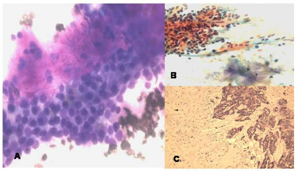
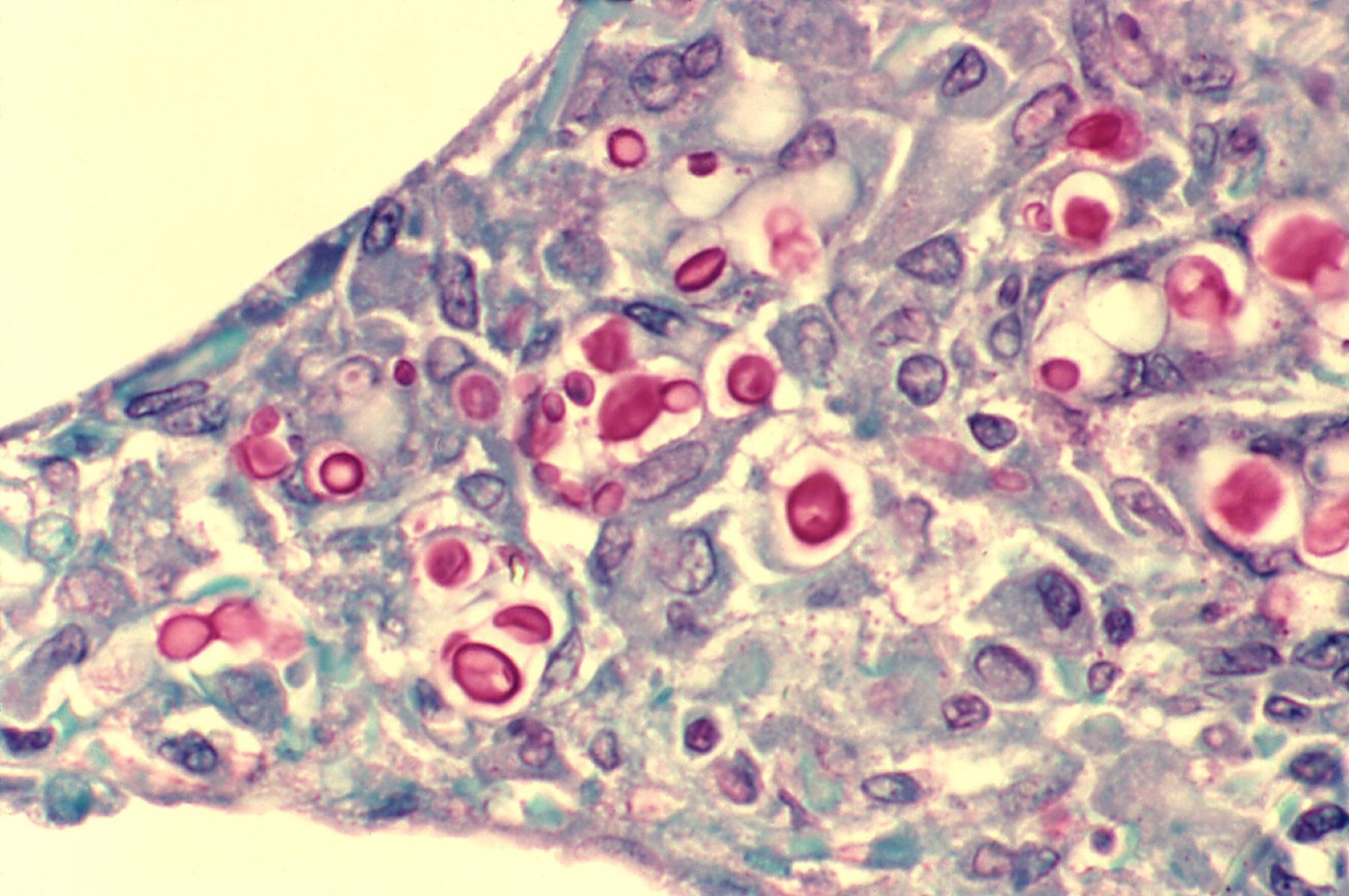
One aspirate, two stains: pleomorphic adenoma of a minor salivary gland. A, the fibrillary stroma is bright magenta (metachromatic) around cohesive epithelial cells on the air-dried Diff-Quik smear (400×). B, the same stroma is pale and the nuclei crisper on the alcohol-fixed Papanicolaou smear (200×). C, histology (H&E, 100×).Image: Saleh Husain A et al., PMC open access (Cytojournal. 2008 Mar 28;5:4. doi: 10.1186/1742-6413-5-4) Figure 3, CC BY.Mucicarmine stain: Cryptococcus neoformans yeasts in lung tissue; the inner layer of each yeast's capsule stains magenta-red. Magnification not stated.Image: CDC/Dr. Edwin P. Ewing, Jr., Wikimedia Commons, Public domain.
Artifacts, Cell Injury and the Nuclear Features of Malignancy in Cytology
Definition. Cytologic interpretation separates three groups of findings. Artifacts are changes introduced by collection, fixation, staining or mounting. Cell injury includes degeneration, cell death, therapy effect and viral cytopathic effect. The criteria of malignancy are morphologic features that, in context, indicate a malignant cell. Members of the American Society of Cytopathology Cell Biology Liaison Working Group classified some of the criteria of malignancy into three groups. Group 1 comprises tissue-level alterations related to anoikis resistance, cell adhesion and loss of polarity. Group 2 reflects genetic instability. Group 3 comprises subcellular structural changes in cytoplasm, nuclear lamina, chromatin and nucleoli that cannot be accounted for by genetic instability. The criteria are extremely varied and appear to reflect many different pathophysiologies. They are not closely related to the histogenetic classification of cancers (Fischer et al. 2010).
Morphology of air-drying. In paired cervical smears, squamous nuclei of air-dried smears more often showed indistinct nuclear borders and hazy chromatin than those of wet-fixed smears. The change was mild in all cases (Jaiwong et al. 2006). Air-drying artifacts were present in 30% of conventional wet-fixed cervical smears and 8% of saline-rehydrated air-dried smears (Kamble, Dombale and Shah 2022).
Coating fixative residue. A coating fixative that is not completely removed before staining gives a bubbling effect that interferes with diagnosis (Sathawane et al. 2022).
Alcohol shrinkage. Alcohol fixatives in liquid-based preservatives and the Cellient system preserve histomorphology but cause shrinkage-related artifacts (Shidham 2019).
Crush artifact in aspirates
High-grade neuroendocrine and basaloid carcinomas. Nuclear crush artifact, nuclear molding, naked nuclei and karyorrhexis were shared features of small cell carcinoma, large cell neuroendocrine carcinoma and basaloid squamous cell carcinoma on lung cytology (Maleki 2011).
Carcinoid tumor. Crush artifact makes a carcinoid tumor resemble small cell carcinoma, and a low Ki-67 index identifies the carcinoid (Rekhtman 2022).
Immunocytochemistry. Immunocytochemistry may help separate crush artifact from neuroendocrine carcinoma or lymphoma, although its interpretation in this setting may be difficult (Dolezal, Kholová and Cai 2024).
Autoimmune thyroiditis. Fragments of crushed cells were present in 51 of 62 aspirates of autoimmune thyroiditis (82%) and support that diagnosis in cellular smears (Gandhi, Aggarwal and Arora 2012).
Staining and mounting artifacts
Artifact
Cause
Appearance or effect
Source
Cornflake artifact
Poor dehydration and drying of the slide after xylene before mounting
Artifact over squamous cells on Papanicolaou slides
Okayama et al. 2022
Cornflake artifact under humid conditions
Drying for 3 minutes before mounting at 80% humidity; drying for 5 minutes at any tested humidity
Ratio of artifacts to normal squamous cells of 0.04 after 3 minutes at 80% humidity; 1.46–6.80 after 5 minutes
Okayama et al. 2026
Blue-green erythrocytes
Exhausted eosin
Red blood cells stain blue-green
Sathawane et al. 2022
Dense, opaque staining
Water absorbed by uncovered stains
Loss of transparency
Sathawane et al. 2022
Destained cytoplasm
Prolonged standing in the final alcohols
Pale cytoplasm
Sathawane et al. 2022
Murky staining and water droplets
Exhausted or water-contaminated alcohols and xylene
Loss of contrast in chromatin and cytoplasm
Sathawane et al. 2022
Red nuclei
Omission of effective bluing
Alum hematoxylin remains red until treated with a weak alkali
Mechanism of cornflake artifact. Smears that were not dried, or were dried after dehydration and before xylene, showed no cornflake artifacts. Smears air-dried after xylene immersion showed them. Residual water in the cells from poor dehydration contributes to the artifact (Okayama et al. 2022).
Mounting without drying. Smears mounted without drying, or after 1 minute of drying, showed no cornflake artifacts at any humidity tested (Okayama et al. 2026).
Ultrasound gel artifact
Cell lysis. Contamination of a breast FNA by ultrasound gel produced a range of changes from cell swelling to leakage of nuclear chromatin and cell lysis. The changes were reproducible ex vivo (Molyneux and Coghill 1994).
Mimic of necrosis. Ultrasound transmission gel can mimic necrosis in ultrasound-guided FNA specimens (Sack and Langer 1994).
Mimic of colloid and apoptosis. In a cadaveric study of thyroid and parotid FNA, two of three gel media produced artifacts mimicking apoptosis, necrosis or colloid on Romanowsky-type (Quik-Diff) and Papanicolaou stains. One gel medium produced no significant artifact and gave equivalent ultrasound images (Royer et al. 2012).
Diff-Quik appearance. Gel artifact also occurs on Diff-Quik-stained thyroid FNA smears (Vennapusa and Joste 2011).
Effect on reporting. Gel artifact may alter the adequacy, the diagnosis or the cytologic appearance of the specimen (Royer et al. 2012).
Contaminants in cytology preparations
Floaters. Cells that float off slides into the alcohols, called floaters, are a source of cross-contamination. Daily filtration of alcohols and xylene reduces them (Sathawane et al. 2022).
Glove starch. Starch powder from surgical gloves may resemble atypical epithelial cells or spores (Lovas et al. 1985). The granules are refractile, glassy and about 5–20 µm, with a central dot or Y-shaped mark. Under polarized light they show Maltese-cross birefringence (Jadhav, Gupta and Ahmed 2010). Maize starch particles lose the Maltese cross when they are transected in paraffin sections (Levison et al. 1988).
Fungal contaminants. Nonpathogenic fungal contaminants were found in 387 of more than 11,000 cytology specimens (3.51%). Fluids were most often affected (210 of 2,000). Fusarium species were the most common contaminant, and Alternaria was the second (Madan et al. 2025).
Mimicry by Fusarium.Fusarium microconidia were misidentified as tachyzoites of Toxoplasma, and in one lavage specimen as Pneumocystis. Culture grew Fusarium (Madan et al. 2025).
Environmental variation. The frequency of fungal contaminants varied with temperature, humidity and time of year (Madan et al. 2025).
Carry-over and environmental contaminants: sources and recognition
Contaminated staining baths. In a histology laboratory study, stainer baths contained abundant tissue contaminants, from 2–3 cells to hundreds of cells. The first xylenes and alcohols were the most heavily contaminated. Thirteen water baths holding 195 L contained one tissue fragment. Blank slides passed through the stainer acquired contaminants at a rate of 8%, most often in the late afternoon (Platt et al. 2009).
Separate staining of high-risk cytology slides. Cytology slides with a high potential for cross-contamination are usually stained separately by dipping in Romanowsky-type stains that are filtered and replaced periodically, usually weekly (Ward, Chadwick and Abraham 2023).
Dropper staining. With a dropper method, a small amount of stain is applied to each slide, and the stain is neither filtered nor reused. Over 5 years, no cross-contamination from staining occurred with this method (Ward, Chadwick and Abraham 2023).
Identification of a floater. The investigation of extraneous tissue from another specimen may include molecular tests for tissue identification. In a set of 2,325 H&E-stained whole-slide images, an image-search tool repeatedly retrieved the source tumor of a floater within the top 3 results (Pantanowitz et al. 2021).
Airborne spores, pollen and plant cells. Papanicolaou smears contained airborne spores of Alternaria, Exserohilum, Aspergillus, Cladosporium, Epicoccum and Curvularia, as well as pollen grains of lily, bulrush, pine, acacia, hazelnut and oak, and vegetable cells. Contamination may be intrinsic or extrinsic to the sample. Pollen grains and vegetable cells may be mistaken for microorganisms or malignant cells (Martínez-Girón et al. 2004).
Sites of Alternaria. In the Madan series, Alternaria was found in seven specimens: five cervical Pap smears and two nasal discharge samples (Madan et al. 2025).
Contaminant
Recognition
Confirmation
Source
Tissue floater from another specimen
Extraneous tissue unrelated to the specimen
Molecular identity testing; image search in digital archives
Pantanowitz et al. 2021
Fusarium
Hyaline septate hyphae 3–8 µm wide; banana-shaped, multicellular macroconidia with foot cells; ovoid to cylindrical unicellular microconidia
Culture
Madan et al. 2025
Airborne fungal spores, including Alternaria
Spores of environmental fungi in Papanicolaou smears
Culture
Martínez-Girón et al. 2004; Madan et al. 2025
Pollen grains and vegetable cells
Structures that may mimic microorganisms or malignant cells
None reported
Martínez-Girón et al. 2004
Artifacts and benign findings that mimic disease
Finding
Mimic
Distinguishing evidence
Source
Ultrasound gel
Necrosis, apoptotic debris or colloid
Material present on both Romanowsky and Papanicolaou smears of image-guided aspirates; reproduced ex vivo
Molyneux and Coghill 1994; Royer et al. 2012
Glove starch
Atypical epithelial cells or spores
Refractile granules of 5–20 µm with a central dot or Y-shaped mark; Maltese-cross birefringence under polarized light
Lovas et al. 1985; Jadhav, Gupta and Ahmed 2010
Fusarium microconidia
Tachyzoites of Toxoplasma or cysts of Pneumocystis
Culture growth of Fusarium
Madan et al. 2025
BK polyomavirus decoy cells
Urothelial carcinoma in situ
Homogeneous basophilic ground-glass nuclear inclusion; SV40 large T antigen immunostaining
Klufah et al. 2020
Radiation repair-like change
Atypical squamous cells
Radiation history, hypocellularity, dense polygonal cytoplasm and low N/C ratio
Petrykowski et al. 2025
Radiation cystitis
Invasive urothelial carcinoma
Vascular ectasia and atypical fibroblasts in the biopsy
Baker and Young 2000
Morphology of cell death in cytology
Feature
Apoptosis
Necrosis
Source
Cell size
Shrinkage
Swelling
Elmore 2007
Nucleus
Pyknosis from chromatin condensation, then karyorrhexis
Karyolysis, pyknosis and karyorrhexis
Elmore 2007
Cytoplasm
Dark eosinophilic
Released after loss of membrane integrity
Elmore 2007
Products
Apoptotic bodies of packed organelles with or without a nuclear fragment
Cellular debris
Elmore 2007
Inflammation
Absent
Usually present
Elmore 2007
Pyknosis. Chromatin condensation produces pyknosis, the most characteristic feature of apoptosis (Elmore 2007).
Degeneration in urine. The Paris System for Reporting Urinary Cytology, second edition, discusses cellular degeneration in the criteria of all its categories (Wojcik, Kurtycz and Rosenthal 2022). Accurate interpretation of degenerated cells remains an incompletely resolved problem (Wojcik 2026).
Degenerative changes from delayed fixation, storage and hypotonic exposure
Delayed fixation of urine. Fifty voided urine specimens were each divided and fixed immediately, after 2 hours or after 4 hours. Cell preservation declined significantly with delay (p<0.0001). The authors recommend fixation of urine immediately after collection (Ahmed and Tom 2011).
Hypotonic exposure. Urothelial cells exposed to water for more than 1 minute showed significant hydropic degeneration that affected interpretation. Ionic, hyperosmolar and low-osmolar contrast agents caused no cytologic change with exposure of up to 5 minutes. At 40 minutes or later, all solutions caused severe degeneration (Frees et al. 2016).
Degeneration and urothelial carcinoma. Degenerative artifacts can obscure key cytologic features. Separation of high-grade urothelial carcinoma from benign mimics is one of the most frequent pitfalls in urinary cytology, particularly in specimens affected by prolonged exposure to urine (Allison and VandenBussche 2025).
Storage of pleural fluid. In EDTA-anticoagulated pleural fluid from 30 patients, morphologic changes appeared by day 2 at room temperature (21 °C) and by day 4 at 4 °C. Cell counts changed from day 3 or 4 onward (Antonangelo et al. 2012).
Radiation effect in cytology specimens
Classic features. Radiation changes include two-toned cytoplasm, cytoplasmic and nuclear vacuolization, smudgy degenerative nuclei, prominent nucleoli, bizarre cells and cytomegaly with a low nuclear-to-cytoplasmic (N/C) ratio (Petrykowski et al. 2025).
Pulmonary samples. In sputum, washings and brushings from eight patients after radiotherapy for bronchogenic carcinoma, nuclear and cytoplasmic vacuolization were the most frequent changes. Metaplastic cells, macrophages and leukocytes increased after radiation (Longatto Filho et al. 1991).
Repair-like change. After lung radiation, touch preparations of two patients were hypocellular and showed polygonal or elongated cells with dense cytoplasm that mimicked atypical squamous cells. At on-site evaluation, one case was interpreted as malignant and the other as atypical (Petrykowski et al. 2025).
Phases in the lung. The acute exudative phase occurs 2–3 months after radiation, the proliferative phase 2–9 months after and the chronic fibrotic phase more than 9 months after. Type II pneumocytes proliferate and can show hyperchromasia and multinucleation (Petrykowski et al. 2025).
Persistence. Radiation changes may resolve within a month after radiation ends or may persist for life (Petrykowski et al. 2025).
Bladder. Radiation cystitis can produce pseudocarcinomatous epithelial proliferations with reactive atypia. Vascular ectasia and atypical fibroblasts in the biopsy prompted review of the radiation history (Baker and Young 2000).
Viral cytopathic effect of BK polyomavirus
Decoy cells. BK polyomavirus-infected urothelial and renal tubular cells appear as decoy cells. Their morphology, SV40 immunostaining and Paris System reporting are described in The Paris System for Reporting Urinary Cytology.
Frequency. Decoy cells were found in 46 of 22,867 urine specimens (0.2%) from 30 patients (Klufah et al. 2020).
Relation to carcinoma. BK polyomavirus DNA was detected in the urine of patients with urothelial carcinoma. Primary carcinomas and metastases were negative for BK polyomavirus (Klufah et al. 2020).
Nuclear features of malignancy in cytology specimens
Urothelial criteria. The Paris System criteria for atypical urothelial cells and high-grade urothelial carcinoma, including N/C ratio thresholds, are given in The Paris System for Reporting Urinary Cytology.
Hyperchromasia. Nuclei are dark because of increased DNA content or condensation (Wojcik 2026).
Nuclear lamina. Wrinkles of the nuclear lamina are present in control and cancer tissue and are obscured on H&E images. Extreme wrinkling is a marker of cancer (Wang et al. 2024).
Nucleoli and chromatin in squamous carcinoma. Metastatic squamous cell carcinoma in lymph node aspirates showed coarse chromatin, prominent nucleoli and dense cytoplasm (Deepthi, Prayaga and Rukmangadha 2022).
Papillary-like nuclear features. Nuclear enlargement, elongation, chromatin clearing and intranuclear pseudoinclusions characterize papillary thyroid carcinoma. In noninvasive follicular thyroid neoplasm with papillary-like nuclear features (NIFTP), these features are usually mild and patchy (Na and Park 2022).
Grooves and pseudoinclusions on Romanowsky stains. Nuclear grooves and intranuclear pseudoinclusions of papillary thyroid carcinoma were detectable on a rapid Giemsa stain (Deepthi, Prayaga and Rukmangadha 2022).
Features of injury and of malignancy compared
Feature
Injury, reactive or treatment effect
Malignancy
Source
N/C ratio
Low in radiation cytomegaly
High; a criterion of high-grade urothelial carcinoma
Petrykowski et al. 2025; Wojcik 2026
Nucleoli
Prominent in radiation effect
Prominent in metastatic squamous cell carcinoma
Petrykowski et al. 2025; Deepthi, Prayaga and Rukmangadha 2022
Hyperchromasia
Present in radiation-induced type II pneumocyte atypia
Required for high-grade urothelial carcinoma
Petrykowski et al. 2025; Wojcik 2026
Vacuolization
Nuclear and cytoplasmic vacuolization after radiation
Absent from the Paris System criteria
Longatto Filho et al. 1991; Wojcik 2026
Nuclear inclusions
Basophilic ground-glass inclusion of BK polyomavirus decoy cells
Intranuclear pseudoinclusions of papillary thyroid carcinoma
Klufah et al. 2020; Na and Park 2022
Nuclear contour
Lamina wrinkles in nonneoplastic tissue
Extreme lamina wrinkling
Wang et al. 2024
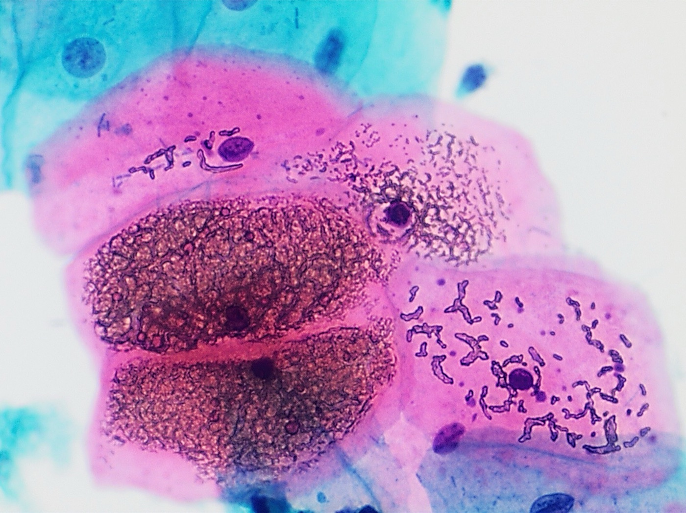
Cornflake artifact on a cervical Pap test: dark brown, refractile granular deposits over superficial squamous cells (center left), caused by air trapped when mounting medium evaporates before coverslipping. Pink superficial and blue-green intermediate cells with bacterial rods also show the Papanicolaou color scheme. Papanicolaou stain, magnification not stated.Image: Mikael Häggström, M.D., Wikimedia Commons, CC0.
References
Ahmed HG, Tom MA. The consequence of delayed fixation on subsequent preservation of urine cells. Oman Med J. 2011;26(1):14–18. doi:10.5001/omj.2011.04. PMID 22043372
Al-Attar MM, Zou T, Khedr S, Zhang M, Dresser K, Fischer AH. Destaining hematoxylin and eosin stains and restaining for immunohistochemistry has diagnostic value for cytology samples. Cytojournal. 2026;23:17. doi:10.25259/Cytojournal_171_2025. PMID 42022038
Allison DB, VandenBussche CJ. Diagnostic challenges in urinary cytology: practical insights from The Paris System for Reporting Urinary Cytology. Cancer Cytopathol. 2025;133(8):e70033. doi:10.1002/cncy.70033. PMID 40762622
Allison KH, Hammond MEH, Dowsett M, et al. Estrogen and progesterone receptor testing in breast cancer: ASCO/CAP guideline update. J Clin Oncol. 2020;38(12):1346–1366. doi:10.1200/JCO.19.02309. PMID 31928404
Antonangelo L, Vargas FS, Acencio MM, et al. Effect of temperature and storage time on cellular analysis of fresh pleural fluid samples. Cytopathology. 2012;23(2):103–107. doi:10.1111/j.1365-2303.2011.00863.x. PMID 21418346
Arai T, Kagawa T, Sasaki Y, et al. Hemosiderin-laden macrophages in bronchoalveolar lavage: predictive role for acute exacerbation of idiopathic interstitial pneumonias. Can Respir J. 2021;2021:4595019. doi:10.1155/2021/4595019. PMID 34966470
Baker PM, Young RH. Radiation-induced pseudocarcinomatous proliferations of the urinary bladder: a report of 4 cases. Hum Pathol. 2000;31(6):678–683. doi:10.1053/hupa.2000.7894. PMID 10872660
Blumenfeld W, Hildebrandt RH. Fine needle aspiration of abdominal fat for the diagnosis of amyloidosis. Acta Cytol. 1993;37(2):170–174. PMID 8465636
Carmack HJ, Lazenby BS, Wilson KJ, Bakkum-Gamez JN, Carranza L. Lost, mislabeled, and mishandled surgical and clinical pathology specimens: a systematic review of published literature. Am J Clin Pathol. 2024;162(4):349–355. doi:10.1093/ajcp/aqae055. PMID 38738289
Chan JK, Kung IT. Rehydration of air-dried smears with normal saline: application in fine-needle aspiration cytologic examination. Am J Clin Pathol. 1988;89(1):30–34. doi:10.1093/ajcp/89.1.30. PMID 3276144
Chantziantoniou N, Donnelly AD, Mukherjee M, Boon ME, Austin RM. Inception and development of the Papanicolaou stain method. Acta Cytol. 2017;61(4-5):266–280. doi:10.1159/000457827. PMID 28384641
Corwin RW, Irwin RS. The lipid-laden alveolar macrophage as a marker of aspiration in parenchymal lung disease. Am Rev Respir Dis. 1985;132(3):576–581. doi:10.1164/arrd.1985.132.3.576. PMID 4037530
Crapanzano JP, Heymann JJ, Monaco S, Nassar A, Saqi A. The state of cell block variation and satisfaction in the era of molecular diagnostics and personalized medicine. Cytojournal. 2014;11:7. doi:10.4103/1742-6413.129187. PMID 24799951
Deepthi B, Prayaga AK, Rukmangadha N. Comparison of modified ultrafast Giemsa stain with the standard May Grunwald Giemsa stain in FNAC of various organs. J Cytol. 2022;39(4):174–179. doi:10.4103/joc.joc_43_22. PMID 36605869
Dolezal D, Kholová I, Cai G. The World Health Organization Reporting System for Lung Cytopathology: a review of the first edition. J Clin Transl Pathol. 2024;4(1):18–35. doi:10.14218/jctp.2023.00068. PMID 38736711
Elmore S. Apoptosis: a review of programmed cell death. Toxicol Pathol. 2007;35(4):495–516. doi:10.1080/01926230701320337. PMID 17562483
Fischer AH, Schwartz MR, Moriarty AT, et al. Immunohistochemistry practices of cytopathology laboratories: a survey of participants in the College of American Pathologists Nongynecologic Cytopathology Education Program. Arch Pathol Lab Med. 2014;138(9):1167–1172. doi:10.5858/arpa.2013-0259-CP. PMID 24840035
Fischer AH, Zhao C, Li QK, et al. The cytologic criteria of malignancy. J Cell Biochem. 2010;110(4):795–811. doi:10.1002/jcb.22585. PMID 20564180
Frees S, Bidnur S, Metcalfe M, et al. Effect of contrast media on urinary cytopathology specimens. Can Urol Assoc J. 2016;10(7-8):228–233. doi:10.5489/cuaj.3874. PMID 27878040
Gandhi A, Aggarwal S, Arora VK. Diagnostic usefulness of fragments of crushed cells in autoimmune thyroiditis. Diagn Cytopathol. 2012;40(Suppl 1):E38–E42. doi:10.1002/dc.21572. PMID 22619158
Giorgadze TA, Shiina N, Baloch ZW, Tomaszewski JE, Gupta PK. Improved detection of amyloid in fat pad aspiration: an evaluation of Congo red stain by fluorescent microscopy. Diagn Cytopathol. 2004;31(5):300–306. doi:10.1002/dc.20131. PMID 15468138
Golde DW, Drew WL, Klein HZ, Finley TN, Cline MJ. Occult pulmonary haemorrhage in leukaemia. Br Med J. 1975;2(5964):166–168. doi:10.1136/bmj.2.5964.166. PMID 1125726
Hammond MEH, Hayes DF, Dowsett M, et al. American Society of Clinical Oncology/College of American Pathologists guideline recommendations for immunohistochemical testing of estrogen and progesterone receptors in breast cancer. Arch Pathol Lab Med. 2010;134(6):907–922. doi:10.5858/134.6.907. PMID 20524868
Heymann JJ, Pineda CM, Booth CN, et al. Tools, techniques, and challenges in preparing cytology specimens for ancillary studies: results of the ASC Optimizing Cytology and Small Biopsy Specimen Processing for Ancillary Studies task force survey. J Am Soc Cytopathol. 2025;14(1):55–63. doi:10.1016/j.jasc.2024.10.001. PMID 39547835
Jadhav KB, Gupta N, Ahmed MB. Maltese cross: starch artifact in oral cytology, divulged through polarized microscopy. J Cytol. 2010;27(1):40–41. doi:10.4103/0970-9371.66698. PMID 21042536
Jaiwong K, Nimmanhaeminda K, Siriaree S, Khunamornpong S. Cytomorphologic comparison between rehydrated air-dried and conventional wet-fixed Pap smears. J Med Assoc Thai. 2006;89(11):1811–1816. PMID 17205859
Kamble SA, Dombale VD, Shah F. Merits and pitfalls of normal saline rehydrated air-dried cervical smears over conventional wet-fixed PAP smears: a comparative study. Indian J Pathol Microbiol. 2022;65(1):100–104. doi:10.4103/IJPM.IJPM_1214_20. PMID 35074972
Kang YJ, Lee HW, Stybayeva G, Hwang SH. Comparison of liquid-based preparations with conventional smears in thyroid fine-needle aspirates: a systematic review and meta-analysis. Cancers (Basel). 2024;16(4):751. doi:10.3390/cancers16040751. PMID 38398142
Klufah F, Mobaraki G, Hausen AZ, Samarska IV. Reactivation of BK polyomavirus in urine cytology is not associated with urothelial cell carcinoma. Viruses. 2020;12(12):1412. doi:10.3390/v12121412. PMID 33302606
Koomen BM, de Boer M, van Dooijeweert C, et al. Nationwide differences in cytology fixation and processing methods and their impact on interlaboratory variation in PD-L1 positivity. Virchows Arch. 2023;482(4):707–720. doi:10.1007/s00428-022-03446-w. PMID 36370167
Koomen BM, van der Starre-Gaal J, Vonk JM, et al. Formalin fixation for optimal concordance of programmed death-ligand 1 immunostaining between cytologic and histologic specimens from patients with non-small cell lung cancer. Cancer Cytopathol. 2021;129(4):304–317. doi:10.1002/cncy.22383. PMID 33108706
Krafts KP, Pambuccian SE. Romanowsky staining in cytopathology: history, advantages and limitations. Biotech Histochem. 2011;86(2):82–93. doi:10.3109/10520295.2010.515492. PMID 21395493
Lazcano O, Speights VO, Bilbao J, Becker J, Diaz J. Combined Fontana-Masson-mucin staining of Cryptococcus neoformans. Arch Pathol Lab Med. 1991;115(11):1145–1149. PMID 1720948
Lazcano O, Speights VO, Strickler JG, Bilbao JE, Becker J, Diaz J. Combined histochemical stains in the differential diagnosis of Cryptococcus neoformans. Mod Pathol. 1993;6(1):80–84. PMID 7678937
Levison DA, Crocker PR, Jones S, Owen RA, Barnard NJ. The varied appearances of starch particles in smears and paraffin sections. Histopathology. 1988;13(6):667–674. doi:10.1111/j.1365-2559.1988.tb02097.x. PMID 3069698
Lindeman NI, Cagle PT, Aisner DL, et al. Updated molecular testing guideline for the selection of lung cancer patients for treatment with targeted tyrosine kinase inhibitors: guideline from the College of American Pathologists, the International Association for the Study of Lung Cancer, and the Association for Molecular Pathology. Arch Pathol Lab Med. 2018;142(3):321–346. doi:10.5858/arpa.2017-0388-CP. PMID 29355391
Longatto Filho A, Shirata NK, Maeda MY, Oyafuso MS. Cytology of pulmonary samples after cancer radiation therapy. Pathologica. 1991;83(1085):317–324. PMID 1923631
Lovas GL, Howell RE, Peters E, Gardner DG. Starch artifacts in oral cytologic specimens. Oral Surg Oral Med Oral Pathol. 1985;60(2):195–196. doi:10.1016/0030-4220(85)90292-0. PMID 3862027
Madan NK, Agrawal M, Teckchandani V, Shaktivel V, Capoor MR, Das S. Non pathogenic fungal contaminants in cytological specimens: beware the imitation. J Cytol. 2025;42(4):199–203. doi:10.4103/joc.joc_45_25. PMID 41341477
Majeed MM, Bukhari MH. Evaluation for granulomatous inflammation on fine needle aspiration cytology using special stains. Patholog Res Int. 2011;2011:851524. doi:10.4061/2011/851524. PMID 21776343
Makde MM, Sathawane P. Liquid-based cytology: technical aspects. Cytojournal. 2022;19:41. doi:10.25259/CMAS_03_16_2021. PMID 35928530
Maleki Z. Diagnostic issues with cytopathologic interpretation of lung neoplasms displaying high-grade basaloid or neuroendocrine morphology. Diagn Cytopathol. 2011;39(3):159–167. doi:10.1002/dc.21351. PMID 21319315
Manosca F, Schinstine M, Fetsch PA, et al. Diagnostic effects of prolonged storage on fresh effusion samples. Diagn Cytopathol. 2007;35(1):6–11. doi:10.1002/dc.20587. PMID 17173298
Martínez-Girón R, Ribas-Barceló A, García-Miralles MT, et al. Airborne fungal spores, pollen grains, and vegetable cells in routine Papanicolaou smears. Diagn Cytopathol. 2004;30(6):381–385. doi:10.1002/dc.10395. PMID 15176023
Meyer KC, Raghu G, Baughman RP, et al. An official American Thoracic Society clinical practice guideline: the clinical utility of bronchoalveolar lavage cellular analysis in interstitial lung disease. Am J Respir Crit Care Med. 2012;185(9):1004–1014. doi:10.1164/rccm.201202-0320ST. PMID 22550210
Molyneux AJ, Coghill SB. Cell lysis due to ultrasound gel in fine needle aspirates; an important new artefact in cytology. Cytopathology. 1994;5(1):41–45. doi:10.1111/j.1365-2303.1994.tb00126.x. PMID 8173030
Na HY, Park SY. Noninvasive follicular thyroid neoplasm with papillary-like nuclear features: its updated diagnostic criteria, preoperative cytologic diagnoses and impact on the risk of malignancy. J Pathol Transl Med. 2022;56(6):319–325. doi:10.4132/jptm.2022.09.29. PMID 36345620
Nay S, Konen FF, Bachhuber F, et al. CSF cytology: core principles, diagnostic pitfalls and emerging digital tools including artificial intelligence and telecytology. Acta Neuropathol. 2026;152(1):64. doi:10.1007/s00401-026-03101-7. PMID 42961673
Okayama K, Ishii Y, Fujii M, Oda M, Okodo M. Causation of cornflake artifacts: possible association of poor dehydration with drying before mounting in Papanicolaou stain. Diagn Cytopathol. 2022;50(10):E301–E305. doi:10.1002/dc.25002. PMID 35712848
Okayama K, Tajima S, Tanaka Y, et al. Humidity conditions involved in the formation of cornflake artifacts in Papanicolaou stains. Acta Cytol. 2026;70(2):263–268. doi:10.1159/000549244. PMID 41150840
Pantanowitz L, Michelow P, Hazelhurst S, et al. A digital pathology solution to resolve the tissue floater conundrum. Arch Pathol Lab Med. 2021;145(3):359–364. doi:10.5858/arpa.2020-0034-OA. PMID 32886759
Papanicolaou GN. A new procedure for staining vaginal smears. Science. 1942;95(2469):438–439. doi:10.1126/science.95.2469.438. PMID 17842594
Petrykowski BL, Rodriguez RV, Munden R, et al. Prominent repair-like changes that mimic atypical squamous cells after radiation treatment in the lung: a report of two cases. Cytojournal. 2025;22:96. doi:10.25259/Cytojournal_54_2025. PMID 41459291
Platt E, Sommer P, McDonald L, Bennett A, Hunt J. Tissue floaters and contaminants in the histology laboratory. Arch Pathol Lab Med. 2009;133(6):973–978. doi:10.5858/133.6.973. PMID 19492892
Porcel JM, Queimaliños D, Alás S, et al. The combination of cytospin Diff-Quik and conventional cytological examination increases the diagnostic yield for malignant pleural effusion. ERJ Open Res. 2025;11(3). doi:10.1183/23120541.00856-2024. PMID 40470149
Qamar I, Rehman S, Mehdi G, Maheshwari V, Ansari HA, Chauhan S. Utility of cytospin and cell block technology in evaluation of body fluids and urine samples: a comparative study. J Cytol. 2018;35(2):79–82. doi:10.4103/JOC.JOC_240_16. PMID 29643653
Rekhtman N. Lung neuroendocrine neoplasms: recent progress and persistent challenges. Mod Pathol. 2022;35(Suppl 1):36–50. doi:10.1038/s41379-021-00943-2. PMID 34663914
Rooper LM, Ali SZ, Olson MT. A minimum fluid volume of 75 mL is needed to ensure adequacy in a pleural effusion: a retrospective analysis of 2540 cases. Cancer Cytopathol. 2014;122(9):657–665. doi:10.1002/cncy.21452. PMID 25060164
Roy-Chowdhuri S, Dacic S, Ghofrani M, et al. Collection and handling of thoracic small biopsy and cytology specimens for ancillary studies: guideline from the College of American Pathologists in collaboration with the American College of Chest Physicians, Association for Molecular Pathology, American Society of Cytopathology, American Thoracic Society, Pulmonary Pathology Society, Papanicolaou Society of Cytopathology, Society of Interventional Radiology, and Society of Thoracic Radiology. Arch Pathol Lab Med. 2020;144(8):933–958. doi:10.5858/arpa.2020-0119-CP. PMID 32401054
Royer MC, Davidson DD, Dimitrov RK, Kuo CY, Kokoska MS. Ultrasound gel causes fine needle aspiration artifact? A clear choice. Acta Cytol. 2012;56(2):146–154. doi:10.1159/000335484. PMID 22378076
Saharti S, Aljhdali H, Ajabnoor R, et al. Cytospin performance when using Paris system for reporting urinary cytology. Cytojournal. 2022;19:47. doi:10.25259/Cytojournal_48_2021. PMID 36128466
Saqi A. The state of cell blocks and ancillary testing: past, present, and future. Arch Pathol Lab Med. 2016;140(12):1318–1322. doi:10.5858/arpa.2016-0125-RA. PMID 27557411
Sathawane P, Kamal MM, Deotale PR, Mankar H. Nuances of the Papanicolaou stain. Cytojournal. 2022;19:43. doi:10.25259/CMAS_03_18_2021. PMID 35928529
Shamsi M, Abdali K, Montazer NR, Kumar PV, Tabatabaee HR. Comparison of Carnoy's solution and 96% ethyl alcohol fixation in bloody Pap smears. Acta Cytol. 2008;52(2):187–190. doi:10.1159/000325477. PMID 18499991
Shidham VB, Kampalath B, England J. Routine air drying of all smears prepared during fine needle aspiration and intraoperative cytology studies: an opportunity to practice a unified protocol offering the flexibility of choosing a variety of staining methods. Acta Cytol. 2001;45(1):60–68. doi:10.1159/000327188. PMID 11213506
Shidham VB. CellBlockistry: chemistry and art of cell-block making: a detailed review of various historical options with recent advances. Cytojournal. 2019;16:12. doi:10.4103/cytojournal.cytojournal_20_19. PMID 31367220
Shidham VB. Collection and processing of effusion fluids for cytopathologic evaluation. Cytojournal. 2022;19:5. doi:10.25259/CMAS_02_14_2021. PMID 35541031
Sigstad E, Dong HP, Davidson B, Berner A, Tierens A, Risberg B. The role of flow cytometric immunophenotyping in improving the diagnostic accuracy in referred fine-needle aspiration specimens. Diagn Cytopathol. 2004;31(3):159–163. doi:10.1002/dc.20108. PMID 15349984
VandenBussche CJ, Rosenthal DL, Olson MT. Adequacy in voided urine cytology specimens: the role of volume and a repeat void upon predictive values for high-grade urothelial carcinoma. Cancer Cytopathol. 2016;124(3):174–180. doi:10.1002/cncy.21634. PMID 26524350
Vennapusa B, Joste NE. Ultrasound gel artifact on Diff-Quik stained thyroid fine-needle aspirate smears. Diagn Cytopathol. 2011;39(4):270–271. doi:10.1002/dc.21387. PMID 21416641
Wang TC, Dollahon CR, Mishra S, et al. Extreme wrinkling of the nuclear lamina is a morphological marker of cancer. NPJ Precis Oncol. 2024;8(1):276. doi:10.1038/s41698-024-00775-8. PMID 39623008
Ward M, Chadwick B, Abraham D. Dropper method for cytology slide staining: an effective and sustainable approach to eliminating cross contamination. Diagn Cytopathol. 2023;51(10):636–638. doi:10.1002/dc.25191. PMID 37403898
Wojcik EM, Kurtycz DFI, Rosenthal DL. We'll always have Paris: The Paris System for Reporting Urinary Cytology 2022. J Am Soc Cytopathol. 2022;11(2):62–66. doi:10.1016/j.jasc.2021.12.003. PMID 35094954
Wojcik EM. The Paris system for reporting urinary cytology: what worked and what still needs to be improved. Histopathology. 2026;88(1):131–136. doi:10.1111/his.70038. PMID 41384697
Wolff AC, Hammond MEH, Hicks DG, et al. Recommendations for human epidermal growth factor receptor 2 testing in breast cancer: American Society of Clinical Oncology/College of American Pathologists clinical practice guideline update. Arch Pathol Lab Med. 2014;138(2):241–256. doi:10.5858/arpa.2013-0953-SA. PMID 24099077
Yang GC, Alvarez II. Ultrafast Papanicolaou stain: an alternative preparation for fine needle aspiration cytology. Acta Cytol. 1995;39(1):55–60. PMID 7531380
Yang GC. Ultrafast Papanicolaou stain is not limited to rapid assessments: application to permanent fine-needle aspiration smears. Diagn Cytopathol. 1995;13(2):160–162. doi:10.1002/dc.2840130217. PMID 8542798