Library › Ovary, fallopian tube and peritoneum pathology: non-neoplastic lesions, epithelial, sex cord-stromal and germ cell tumors, staging and peritoneal lesions
Ovary, fallopian tube and peritoneum pathology: non-neoplastic lesions, epithelial, sex cord-stromal and germ cell tumors, staging and peritoneal lesions
Last reviewed 2026-11-10 by a board-certified pathologist. 277 references.
Non-Neoplastic Ovary and Fallopian Tube: Cysts, Endometriosis, Torsion, Salpingitis, Ectopic Pregnancy and Tubal Precursor Lesions
Definition. Non-neoplastic adnexal lesions are cysts, stromal proliferations, pregnancy-related changes, inflammatory and infectious processes, ectopic pregnancy and endometriosis of the ovary, fallopian tube and broad ligament. Several of them simulate neoplasms on clinical, gross or microscopic examination (Clement 1993; Medeiros and Bell 2010). The distal fallopian tube also contains the precursor lesions of tubo-ovarian high-grade serous carcinoma: the p53 signature, the serous tubal intraepithelial lesion (STIL) and serous tubal intraepithelial carcinoma (STIC) (Lee et al. 2007; Visvanathan et al. 2011). The WHO Classification of Female Genital Tumours, 5th edition (2020), is the current classification (Köbel and Kang 2022; CAP Ovary 1.5.0.0).
Normal ovary and fallopian tube: structures that mimic lesions
Tubal epithelium. The fallopian tube mucosa is lined mainly by a single layer of secretory, ciliated and intercalated cells over a stromal core (Bogaerts et al. 2022). Early serous neoplasia arises in the secretory cells (Lee et al. 2007).
Walthard cell nests. Walthard nests are nests of transitional-type epithelium on the tubal serosa and paratubal tissue. All Walthard nests in one series expressed GATA3, 3 of 13 showed occasional PAX8 expression, and all lacked PAX2 and SALL4 (Roma and Masand 2014). In a second series, 89.5% of Walthard nests expressed GATA3 and all expressed p63 (Esheba et al. 2009).
Walthard nests and Brenner tumor. Brenner tumors share the GATA3-positive, PAX8-negative profile of Walthard nests, and 43% of patients with a Brenner tumor had Walthard nests in the tube or periovarian tissue (Roma and Masand 2014). Walthard nests were found in 28% of controls, 50% of patients with Brenner tumors and 59% of patients with mucinous tumors (Seidman and Khedmati 2008). A Walthard nest is an incidental microscopic finding; a Brenner tumor forms an ovarian mass (Mucinous, Endometrioid, Clear Cell, Seromucinous and Brenner Tumors of the Ovary).
Hilus cells. Hilus cell nests occur in the ovarian hilum. They were four times as frequent in Stein-Leventhal ovaries as in control ovaries (Hughesdon 1982). Hilus cell hyperplasia is one of the pregnancy-associated lesions that mimic a neoplasm (Clement 1993). Hilus cells are ultrastructurally similar to testicular Leydig cells. Reinke crystals, acidophilic rod-like cytoplasmic inclusions, occur in hilus cells; they are pathognomonic of hilus cell tumor but are found in only about half of these tumors (Malakzai et al. 2021).
Cortical inclusion cysts. Two types of cortical inclusion cyst occur. Ciliated tubal-type cysts were PAX8 positive in 20 of 21 cases and were found only after menarche. Flat cysts of ovarian surface type were PAX8 negative and calretinin positive in 4 of 5 cases (Banet and Kurman 2015). Progesterone receptor was expressed in 66% of ciliated cysts and in no flat cysts (Banet and Kurman 2015). No p53 signatures were found in cortical inclusion cysts of BRCA carriers (Folkins et al. 2008).
Ovarian cysts and stromal lesions
Follicle cyst. The wall of a follicle cyst is lined by an inner granulosa cell layer and an outer theca interna layer, with surrounding ovarian stroma (Chae and Rheu 2013).
Autonomous follicle cyst in childhood. Autonomous functional follicle cysts are the most common cause of gonadotropin-independent precocious puberty in girls. Unilocular cysts <1 cm in prepubertal girls are clinically insignificant, whereas cysts with precocious pseudopuberty generally measure >2 cm. The differential diagnosis includes granulosa cell tumor and McCune-Albright syndrome (Chae and Rheu 2013).
Corpus luteum and hemorrhagic corpus luteum cyst
Formation. After ovulation the follicle collapses and the granulosa layer luteinizes while it is still avascular. Vessels then penetrate the granulosa layer and fill the central cavity with blood. Excessive bleeding enlarges the corpus luteum into a hemorrhagic corpus luteum cyst, which may rupture (Medvediev et al. 2020).
Evolution. The clot first forms a fine fibrin network in the central cavity and then organizes. After luteolysis the corpus luteum becomes a corpus albicans (Medvediev et al. 2020).
Rupture. Rupture causes hemoperitoneum, especially with anticoagulant therapy or congenital bleeding disorders. Right-sided rupture predominated in reported series (65.9% and 81.25%) (Medvediev et al. 2020).
Polycystic ovary syndrome and stromal hyperthecosis
Diagnostic criteria. The 2023 international guideline diagnoses polycystic ovary syndrome (PCOS) with two of three features: clinical or biochemical hyperandrogenism, ovulatory dysfunction, and polycystic ovaries on ultrasound. Anti-Müllerian hormone may replace ultrasound in adults. Other causes must be excluded (Teede et al. 2023). These criteria extend the 2003 Rotterdam consensus, which states that no single criterion is sufficient for diagnosis (Rotterdam ESHRE/ASRM 2004).
Ultrasound threshold. A follicle number per ovary of ≥20 in at least one ovary is the threshold for polycystic ovarian morphology in adults. An ovarian volume of ≥10 mL or ≥10 follicles per section is used with older equipment (Teede et al. 2023). PCOS prevalence is 10–13% worldwide under the Rotterdam criteria (Teede et al. 2023).
Morphology. Compared with control ovaries, Stein-Leventhal ovaries had twice the cross-sectional area, the same number of primordial follicles, twice the number of ripening and atretic follicles, a tunica thickened by 50% and more collagenized, and five times as much subcortical stroma. Lutein cell nests were present in four-fifths of cases (Hughesdon 1982).
Stromal hyperthecosis. Hyperthecosis combines nests of luteinized stromal cells with marked stromal increase. Hughesdon regarded it as a late stage of Stein-Leventhal morphology (Hughesdon 1982). In postmenopausal women it presents with virilization and high serum testosterone, almost always involves both ovaries, and requires exclusion of androgen-producing neoplasms (Boudaouara et al. 2025).
Stromal hyperplasia. Ovarian stromal hyperplasia is a nodular or diffuse proliferation of the ovarian stroma without the luteinized stromal cells of hyperthecosis. Stromal hyperplasia and hyperthecosis are uncommon non-neoplastic causes of ovarian hyperandrogenism, with an incidence of 1 in 300 to 1 in 1,000 cases of hirsutism. In postmenopausal women, laparoscopic bilateral salpingo-oophorectomy is both diagnostic and therapeutic (Lozoya Araque et al. 2020).
Pregnancy-associated ovarian lesions
Six non-neoplastic ovarian lesions of pregnancy can simulate a neoplasm: pregnancy luteoma, hyperreactio luteinalis, large solitary luteinized follicle cyst of pregnancy and puerperium, intrafollicular granulosa cell proliferations, hilus cell hyperplasia and ectopic decidua. Each regresses after pregnancy or is treated conservatively (Clement 1993).
Lesion
Gross
Microscopic
Clinical features
Main mimic
Source
Pregnancy luteoma
Solid, brown, friable mass; bilateral in 25–50%
Large epithelioid luteinized cells with eosinophilic cytoplasm; inhibin A and melan-A positive; AE1/AE3 negative
Found at cesarean section; maternal virilization in 25–65%; regresses after delivery
Sertoli-Leydig cell tumor, Krukenberg tumor
Stewart and Twidale 2024
Hyperreactio luteinalis
Bilateral multicystic ovaries, 8.5–29 cm
Multiple follicle cysts lined by granulosa cells and theca cells with abundant eosinophilic cytoplasm; stromal edema; no solid granulosa growth
Pregnancy and gestational trophoblastic disease; regresses spontaneously
Cystic granulosa cell tumor, especially juvenile type
Watkins et al. 2021
Large solitary luteinized follicle cyst of pregnancy and puerperium
Unilocular thin-walled cyst; 15 cm and 14 cm in two cases
One to several layers of luteinized cells with focal bizarre nuclei; nests of luteinized cells in an edematous wall; reticulin around cell groups
Pregnancy or puerperium; benign
Unilocular cystic granulosa cell tumor
Lomme et al. 2011
Hyperreactio luteinalis. In 10 cases, patients were 22 to 30 years old, most cases were found incidentally at cesarean section, and ovaries were bilateral and multicystic in 8 (Watkins et al. 2021). Mitoses were more frequent in the granulosa layer (mean 2.6) than in the theca layer (mean 1 per 10 high-power fields). The absence of pale or vacuolated cytoplasm and of solid granulosa growth separates hyperreactio luteinalis from juvenile granulosa cell tumor (Watkins et al. 2021).
Large solitary luteinized follicle cyst. Occasional mitoses do not exclude the diagnosis. Adult granulosa cell tumor lacks bizarre luteinized cells and shows nuclear grooves and Call-Exner bodies. Juvenile granulosa cell tumor is mitotically active (Lomme et al. 2011).
Pregnancy luteoma and the fetus. When the mother is virilized, about 80% of female fetuses are also virilized. Without maternal virilization the fetus is not affected (Stewart and Twidale 2024).
Adnexal torsion, massive ovarian edema and ovarian fibromatosis
Torsion. Adnexal torsion is the fifth most common gynecologic emergency. In adolescents the most common associated lesions are benign functional cysts and benign teratomas, and up to 46% of torsed ovaries in children and adolescents have no associated mass (ACOG 2019). No clinical or imaging finding confirms torsion before surgery. ACOG recommends detorsion with preservation of the adnexa regardless of its appearance (ACOG 2019).
Massive ovarian edema. In 11 cases, patients were 13 to 39 years old, most presented with abdominal pain, torsion was present in 5, and the ovary measured up to 15 cm with a watery cut surface. Edematous stroma surrounded residual normal ovarian structures; lutein cells were present in the stroma in 4 (Young and Scully 1984).
Ovarian fibromatosis. In 14 cases the ovary measured up to 12 cm and was firm and white. Spindle cells with dense collagen surrounded normal follicular structures (Young and Scully 1984). The overlap of age, symptoms and histology with massive edema suggests that the two lesions are related (Young and Scully 1984).
Paratubal and broad ligament cysts
Origin. Paratubal cysts are remnants of the paramesonephric or mesonephric ducts and make up an estimated 5–20% of adnexal masses (Atileh et al. 2020).
Parovarian cysts. Of 8 large intraligamentous parovarian cysts, 7 were classified as paramesonephric in origin on surgical and histologic grounds. Earlier reports had proposed a mesothelial origin for most of these cysts (Stock 1987).
Papillary cystadenoma of the broad ligament. Clear cell papillary cystadenoma of the broad ligament is a manifestation of von Hippel-Lindau syndrome. Its recognition prompts referral for genetic risk assessment (Garg et al. 2018).
Endometriosis
Definition and frequency. Endometriosis is ectopic endometrial stroma and epithelium. It affects about 10% of women of reproductive age (Anglesio et al. 2017).
CD10 for the stroma. CD10 stained ectopic endometrial stroma in 22 of 25 cases of endometriosis, with little staining of other tissues. It confirms endometriosis when morphology is doubtful (Sumathi and McCluggage 2002).
Variant appearances. Glands may be sparse or absent (stromal endometriosis). Foamy and pigmented histiocytes, fibrosis, elastosis, smooth muscle metaplasia, myxoid change and decidual change can obscure the stroma. Necrotic pseudoxanthomatous nodules, polypoid growth, bulky disease and vascular, lymphatic or perineural invasion can suggest a neoplasm (Clement 2007).
Polypoid endometriosis. In 24 cases, polypoid masses up to 14 cm involved the colon, ovary, uterine serosa, cervix, vagina and other sites. Seven patients were on unopposed estrogen. Periglandular stromal hypercellularity, stromal atypia and intraglandular stromal papillae were absent in all but two cases, which separates polypoid endometriosis from adenosarcoma (Parker et al. 2004).
Atypical endometriosis. Cytologic atypia and hyperplasia are separate findings and should be reported separately because their premalignant potential likely differs (Clement 2007). Atypical endometriosis was present in 1.7% of ovarian endometriosis. It was in contiguity with carcinoma in 13 cases, most often clear cell and endometrioid carcinoma (Fukunaga et al. 1997).
Somatic mutations. Deep infiltrating endometriosis without cancer carried cancer driver mutations in ARID1A, PIK3CA, KRAS or PPP2R1A in 10 of 39 patients (26%). The mutations were confined to the epithelium (Anglesio et al. 2017).
Cancer risk. Self-reported endometriosis was associated with clear cell carcinoma (odds ratio 3.05), low-grade serous carcinoma (2.11) and endometrioid carcinoma (2.04). It was not associated with mucinous carcinoma, high-grade serous carcinoma or borderline tumors (Pearce et al. 2012).
Inflammatory and infectious lesions of the fallopian tube
Pelvic inflammatory disease and tubo-ovarian abscess
Definition. Pelvic inflammatory disease (PID) is a spectrum of upper genital tract inflammation that includes endometritis, salpingitis, tubo-ovarian abscess and pelvic peritonitis (Workowski et al. 2021).
Organisms.Neisseria gonorrhoeae and Chlamydia trachomatis are often implicated, but only about 50% of women with acute PID test positive for either. Vaginal flora such as anaerobes, Gardnerella vaginalis, Haemophilus influenzae, enteric gram-negative rods and Streptococcus agalactiae have also been associated with PID (Workowski et al. 2021).
Histologic criteria. Endometrial biopsy with histopathologic evidence of endometritis is one of the more specific criteria for PID. Endometritis may be the only sign of PID in some women, so endometrial biopsy is warranted at laparoscopy without visible salpingitis (Workowski et al. 2021).
Intrauterine devices. The PID risk associated with an intrauterine device (IUD) is confined mainly to the first 3 weeks after insertion (Workowski et al. 2021).
Actinomyces. Pelvic and ovarian actinomycosis is associated with long-term IUD use. It forms solid or cystic adnexal masses that invade adjacent tissue, can raise CA125 and is often mistaken for ovarian carcinoma. Sulfur granules within suppurative inflammation establish the diagnosis (Cao et al. 2023).
Tuberculous salpingitis and hydrosalpinx
Tuberculous salpingitis. Female genital tuberculosis usually spreads from a focus elsewhere in the body. The tubes are affected, typically bilaterally, in 90–100% of cases, the endometrium in 50–80% and the ovaries in 20–30% (Tzelios et al. 2022). Hysterosalpingography can show a rigid pipe-stem tube. Mycobacterial culture is the reference standard, and endometrial biopsies are most useful in the late secretory phase (Tzelios et al. 2022).
Hydrosalpinx. Hydrosalpinx follows tubal occlusion, mainly of the distal fimbriated end, and is caused mostly by PID; endometriosis, pelvic adhesions and previous surgery also contribute. It is present in about 30% of cases of tubal disease. It lowers implantation success in in vitro fertilization, and salpingectomy is offered before IVF (Aljassim et al. 2026).
Xanthomatous lesions and salpingitis isthmica nodosa
Pseudoxanthomatous salpingiosis. Lipofuscin-laden macrophages accumulate in the tubal lamina propria. It is associated with longstanding endometriosis and is thought to follow leakage of blood from ovarian endometriosis into the tube (Seidman et al. 1993). Xanthogranulomatous salpingitis is a separate form of chronic inflammation (Kostopoulou et al. 2008).
Salpingitis isthmica nodosa. Salpingitis isthmica nodosa (SIN) is nodular thickening of the proximal tube (Skibsted et al. 1991). It is diagnosed by the presence of isthmic diverticula (Jenkins et al. 1993). Its incidence in fertile women is 0.6–11%, and it is more common with ectopic pregnancy and infertility (Jenkins et al. 1993). Re-examination of salpingectomy specimens doubled the number of cases detected (24 compared with 12). Women with SIN had more histologic salpingitis and a higher risk of two or more tubal pregnancies (Skibsted et al. 1991).
Tubal ectopic pregnancy
Definition. Ectopic pregnancy is a pregnancy outside the uterine cavity. The fallopian tube is the most common site. Early tubal pregnancy is treated with minimally invasive surgery or with methotrexate; tubal pregnancy in an unstable patient requires prompt surgery (ACOG 2018).
Tubal disease and recurrence. In 15 tubes with a recurrent ipsilateral tubal pregnancy after conservative surgery, residual histologic evidence of the earlier implantation site was present in only 5 of 16 sites. Chronic salpingitis, follicular salpingitis and SIN were the main findings associated with recurrence (Stock 1990).
Persistent ectopic pregnancy. Of 2498 patients treated by salpingostomy, 52 (2.08%) had persistent ectopic pregnancy. It was defined as a postoperative day 7 serum β-hCG that fell less than 15% from the day 4 level, or rose or failed to fall for at least a week. Persistent trophoblast was more frequent after salpingostomy than after salpingectomy (Zhang et al. 2017).
Risk-reducing salpingo-oophorectomy and tubal precursor lesions
Origin of high-grade serous carcinoma in the distal tube
Fimbrial origin. In BRCA-positive women undergoing salpingo-oophorectomy, all 5 carcinomas were tubal, 3 were in the fimbria, and no ovarian carcinoma was found. One carcinoma was found only after further sectioning of the fimbria (Medeiros et al. 2006). In 122 BRCA-positive women, 7 (5.7%) had an early malignancy, all in the fimbrial or ampullary tube and all in women aged 44 years or older (Callahan et al. 2007).
Association with pelvic carcinoma. With complete adnexal examination, tubal intraepithelial carcinoma was present in 5 of 5 tubal, 4 of 6 peritoneal and 20 of 30 ovarian serous carcinomas. Of these lesions, 93% involved the fimbria (Kindelberger et al. 2007).
Clonality. Identical TP53 mutations were present in 27 of 29 pairs of STIC and concurrent high-grade serous carcinoma (Kuhn et al. 2012). Evolutionary analysis of exome sequencing placed p53 signatures and STIC upstream of ovarian carcinoma, with a window of about 7 years between STIC and ovarian carcinoma (Labidi-Galy et al. 2017).
Gross handling: the SEE-FIM protocol
Method. In the SEE-FIM protocol (Sectioning and Extensively Examining the FIMbriated end), the tube is dissected from the ovary. The fimbrial end, about the distal 2 cm, is amputated and sliced longitudinally into 4 or more slices. The rest of the tube is sliced transversely. All tissue is processed (Singh et al. 2016).
CAP requirements. The ovaries and tubes are submitted entirely in patients with known BRCA1/BRCA2 or other predisposing variants, or at suspected increased risk, even when grossly normal. This detailed examination increases detection of precursor lesions or early carcinoma about 4-fold. The isthmus and ampulla are cut transversely at 2- to 3-mm intervals. Fixation for 1 to 2 hours before sectioning may prevent epithelial sloughing. In high-grade serous carcinoma without a gross fimbrial lesion, complete microscopic examination of each tube by SEE-FIM is recommended (CAP Ovary 1.5.0.0).
Effect of a protocol. A surgical-pathologic protocol of complete removal and serial sectioning of tubes and ovaries detected 7 occult malignancies in 67 BRCA carriers. All 7 were in specimens processed by complete or partial protocol (Powell et al. 2005).
Meta-analysis. Across 39 studies of 6833 BRCA1/2 carriers, pooled STIC incidence at risk-reducing salpingo-oophorectomy was 2.8%. Incidence was 2.8% with SEE-FIM and 2.7% without it (P = .92), and 3.2% with total embedding compared with 1.7% without (P = .24) (Bogaerts et al. 2022).
Benign salpingectomy. Systematic examination of 522 tubes removed for benign indications in women at low risk found 4 STICs. Three would have been missed by a single random section, and the fimbriae of most specimens fit in 1 or 2 cassettes per tube (Rabban et al. 2014). STIC is found in <0.01% of salpingectomies for benign indications (Bogaerts et al. 2022).
Spectrum of tubal lesions: p53 signature, STIL and STIC
p53 signature. A p53 signature is a stretch of morphologically normal secretory cells, at least 12 consecutive cells long, with strong nuclear p53 expression (Folkins et al. 2008; Visvanathan et al. 2011). p53 signatures predominate in the fimbria (80–100%) and show DNA damage by γ-H2AX staining. TP53 mutations were found in 8 of 14 fully analyzed signatures (Lee et al. 2007). p53 signatures occurred in 38% of tubes from BRCA-positive women and in 33% from women of unknown BRCA status (Folkins et al. 2008).
Proliferation index. p53 signatures had a MIB-1 index of 0–30%. STICs had an index above 45%, usually above 70%. Intermediate lesions had an index of 40–75% with preserved polarity (Jarboe et al. 2008).
STIC morphology. The most frequently reported features of STIC are loss of cell polarity, nuclear pleomorphism, high nuclear to cytoplasmic ratio, mitotic activity, pseudostratification and prominent nucleoli (Bogaerts et al. 2022). In the reproducibility study, at least 1 mitotic figure and epithelial stratification were the criteria with the best agreement (Visvanathan et al. 2011).
Reproducibility. Interobserver agreement for STIC compared with not STIC was fair on morphology alone (κ = 0.39). It rose to substantial (κ = 0.73) with an algorithm that combines morphology with p53 and Ki-67 (Visvanathan et al. 2011). Six pathologists who completed web-based training on the algorithm reached κ = 0.67 (Vang et al. 2012).
Category
Morphology
p53 immunostain
Ki-67 index
Source
STIC
Unequivocal or suspicious for STIC
Abnormal (moderate to strong nuclear expression in >75% of lesional cells, ≥12 cells when no atypia, or complete absence)
≥10%
Visvanathan et al. 2011; Vang et al. 2012
STIL
Atypia (unequivocal or suspicious) without the full STIC combination of abnormal p53 and Ki-67 ≥10%
Abnormal or wild-type
<10% or ≥10%
Visvanathan et al. 2011; Vang et al. 2012
p53 signature
Not suspicious; normal-appearing secretory cells
Abnormal, ≥12 consecutive cells
Low
Visvanathan et al. 2011; Folkins et al. 2008
Normal or reactive
Suspicious or not suspicious
Wild-type
<10%
Visvanathan et al. 2011
STIL is the algorithm's atypical intermediate category. Agreement among 6 trained pathologists was 76% for the 4 categories and 93% for STIC compared with non-STIC (Vang et al. 2012).
Algorithm rule. Abnormal p53 combined with a high Ki-67 index is required for a final diagnosis of STIC when morphology is unequivocal or suspicious (Visvanathan et al. 2011). Abnormal p53 includes complete absence of staining as well as overexpression. Strong diffuse staining correlates with TP53 missense mutations and complete absence with null mutations (Kuhn et al. 2012).
Frequency of lesions at RRSO. Pooled incidence in risk-reducing specimens was 16.2% for p53 signatures and 1.6% for STIL (Bogaerts et al. 2022). TP53 is mutated in about 92% of STICs by sequencing (Bogaerts et al. 2022).
2023 Delphi consensus. An international panel of 34 gynecologic pathologists from 11 countries rated 64 statements on STIC diagnosis. Twenty-seven statements (42%) reached consensus, covering processing and macroscopy, microscopy, immunohistochemistry, interpretation and reporting, and the final set was approved by 85% of the panel (Bogaerts et al. 2023). The 2022 systematic review recommends SEE-FIM grossing, review by a subspecialized gynecologic pathologist, selective p53 and Ki-67 staining and a second opinion (Bogaerts et al. 2022).
Clinical significance of isolated STIC
CAP reporting and staging. STIC found incidentally or in a risk-reducing salpingo-oophorectomy or opportunistic salpingectomy does not need synoptic reporting. When a staging procedure was done, STIC without invasion or extratubal spread may be staged pT1a if it involves one tube, pT1b if it involves both tubes and pT1c3 if peritoneal washings or ascites contain malignant cells, with an annotation that no invasive carcinoma is present. Ascites without malignant cells does not affect stage (CAP Ovary 1.5.0.0).
Peritoneal carcinoma risk. In an individual patient data meta-analysis of 3121 BRCA carriers, 115 had STIC at risk-reducing salpingo-oophorectomy. The hazard ratio for later peritoneal carcinomatosis was 33.9 with STIC. Risks at 5 and 10 years were 10.5% and 27.5% with STIC and 0.3% and 0.9% without STIC (Steenbeek et al. 2022).
Earlier review. Of 78 patients with isolated STIC, 3 BRCA carriers (4.5%) developed primary peritoneal carcinoma at 43 to 72 months (Patrono et al. 2015).
Management. No evidence supports adjuvant therapy for isolated STIC, and it is not recommended (Renz et al. 2025). STIC found at risk-reducing surgery is reported as originating in the fallopian tube (Renz et al. 2025).
Timing of surgery. Guidelines advise risk-reducing salpingo-oophorectomy at 35–40 years for BRCA1 carriers and 40–45 years for BRCA2 carriers (Renz et al. 2025). ACOG supports complete bilateral salpingectomy at obstetric, gynecologic and some non-gynecologic abdominopelvic surgery for primary prevention of epithelial ovarian cancer (ACOG 2026).
Differential diagnosis of non-neoplastic adnexal masses
Finding
Favors
Alternative diagnosis (distinguishing features)
Source
Bilateral multicystic ovaries in pregnancy, luteinized theca cells, edema
Solid brown mass at cesarean section, inhibin and melan-A positive, regresses after delivery
Pregnancy luteoma
Sertoli-Leydig cell tumor (usually unilateral; virilization in 40–80%)
Stewart and Twidale 2024
Polypoid endometriotic mass without stromal hypercellularity or atypia
Polypoid endometriosis
Adenosarcoma
Parker et al. 2004
Adnexal mass with sulfur granules in a long-term IUD user
Actinomycosis
Ovarian carcinoma
Cao et al. 2023
GATA3-positive, PAX8-negative transitional nest on tubal serosa
Walthard nest
Brenner tumor (mass)
Roma and Masand 2014
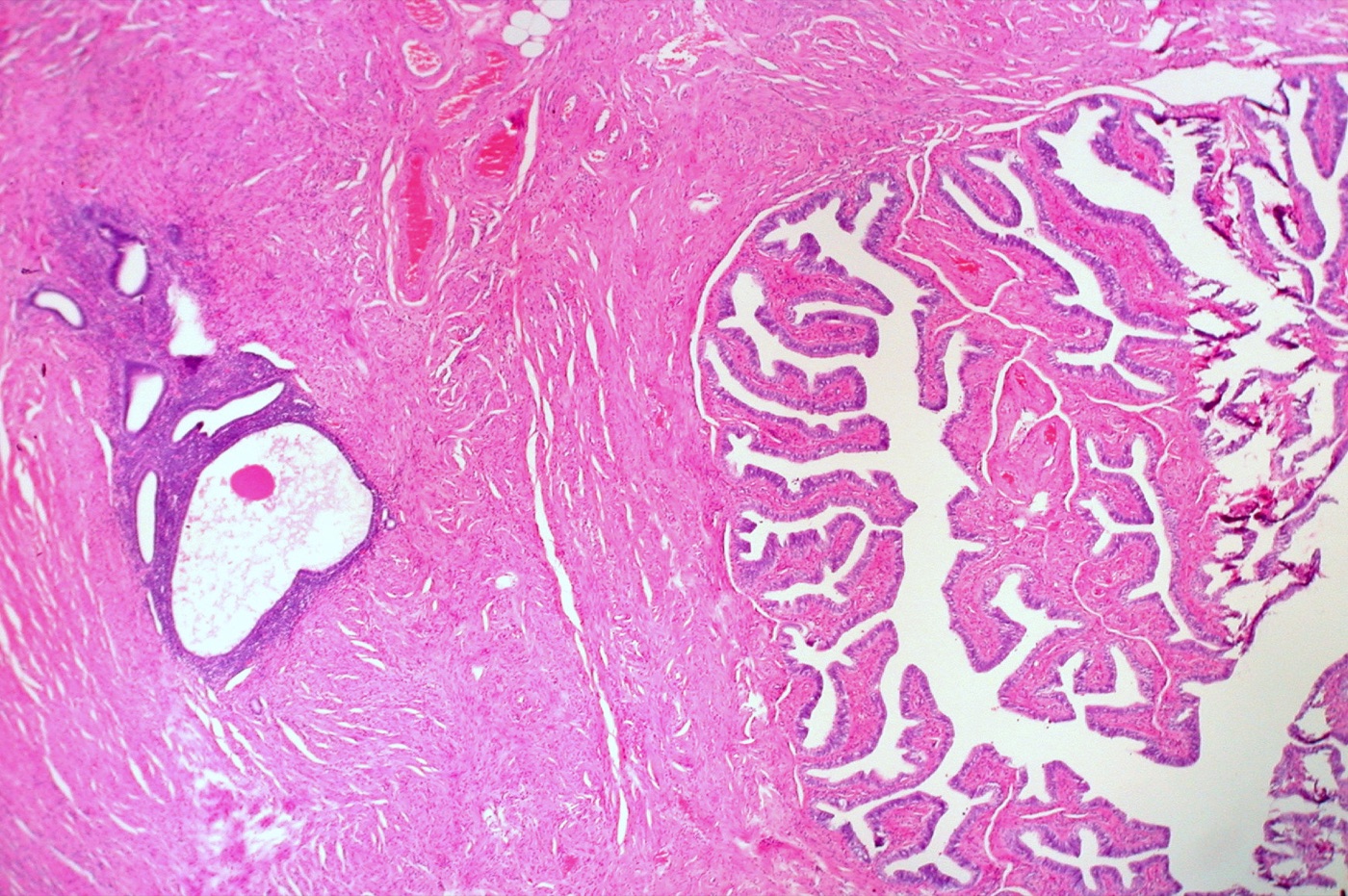
Endometriosis in the wall of the fallopian tube: a focus of endometrial-type glands, some cystically dilated, surrounded by a cuff of dense, cellular endometrial-type stroma (left), within the smooth muscle of the tubal wall; the normal plicae of the tubal lumen are at right. Glands plus stroma make the diagnosis. H&E, low power (magnification not stated).Image: Ed Uthman, Wikimedia Commons, CC BY 2.0.
Ovarian and Tubal Serous Tumors: Serous Borderline Tumor, Low-Grade and High-Grade Serous Carcinoma, BRCA and HRD Testing and Chemotherapy Response
Definition. Serous tumors of the ovary, fallopian tube and peritoneum range from benign serous cystadenoma and adenofibroma through serous borderline tumor to low-grade serous carcinoma (LGSC) and high-grade serous carcinoma (HGSC). LGSC and HGSC have been separate histotypes since the WHO 4th edition (2014). They arise by different pathways: LGSC carries MAPK pathway mutations (KRAS, BRAF, NRAS), and HGSC carries TP53 mutations in almost every case (Köbel and Kang 2022). The WHO 5th edition (2020) uses the combined term tubo-ovarian high-grade serous carcinoma, reflecting the evidence that most HGSCs arise from serous tubal intraepithelial carcinoma (STIC) in the fallopian tube (Köbel and Kang 2022; CAP Ovary 1.5.0.0).
Pathogenesis of serous carcinoma: the dualistic model
Type I and type II tumors. The dualistic model divides ovarian carcinomas into type I tumors, which develop from benign extraovarian lesions that implant on the ovary, and type II tumors. Type II tumors are mainly HGSCs, many of which develop from intraepithelial carcinoma of the fallopian tube and disseminate early (Kurman and Shih 2016).
Low-grade pathway. LGSC evolves slowly from serous adenofibroma or serous borderline tumor. It has frequent KRAS, BRAF or ERBB2 mutations, lacks TP53 mutation and is genetically stable (Vang et al. 2009; Singer et al. 2003).
High-grade pathway. HGSC shows TP53 mutation in 96% of tumors, low-frequency recurrent mutations in NF1, BRCA1, BRCA2, RB1 and CDK12, and extensive copy-number change. Homologous recombination is defective in about half of HGSCs (Cancer Genome Atlas Research Network 2011).
Serous cystadenoma, adenofibroma and serous borderline tumor
Serous cystadenoma and KRAS or BRAF mutation. Identical KRAS or BRAF mutations were found in serous cystadenoma epithelium adjacent to serous borderline tumor. Serous cystadenomas without a borderline component lacked these mutations, which places the mutation early in the transition from cystadenoma to borderline tumor (Vang et al. 2009).
Serous papillary adenofibroma. In 13 serous papillary adenofibromas (median age 47 years), the tumors were cystic or cystic and solid with small papillae. Ultrasound showed papillae in 2 of 13 cases. ER was positive in 12 and PR in all 13, and Ki-67 was ≤1% or negative in 9 (Zhao et al. 2025).
LGSC with macropapillary invasion. Some LGSCs invade as inverted macropapillae, a pattern that can be misdiagnosed as serous adenofibroma (Vang et al. 2009).
Morphology of serous borderline tumor
Definition and terminology. Serous borderline tumor shows epithelial proliferation with hierarchical papillary branching and low-grade cytology in at least 10% of the tumor volume, without stromal invasion (CAP Ovary 1.5.0.0). Serous borderline tumor is the term used in the CAP protocol and the WHO 5th edition; older papers use atypical proliferative serous tumor (Zeppernick et al. 2014; CAP Ovary 1.5.0.0). It is the precursor of LGSC (Zeppernick et al. 2014). Serous borderline tumors can arise in serous cystadenomas (Vang et al. 2009).
Typical pattern. Typical serous borderline tumors show hierarchical branching papillae that end in small papillae or tufts. The lining cells are columnar and often ciliated (Burks et al. 1996).
Eosinophilic cells. Cells with abundant eosinophilic cytoplasm and bland nuclei were present in all 25 BRAF-mutated borderline tumors. They express p16, have a low Ki-67 index and show features of oncogene-induced senescence (Zeppernick et al. 2014).
Micropapillary pattern of serous borderline tumor
Definition. The micropapillary pattern shows a filigree of highly complex micropapillae arising directly from large bulbous papillae. The micropapillae are covered by round to cuboidal cells with a high nuclear to cytoplasmic ratio (Burks et al. 1996). Of 26 such tumors, 24 (92%) contained areas of typical serous borderline tumor (Burks et al. 1996).
Size criterion. The micropapillary/cribriform variant has micropapillae without fibrovascular cores, at least 5 times longer than wide, arising directly from broad papillae ("Medusa head"), and/or small cribriform spaces, in an area measuring at least 5 mm (CAP Ovary 1.5.0.0). Borderline tumors with micropapillary foci or microinvasion are sampled extensively (CAP Ovary 1.5.0.0).
Microinvasion in serous borderline tumor
Current definition. Serous borderline tumor with microinvasion has invasive foci <5 mm in greatest dimension in any single focus, seen as single cells or small clusters of plump eosinophilic cells, or small papillary clusters in lacunar spaces without a stromal reaction. Microinvasive LGSC resembles LGSC but has only foci <5 mm; these tumors usually behave like serous borderline tumors at stages I–II (CAP Ovary 1.5.0.0).
Prognosis. In a series of 276 patients, stromal microinvasion correlated with adverse outcome independent of stage, micropapillary architecture and implant type (Longacre et al. 2005). Micropapillary stromal invasion may carry a risk closer to LGSC and is reported specifically; size limits of 3 mm or 5 mm were used historically (McKenney et al. 2006).
Peritoneal implants and lymph node involvement in serous borderline tumor
Implant prognosis. In 4129 patients from 97 reports, survival was 95.3% with non-invasive implants and 66% with invasive implants (Seidman and Kurman 2000).
Classification of invasive implants. Some centers consider invasive implants to be LGSC, and WHO nomenclature changed accordingly in 2014. ESGO considers it premature to define serous borderline tumors with invasive implants as LGSC (Renz et al. 2025).
Current CAP terminology. Implants are non-invasive by definition, and former "invasive implants" are reported as extraovarian LGSC. When an ovarian tumor that appears to be a serous borderline tumor has such deposits, it is sampled further and categorized as LGSC (CAP Ovary 1.5.0.0).
Peritoneal fluid. Borderline tumor cells in peritoneal fluid are reported as atypia of undetermined significance under the International System for Reporting Serous Fluid Cytopathology. In a T1 tumor they stage it pT1c3 (CAP Ovary 1.5.0.0). Washings are covered in Pelvic washing cytology and ovarian cyst FNA.
Lymph nodes. Lymph node involvement was associated with 98% survival at 6.5 years (Seidman and Kurman 2000).
Progression. Progression to LGSC occurred in 6.8% of 276 patients, at 7 to 288 months, and was associated with lower survival (Longacre et al. 2005).
Treatment. Borderline tumors at all stages are treated with surgery. Chemotherapy has not been shown to be beneficial. Spontaneous regression of peritoneal implants has been observed (Renz et al. 2025).
Primary peritoneal serous borderline tumor. This rare tumor is often asymptomatic and is frequently associated with endosalpingiosis. Recurrence is low and no cancer-related deaths have been reported (Iannone et al. 2025). A serous tumor is designated primary peritoneal only when entirely submitted ovaries and tubes show no involvement and no STIC (CAP Ovary 1.5.0.0).
Low-grade serous carcinoma
Morphology. The invasive component consists of micropapillae and small round nests that infiltrate the stroma haphazardly and are often surrounded by a clear space or cleft. The nuclei are small, uniform and round to oval with even chromatin. Psammoma bodies are common and may be numerous. An inverted macropapillary pattern shows medium-sized round papillae surrounded by clear spaces (Vang et al. 2009).
Nuclear criterion. Low-grade nuclei show mild to moderate atypia. Variation of ≥3:1 in nuclear size and shape indicates HGSC (Malpica et al. 2004; Vang et al. 2009).
Molecular features. About 50% of LGSCs carry MAPK pathway mutations, including BRAF and KRAS, and they are TP53 wild type (Renz et al. 2025).
Clinical behavior. LGSC is less sensitive to chemotherapy than HGSC (Gershenson et al. 2022).
Targeted therapy. In the GOG 281/LOGS trial of 260 patients with recurrent LGSC, trametinib prolonged median progression-free survival to 13.0 months, compared with 7.2 months for standard of care (hazard ratio 0.48) (Gershenson et al. 2022). Avutometinib (a RAF/MEK clamp) with defactinib (a FAK inhibitor) received FDA accelerated approval for KRAS-mutated recurrent LGSC (Grisham et al. 2026b). In the RAMP 201 study of 115 patients, median progression-free survival was 19.6 months with a KRAS mutation and 12.7 months without (Grisham et al. 2026a).
High-grade serous carcinoma
Architecture. HGSC shows papillary, glandular, nested and solid growth. Papillae are large and complex, lined by stratified epithelium with irregular slit-like spaces. Occasional HGSCs have a micropapillary pattern (Vang et al. 2009).
Cytology. Nuclei are enlarged and pleomorphic with prominent nucleoli and abundant mitoses, including atypical forms. Bizarre tumor giant cells are common.
SET features. Solid, pseudoendometrioid and transitional cell carcinoma-like (SET) patterns occur in HGSC. BRCA1-associated HGSCs more often showed SET features, higher mitotic indexes, more tumor-infiltrating lymphocytes and geographic or comedo necrosis (Soslow et al. 2012).
Intermediate nuclei. About 4% of serous carcinomas have intermediate ("grade 2") nuclei, and 10 of 11 such tumors carried TP53 mutations (Vang et al. 2009). The term "moderately differentiated" serous carcinoma is no longer used; these tumors are high grade (Renz et al. 2025).
TP53 as a defining feature. On review of 14 TCGA HGSCs without TP53 mutation, 13 were either not pure HGSC or another diagnosis. Absence of TP53 alteration is essentially inconsistent with de novo HGSC (Vang et al. 2016).
Comparison of serous borderline tumor, LGSC and HGSC
Feature
Serous borderline tumor
Low-grade serous carcinoma
High-grade serous carcinoma
Source
Invasion
None, or microinvasion (<5 mm)
Destructive stromal invasion by micropapillae and nests with clefts
Destructive invasion; papillary, glandular, solid
CAP Ovary 1.5.0.0; Vang et al. 2009
Lining cells and nuclei
Columnar, often ciliated cells in hierarchical papillae
Uniform, small nuclei; <3:1 variation
Pleomorphic; ≥3:1 variation
Malpica et al. 2004; Vang et al. 2009
Mitoses per 10 HPF
Not used for classification
Mean 4 (range 1–12)
Median 38 (range 14–137)
Vang et al. 2009
Psammoma bodies
Not graded
Common, often numerous
Less frequent
Vang et al. 2009
KRAS/BRAF
61%
68% (micropapillary carcinoma)
0 of 72
Singer et al. 2003
TP53 mutation
2 of 25 (8%)
1 of 12 (8.3%); rare
96%
Singer et al. 2005; Vang et al. 2009; Cancer Genome Atlas Research Network 2011
p53 immunostain
Not tested in the cited series
Wild type
Abnormal (overexpression, complete absence or cytoplasmic)
Köbel et al. 2016; CAP Gynecologic Biomarker 1.3.0.0
Cytoplasmic
Cytoplasmic staining
Loss-of-function mutation; 2.3% of HGSCs
Köbel et al. 2016
Wild type
Variable nuclear staining of differing intensity
No detectable mutation, or a nonexpressed loss-of-function mutation in 4.1% of HGSCs
Köbel et al. 2016
Accuracy. Deleterious TP53 mutations were found in 169 of 171 HGSCs (99%). Optimized p53 staining reached a specificity of 1.00 for TP53 mutation (Köbel et al. 2016).
Use in serous typing. Abnormal p53 excludes LGSC with 100% specificity (Köbel and Kang 2022).
Sources of p53 misinterpretation. In a proficiency test, the p53 error rate was 8.8%, and 84% of errors resulted from weak staining (Lee et al. 2016). Local optimization with low-expressing control tissue such as tonsil is required for accurate reading of the complete-absence and cytoplasmic patterns (Köbel et al. 2021).
Low-grade versus high-grade serous carcinoma
Two tumor types. Serous carcinomas are not graded. Low-grade and high-grade serous carcinoma are distinct tumor types that differ in risk factors, precursors, chemotherapy response and genetic events. They are separated mainly by nuclear variability, with at least 3-fold variation in nuclear size in HGSC. In difficult cases block-type p16, abnormal p53 and mitotic activity help (CAP Ovary 1.5.0.0).
Origin of the division. The MD Anderson system that introduced this division uses nuclear atypia as the primary criterion and mitotic rate as a secondary feature: low-grade tumors have mild to moderate atypia and up to 12 mitoses per 10 HPF, and high-grade tumors have marked atypia and more than 12 mitoses per 10 HPF (Malpica et al. 2004).
Reproducibility. Among 9 pathologists classifying 80 serous carcinomas, the overall κ was 0.909 (Malpica et al. 2007).
Primary site assignment of high-grade serous carcinoma
Regardless of size of ovarian and peritoneal disease
Singh et al. 2016; CAP Ovary 1.5.0.0
Mucosal HGSC in the tube, with or without STIC
Fallopian tube
Regardless of size of ovarian and peritoneal disease
Singh et al. 2016; CAP Ovary 1.5.0.0
Part or all of the tube inseparable from the tubo-ovarian mass
Fallopian tube
Regardless of size of ovarian and peritoneal disease
Singh et al. 2016; CAP Ovary 1.5.0.0
Both tubes separate from the mass, with no STIC or mucosal HGSC in either tube
Ovary
Both tubes fully examined by SEE-FIM
Singh et al. 2016; CAP Ovary 1.5.0.0
Both tubes and both ovaries fully examined, with no gross or microscopic STIC or HGSC
Peritoneal
Only in specimens removed at primary surgery before chemotherapy
Singh et al. 2016; CAP Ovary 1.5.0.0
Tubes and ovaries not available for complete examination: small biopsy or cytology, prior salpingo-oophorectomy with incomplete tubal examination, some treated specimens
Tubo-ovarian
Uterine serous carcinoma is the main mimic on small biopsies
Singh et al. 2016; CAP Ovary 1.5.0.0
After chemotherapy with no residual disease
Tubo-ovarian
Residual disease after chemotherapy is assigned by the criteria above
Singh et al. 2016
The CAP protocol adopts the WHO 5th edition criteria, and site assignment as "undesignated" should be avoided (CAP Ovary 1.5.0.0).
Validation. With the new criteria, 79% of chemotherapy-naive and 68% of post-neoadjuvant HGSCs were assigned to the tube. Traditional criteria had classified 73% and 63% as ovarian primaries. Four pathologists agreed on the primary site in 48 of 50 cases (96%) (Singh et al. 2015).
Effect on terminology. With current site assignment, primary peritoneal HGSC is exceedingly rare (Köbel and Kang 2022). The term tubo-ovarian HGSC separates this disease from uterine serous carcinoma and ovarian LGSC (Singh et al. 2016).
Hereditary syndromes and germline testing
Prevalence of germline variants. Of 1915 unselected women with ovarian carcinoma, 15% carried BRCA1 or BRCA2 mutations, 3.3% carried mutations in BRIP1, RAD51C, RAD51D, PALB2 or BARD1, and 0.4% carried mismatch repair gene mutations (Norquist et al. 2016).
Family history. Of women with high-grade non-mucinous ovarian cancer and germline BRCA1/2 mutations, 44% have no family history of breast or ovarian cancer (Renz et al. 2025). BRCA1-associated cancers present at a median age in the mid-40s (Renz et al. 2025).
Gene or syndrome
Ovarian or tubo-ovarian cancer risk
Associated tumors
Source
BRCA1
44% to age 80
Breast cancer
Kuchenbaecker et al. 2017
BRCA2
17% to age 80
Breast cancer
Kuchenbaecker et al. 2017
RAD51C
11% to age 80 (relative risk 7.55)
Tubo-ovarian carcinoma; breast cancer
Yang et al. 2020a
RAD51D
13% to age 80 (relative risk 7.60)
Tubo-ovarian carcinoma; breast cancer
Yang et al. 2020a
BRIP1
Odds ratio 8.13 (Icelandic frameshift variant)
Ovarian cancer
Rafnar et al. 2011
PALB2
5% to age 80 (relative risk 2.91)
Breast cancer; pancreatic cancer
Yang et al. 2020b
Lynch syndrome
20% (MLH1), 24% (MSH2), 1% (MSH6) to age 70
Endometrial cancer 54% (MLH1); colorectal cancer 41–48% (MLH1, MSH2)
Bonadona et al. 2011
BRCA and homologous recombination deficiency testing
Guideline recommendations for BRCA and HRD testing
ASCO 2020. All women with epithelial ovarian cancer should be offered germline testing for BRCA1, BRCA2 and other ovarian cancer susceptibility genes, irrespective of clinical features or family history. Women without a germline BRCA1/2 pathogenic or likely pathogenic variant should have somatic tumor testing for BRCA1/2 (Konstantinopoulos et al. 2020).
Germline and somatic yield. Germline BRCA1/2 mutations are found in 13–15% of women with ovarian cancer, and somatic tumor testing finds about another 5–7% (Konstantinopoulos et al. 2020). Tumor testing is less sensitive than germline testing: up to 5% of germline mutations are missed if tumor results decide whether germline sequencing is done (Konstantinopoulos et al. 2020).
Mismatch repair. Women with clear cell, endometrioid or mucinous ovarian cancer should be offered somatic tumor testing for mismatch repair deficiency (Konstantinopoulos et al. 2020).
HRD assays in ASCO. ASCO made no recommendation supporting routine tumor testing with homologous recombination deficiency (HRD) assays (Konstantinopoulos et al. 2020).
ESMO 2020. Trial evidence supports BRCA mutation testing and two commercial assays that also measure genomic instability for identifying HGSCs with different magnitudes of PARP inhibitor benefit. Existing tests lack negative predictive value (Miller et al. 2020).
European consensus 2022. Germline non-BRCA homologous recombination repair and mismatch repair gene mutations predict familial risk. They do not predict PARP inhibitor sensitivity in the first-line setting (Vergote et al. 2022).
Tissue before neoadjuvant chemotherapy. Histologic confirmation before neoadjuvant chemotherapy ensures enough tissue for HRD and somatic BRCA testing (Renz et al. 2025).
CAP biomarker template. The CAP gynecologic biomarker template (version 1.3.0.0) has no reporting element for BRCA1/2, HRD or homologous recombination status. Its elements include ER, PR, HER2, mismatch repair, p53, PD-L1, folate receptor alpha and MSI (CAP Gynecologic Biomarker 1.3.0.0).
Tissue selection for HRD testing
Recommended tissue. For HGSC tested with the Myriad myChoice assay, samples with tumor cell content ≥30% and tumor area ≥0.5 cm² on the H&E section are recommended. With appropriate selection, success rates reach 98% (Romey et al. 2024).
Test
Specimen
What it detects
Clinical use
Source
Germline multigene panel
Blood or saliva
Inherited BRCA1/2, RAD51C, RAD51D, BRIP1, PALB2 and mismatch repair variants
All epithelial ovarian cancers; cascade testing of relatives
Konstantinopoulos et al. 2020
Tumor BRCA1/2 sequencing
Tumor tissue
Germline plus somatic BRCA1/2 variants
Women without a germline BRCA1/2 variant
Konstantinopoulos et al. 2020
Genomic instability (HRD) assay
Tumor tissue, ≥30% tumor cells and ≥0.5 cm² tumor area
BRCA status plus a genomic instability score
Magnitude of PARP inhibitor benefit in first-line HGSC
Miller et al. 2020; Romey et al. 2024
Mismatch repair immunohistochemistry
Tumor tissue
Loss of MLH1, PMS2, MSH2 or MSH6
Clear cell, endometrioid and mucinous carcinoma; Lynch screening
Konstantinopoulos et al. 2020
PARP inhibitor and antibody-drug conjugate trials in ovarian carcinoma
Trial
Population
Biomarker result
Source
SOLO1 (olaparib maintenance)
Newly diagnosed stage III–IV high-grade serous or endometrioid carcinoma with BRCA1/2 mutation
Progression-free at 3 years 60% vs 27%; hazard ratio 0.30
Moore et al. 2018
PAOLA-1 (olaparib plus bevacizumab)
Newly diagnosed advanced high-grade carcinoma regardless of BRCA status
Hazard ratio 0.33 in HRD-positive tumors and 0.43 in HRD-positive tumors without BRCA mutation
Ray-Coquard et al. 2019
PRIMA (niraparib maintenance)
Newly diagnosed advanced carcinoma after response to platinum
Median progression-free survival, HRD-positive: 21.9 vs 10.4 months; overall: 13.8 vs 8.2 months
González-Martín et al. 2019
MIRASOL (mirvetuximab soravtansine)
Platinum-resistant high-grade serous carcinoma with high folate receptor alpha
Overall survival 16.46 vs 12.75 months; hazard ratio 0.67
Moore et al. 2023
CAP scoring of folate receptor alpha. With the FDA-approved VENTANA FOLR1 (FOLR-2.1) RxDx companion assay, positivity is ≥75% of viable tumor cells with moderate to strong membrane staining. Moderate to strong staining in <75% of cells, weak staining in any proportion or no staining is negative. Mirvetuximab soravtansine received FDA accelerated approval on November 14, 2022, for FRα-positive platinum-resistant epithelial ovarian, tubal or primary peritoneal cancer after 1–3 prior regimens (CAP Gynecologic Biomarker 1.3.0.0; FDA 2022). Regular approval for the same indication followed on March 22, 2024 (FDA 2024).
Chemotherapy response score after neoadjuvant chemotherapy
CAP requirement. The CRS is required only for HGSC after neoadjuvant chemotherapy. It is assessed on the omental section showing the least response, and 4 to 6 omental sections from the most abnormal areas are recommended. The categories are CRS1, no definite or minimal response; CRS2, moderate response; and CRS3, marked response with no or minimal residual cancer (CAP Ovary 1.5.0.0).
Development. The chemotherapy response score (CRS) was developed in interval debulking specimens of stage IIIC–IV tubo-ovarian HGSC. Applied to the omentum, it was reproducible (κ = 0.67 in validation) and predicted progression-free survival. Applied to the adnexa, it did not stratify prognosis (Böhm et al. 2015).
Platinum sensitivity. CRS 3 had a 94.3% negative predictive value for progression within 6 months (Böhm et al. 2015).
CRS
Response
Histologic definition
Source
1
No or minimal response
Mainly viable tumor with no or minimal regression-associated fibroinflammatory change limited to a few foci; cases in which regression and tumor-associated desmoplasia or inflammation cannot be separated
Böhm et al. 2015; Lawson et al. 2020
2
Appreciable response amid viable tumor
Viable tumor readily identifiable; multifocal or diffuse regression-associated change with tumor in sheets, streaks or nodules, up to extensive regression with multifocal residual tumor
Böhm et al. 2015; Lawson et al. 2020
3
Complete or near-complete response
No residual tumor, or minimal irregularly scattered tumor foci as individual cells, cell groups or nodules up to 2 mm; recording "no residual tumor" or "microscopic residual tumor present" is advisable
Böhm et al. 2015; Lawson et al. 2020; CAP Ovary 1.5.0.0
Regression-associated fibroinflammatory change is fibrosis with macrophages, including foam cells, mixed inflammatory cells and psammoma bodies, as distinct from tumor-related inflammation or desmoplasia (CAP Ovary 1.5.0.0).
Validation. In an individual patient data meta-analysis of 877 patients from 16 sites, CRS 3 occurred in 28%. CRS 3 compared with CRS 1–2 had hazard ratios of 0.55 for progression-free survival and 0.65 for overall survival. Patients with germline BRCA1/2 mutations were more likely to achieve CRS 3 (Cohen et al. 2019; corrigendum Cohen et al. 2021).
Adnexal score. Adnexal CRS 1–2 carried almost four times the risk of platinum-resistant disease compared with adnexal CRS 3 (Lawson et al. 2020).
Differential diagnosis of serous tumors
Diagnostic question
Discriminating features
Source
Serous borderline tumor with micropapillary pattern versus LGSC
Destructive stromal invasion by micropapillae and nests with clefts defines LGSC; foci of invasion <5 mm define microinvasion
CAP Ovary 1.5.0.0; Vang et al. 2009
LGSC versus HGSC
Nuclear variation ≥3:1, high mitotic rate and abnormal p53 indicate HGSC; abnormal p53 excludes LGSC
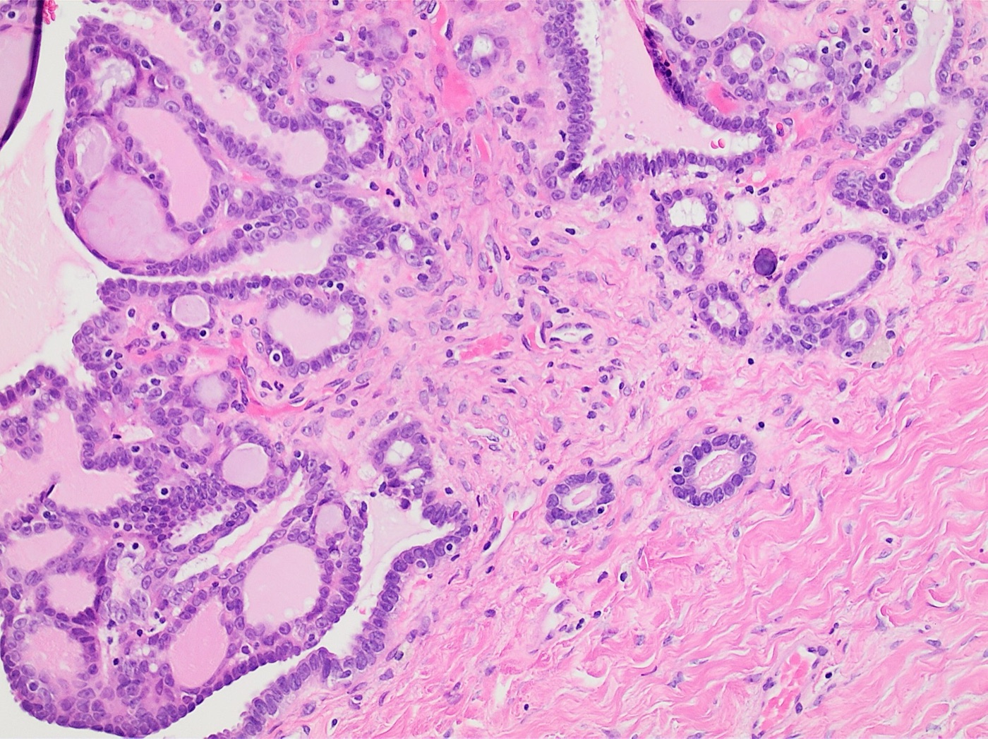
Serous borderline tumor of the ovary: hierarchically branching papillae lined by stratified cuboidal to columnar cells with mild to moderate atypia and tufting (left and top), with small rounded gland-like invaginations of the same epithelium into the fibrous wall (center right). The invaginations are smooth-contoured and lack the irregular small nests, single cells and desmoplasia of stromal invasion, and a psammoma body lies at center right. H&E, medium power (magnification not stated).Image: Mikael Häggström, M.D., Wikimedia Commons, CC0.
Mucinous, Endometrioid, Clear Cell, Seromucinous and Brenner Tumors of the Ovary
Definition. The non-serous epithelial tumors of the ovary are mucinous, endometrioid, clear cell, seromucinous and Brenner tumors, each with benign, borderline and malignant forms, together with mesonephric-like adenocarcinoma, carcinosarcoma, undifferentiated and dedifferentiated carcinoma and mixed carcinoma. The WHO 5th edition (2020) lists five principal ovarian carcinoma histotypes: high-grade serous, low-grade serous, mucinous, endometrioid and clear cell carcinoma. In descending order of frequency they are high-grade serous, endometrioid, clear cell, low-grade serous and mucinous carcinoma (Köbel and Kang 2022). Endometrioid and clear cell carcinomas arise from endometriosis (Köbel and Kang 2022).
Pathologists agreed with a reference diagnosis of seromucinous carcinoma in only 39–56% of cases, and integrated morphology, immunophenotype and genotype reclassified 72% as endometrioid, 25% as low-grade serous and 3% as mucinous carcinoma (Rambau et al. 2017).
Mucinous tumors of the ovary
Mucinous borderline tumor and intraepithelial carcinoma
Definition. Mucinous borderline tumor shows proliferation of gastrointestinal-type mucinous epithelium with low-grade nuclear atypia in at least 10% of the tumor, with stratification, tufting and villous or filiform papillae. Lesser proliferation is classified as mucinous cystadenoma with focal epithelial proliferation (CAP Ovary 1.5.0.0).
Intraepithelial carcinoma and microinvasion. Intraepithelial carcinoma shows excessively stratified epithelium with high nuclear grade and frequent mitoses confined to the epithelium, and it prompts further sampling for invasion. Microinvasion consists of foci <5 mm, usually small nests or single cells with a desmoplastic stromal response (CAP Ovary 1.5.0.0).
Frequency of true primaries. Of 52 consecutive mucinous carcinomas involving the ovaries, 40 (77%) were metastatic and 12 (23%) were primary. Pure primary mucinous carcinoma made up 2.4% of primary ovarian carcinomas (Seidman et al. 2003).
Outcome of intraepithelial carcinoma. Intestinal-type borderline tumors with epithelial atypia only were clinically benign at stage I. Of 69 patients with borderline tumors containing intraepithelial carcinoma, 2 had fatal recurrences; both tumors were incompletely staged and one had ruptured (Lee and Scully 2000).
Molecular progression. Mucinous carcinoma arises from benign and borderline precursors in the ovary. TP53 mutation and copy-number change, including a 9p13 amplicon, accompany progression (Cheasley et al. 2019).
Mucinous carcinoma: expansile and infiltrative invasion
Two patterns. Mucinous carcinomas are classified as expansile (confluent, non-destructive) or infiltrative (destructive stromal invasion), as proposed by Lee and Scully (Renz et al. 2025). Expansile growth is at least 5 mm of back-to-back glands with minimal intervening stroma, without desmoplasia or stromal invasion. Infiltrative growth consists of individual glands and cell clusters that provoke a stromal, often desmoplastic, response (CAP Ovary 1.5.0.0).
Outcome by pattern. All 12 carcinomas with expansile invasion were stage I, and none of the 10 with follow-up recurred. Five of 6 infiltrative carcinomas above stage I with follow-up were fatal (Lee and Scully 2000).
Lymph nodes. No patient with apparent stage I expansile carcinoma had lymph node metastases; 3 of 10 with infiltrative carcinoma did (Muyldermans et al. 2013).
Small infiltrative foci. Microfoci (<5 mm) of infiltrative pattern or nuclear grade 3 within an otherwise expansile carcinoma did not change prognosis. Infiltrative carcinoma had lower overall and progression-free survival than borderline tumor and expansile carcinoma (Genestie et al. 2021).
Treatment relevance. Guidelines suggest avoiding chemotherapy in stage I mucinous carcinoma of expansile type (Renz et al. 2025). Homologous recombination deficiency and mismatch repair deficiency do not occur in mucinous carcinoma (Köbel and Kang 2022).
Mural nodules and tumors associated with ovarian mucinous neoplasms
Sarcoma-like mural nodules. These nodules are multiple, small, sharply demarcated from the mucinous tumor and composed of a heterogeneous cell population. They coexpress vimentin and cytokeratin and behave favorably. True sarcomatous nodules and anaplastic carcinoma carry a worse prognosis (Bagué et al. 2002).
Anaplastic carcinoma. Foci of anaplastic carcinoma were present in 13 of 19 infiltrative mucinous carcinomas (Rodríguez and Prat 2002).
Teratoma-associated mucinous tumors. Of 44 mucinous tumors associated with mature cystic teratoma, all were unilateral and had gastrointestinal-type differentiation. Those with pseudomyxoma ovarii were usually CK7-negative and CK20-positive (86%) and CDX2-positive (79%) (Vang et al. 2007).
Primary versus metastatic mucinous tumors in the ovary
Size and laterality of primary and metastatic mucinous tumors
Seidman rule. All bilateral mucinous carcinomas and unilateral carcinomas <10 cm are classified as metastatic, and unilateral carcinomas ≥10 cm as primary. The rule correctly classified 90% of tumors in routine practice (Seidman et al. 2003).
Exceptions. Metastatic colorectal and endocervical carcinomas produced the most large unilateral exceptions (Yemelyanova et al. 2008).
Primary site of metastasis
Correctly classified by the 10-cm rule
Correctly classified with a 13-cm cutoff
Source
Primary ovarian tumors
100%
98%
Yemelyanova et al. 2008
Colorectum
74%
80%
Yemelyanova et al. 2008
Appendix, low-grade tumors
79%
79%
Yemelyanova et al. 2008
Appendix, carcinomas
100%
100%
Yemelyanova et al. 2008
Pancreaticobiliary tract
95%
100%
Yemelyanova et al. 2008
Stomach
80%
100%
Yemelyanova et al. 2008
Endocervix
55%
70%
Yemelyanova et al. 2008
CK7 and SATB2 in primary and metastatic mucinous tumors
Panel performance. CK7 was the most accurate single marker (91.7% with a diffuse/non-diffuse cutoff), followed by SATB2 (88.8% with a present/absent cutoff). CK7 combined with SATB2, scored as absent, focal or diffuse, reached 95.3% accuracy and outperformed CK7, CK20 and CDX2 (87.5%) (Meagher et al. 2019).
Marker frequencies. CK20, CDX2 and SATB2 were expressed in 87%, 98% and 90% of lower gastrointestinal primaries. CK7 was expressed in 97% and PAX8 in 45% of ovarian mucinous neoplasms (Meagher et al. 2019).
Exceptions to the expected profile. SATB2 was expressed in 13% of ovarian mucinous carcinomas and 15% of ovarian endometrioid carcinomas, often in squamous morules. Diffuse PAX8 was seen in 6% of appendiceal neoplasms (Meagher et al. 2019).
CAP statement. CAP regards an immunohistochemical panel as of limited value because patterns vary. Primary mucinous adenocarcinoma may be PAX8 and/or PAX2 and CK7 positive and SATB2 and CDX2 negative. DPC4 (SMAD4) is intact in most primary ovarian mucinous carcinomas and lost in mucin-producing pancreaticobiliary tumors (CAP Ovary 1.5.0.0).
Feature
Favors primary ovarian mucinous tumor
Favors metastasis
Source
Laterality
Unilateral; bilateral rare
Bilateral in more than 75%
Seidman et al. 2003; CAP Ovary 1.5.0.0
Size
≥10 cm (≥13 cm optimized); size >10–12 cm frequent
<10 cm
Seidman et al. 2003; Yemelyanova et al. 2008; CAP Ovary 1.5.0.0
Surface involvement
Rare
Possible
CAP Ovary 1.5.0.0
Nodular growth
Rare
Frequent
CAP Ovary 1.5.0.0
Hilar involvement
Rare
Frequent
CAP Ovary 1.5.0.0
Lymphovascular invasion
Rare
Possible
CAP Ovary 1.5.0.0
Growth pattern
Expansile frequent; infiltrative possible
Infiltrative frequent; single-cell infiltration possible
CAP Ovary 1.5.0.0
Precursor components
Benign and borderline components in the same tumor
Absent
Cheasley et al. 2019
CK7
Diffuse
Absent or focal (lower gastrointestinal)
Meagher et al. 2019
SATB2
Absent
Present (lower gastrointestinal)
Meagher et al. 2019
PAX8
Present in 45%
Usually absent; diffuse in 6% of appendiceal neoplasms
Meagher et al. 2019
DPC4 (SMAD4)
Intact
Lost in pancreaticobiliary tumors
CAP Ovary 1.5.0.0
Pseudomyxoma peritonei
Rare; occurs with teratoma-associated tumors
Appendiceal mucinous neoplasm
Lee and Scully 2000; Vang et al. 2007
Grading of mucinous and endometrioid carcinoma
Which tumors are graded. Histologic grade is required for endometrioid and mucinous carcinomas, immature teratomas and Sertoli-Leydig cell tumors. Clear cell carcinoma, malignant Brenner tumor, undifferentiated and dedifferentiated carcinoma, mesonephric-like carcinoma and carcinosarcoma are not graded. Serous carcinoma is classified as low-grade or high-grade serous carcinoma, two tumor types. For mixed tumors, the highest grade is reported with a comment on the others (CAP Ovary 1.5.0.0).
FIGO grading of endometrioid carcinoma. Endometrioid carcinoma is graded with the FIGO system used in the endometrium (Endometrial Carcinoma: Histologic Types and Molecular Classification). Grade 1 has 5% or less non-squamous solid growth, grade 2 has 6% to 50% and grade 3 has more than 50%. Notable nuclear atypia in most tumor cells of a grade 1 or 2 tumor, evident at low power and discordant with the architecture, raises the grade by one. Before upgrading, high-grade serous carcinoma is considered (CAP Ovary 1.5.0.0).
Mucinous carcinoma. CAP recommends the Silverberg system for mucinous carcinoma and allows FIGO or growth pattern-based grading. Silverberg grade is prognostic and FIGO grade is not (CAP Ovary 1.5.0.0).
Shimizu-Silverberg system. The system scores architecture (glandular 1, papillary 2, solid 3), nuclear pleomorphism (slight 1, moderate 2, marked 3) and mitoses per 10 high-power fields of 0.345 mm² (0–9 = 1, 10–24 = 2, ≥25 = 3). A total of 3–5 is grade 1, 6–7 grade 2 and 8–9 grade 3 (Shimizu et al. 1998; CAP Ovary 1.5.0.0).
System
Applies to
Criteria
Grades
Source
FIGO
Endometrioid carcinoma (recommended); mucinous carcinoma (may be used)
≤5%, 6–50%, >50% non-squamous solid growth; one-step upgrade for notable nuclear atypia in most cells
1, 2, 3
CAP Ovary 1.5.0.0
Shimizu-Silverberg
Mucinous carcinoma (recommended); endometrioid carcinoma (may be used)
Architecture, pleomorphism and mitoses in 10 HPF of 0.345 mm², each scored 1–3
1 (3–5), 2 (6–7), 3 (8–9)
Shimizu et al. 1998; Silverberg 2000; CAP Ovary 1.5.0.0
Growth pattern-based
Mucinous carcinoma only
Infiltrative growth ≤10% or >10%
Low, high
Busca et al. 2020; CAP Ovary 1.5.0.0
Two-tier (MD Anderson)
Serous carcinoma; separates two tumor types (LGSC, HGSC) without a grade
Nuclear atypia; mitoses >12 per 10 HPF as a secondary feature
Borderline and invasive criteria. In 56 stage I proliferative endometrioid tumors, confluent glandular growth was the most common form of stromal invasion and the best criterion for carcinoma. Invasion of <5 mm was classified as microinvasion and ≥5 mm as carcinoma (Bell and Kurman 2000).
Associated findings. Squamous differentiation was present in 24 and mucinous differentiation in 20 of the 56 tumors. Areas of borderline tumor were present in 13 of 15 carcinomas (Bell and Kurman 2000).
High-grade endometrioid carcinoma and HGSC. Agreement between 5 pathologists on grade 3 endometrioid carcinoma compared with HGSC was 69% on morphology alone and 96% with WT1 and p53 status (Assem et al. 2018).
Molecular classification of ovarian endometrioid carcinoma
Mutations. In 112 endometrioid carcinomas, mutations occurred in CTNNB1 (43%), PIK3CA (43%), ARID1A (36%), PTEN (29%), KRAS (26%) and TP53 (26%). CTNNB1 and TP53 mutations were mutually exclusive. CTNNB1-mutated tumors were 89% stage I–II with excellent outcome, and TP53-mutated tumors were often advanced with inferior survival (Hollis et al. 2020).
Endometrial-type molecular groups. In 511 endometrioid carcinomas, 3.5% were POLE-mutated, 13.7% mismatch repair-deficient, 9.6% p53-abnormal and 73.2% no specific molecular profile. Median overall survival was shortest for p53-abnormal tumors (4.7 years) (Krämer et al. 2020).
β-catenin. Nuclear β-catenin expression is specific for endometrioid carcinoma and is present in about 50%, mostly low-grade tumors with squamous differentiation (Köbel and Kang 2022).
Clear cell tumors of the ovary
Morphology. Clear cell carcinoma shows tubulocystic and papillary patterns. Clear cytoplasm in an HGSC without these patterns does not indicate clear cell carcinoma (Vang et al. 2009).
Endometriosis. Endometriosis was associated with 54% of clear cell carcinomas (Fukunaga et al. 1997).
Napsin A. Napsin A was positive in 71 of 86 clear cell carcinomas (83%) and all 13 clear cell adenofibromas. It was negative in 30 serous carcinomas, 19 endometrioid carcinomas, 10 mucinous carcinomas and 3 yolk sac tumors (Yamashita et al. 2015).
HNF1B. Nuclear HNF1B was expressed in all 30 clear cell tumors, including benign and borderline tumors, and rarely in other epithelial tumors (Kato et al. 2006).
ARID1A.ARID1A mutations were present in 46% of clear cell and 30% of endometrioid carcinomas and in no HGSC. Mutation and BAF250a loss were present in contiguous atypical endometriosis (Wiegand et al. 2010).
PIK3CA and other genes.PIK3CA mutations were present in 33% of clear cell carcinomas (46% of affinity-purified tumors), TP53 in 15% and KRAS in 7% (Kuo et al. 2009).
Mismatch repair. Mismatch repair deficiency was reported in 11.5% of clear cell ovarian cancers (Konstantinopoulos et al. 2020). It does not occur in prototypical clear cell carcinoma, and MMR-deficient tumors with diffuse stromal inflammation are more consistent with endometrioid carcinoma (Köbel and Kang 2022).
Seromucinous tumors
Composition. Seromucinous tumors contain serous and endocervical-type mucinous epithelium together with endometrioid, indifferent and squamous epithelium (Kurman and Shih 2016).
Immunophenotype. They frequently express ER and PR, infrequently express WT1 and lack CK20 and CDX2. They are often associated with endometriosis and frequently lose ARID1A expression (Kurman and Shih 2016).
Seromucinous carcinoma. Seromucinous carcinomas carried KRAS (70%), PIK3CA (37%), PTEN (19%) and ARID1A (16%) alterations and no CTNNB1 mutation (Rambau et al. 2017). In the WHO 5th edition they are classified as endometrioid carcinoma with mucinous differentiation (Köbel and Kang 2022).
Brenner tumors
Immunophenotype. All 32 Brenner tumors in one series were diffusely GATA3-positive and PAX8-, PAX2- and SALL4-negative (Roma and Masand 2014). In another series, all benign Brenner tumors were p63-positive, and the single malignant Brenner tumor was GATA3-negative (Esheba et al. 2009).
Histogenesis. Brenner tumor epithelium shares AKR1C3 and androgen receptor expression with tubal transitional metaplasia and Walthard nests, which supports a tubal origin (Kuhn et al. 2013).
Malignant Brenner tumor and transitional cell carcinoma. Borderline Brenner tumors are cystic, highly papillary tumors of transitional epithelium without stromal invasion; metastatic urothelial carcinoma is excluded. Malignant Brenner tumor shows stromal invasion with a benign or borderline Brenner component. Most tumors formerly called ovarian transitional cell carcinoma are a variant of HGSC with TP53 mutation, occasionally endometrioid carcinoma (CAP Ovary 1.5.0.0).
Mesonephric-like adenocarcinoma, carcinosarcoma, undifferentiated and mixed carcinoma
Mesonephric-like adenocarcinoma. This WHO 5th edition entity resembles cervical mesonephric carcinoma and is thought to arise by transdifferentiation of other Müllerian tumors. It is p53 normal and MMR proficient and frequently carries KRAS mutation (Köbel and Kang 2022). The tumor cells express GATA3, TTF-1, luminal CD10 and PAX8 and lack ER, PR and WT1 (CAP Ovary 1.5.0.0).
Carcinosarcoma. Carcinosarcoma is considered a metaplastic carcinoma, most frequently of HGSC, and is classified as a malignant epithelial tumor (Köbel and Kang 2022). Localized or regional carcinosarcoma had a higher early mortality than HGSC (hazard ratio 3.81) (Peres et al. 2019).
Undifferentiated and dedifferentiated carcinoma. These tumors arise from endometrioid carcinoma, most often in an MMR-deficient background, and acquire mutually exclusive SWI/SNF alterations in the undifferentiated component: ARID1A with ARID1B, SMARCA4 (BRG1) or SMARCB1 (INI1). They are extremely aggressive (Köbel and Kang 2022). Concurrent ARID1A and ARID1B inactivation was found in 12 of 24 BRG1- and INI1-intact dedifferentiated carcinomas (Coatham et al. 2016).
Mixed carcinoma. True mixed carcinoma is rare; mixed endometrioid and clear cell carcinoma in endometriosis is the most common combination, and most mixed tumors share a clonal origin (Köbel and Kang 2022).
Immunohistochemical panel for ovarian carcinoma histotypes
A four-marker panel of WT1, p53, napsin A and PR distinguishes the five principal histotypes with almost 90% precision. PAX8 can be added as a Müllerian marker (Köbel and Kang 2022).
Marker
High-grade serous
Low-grade serous
Endometrioid
Clear cell
Mucinous
Source
WT1
Diffuse
Diffuse
Positive in 10–15%
Absent
Absent
Köbel and Kang 2022
p53
Abnormal in 99%
Wild type
Usually wild type; abnormal in 9.6–11%
Usually wild type; TP53 mutation in 15%
TP53 mutation in 58–71% of carcinomas
Köbel et al. 2016; Krämer et al. 2020; Leskela et al. 2020; Kuo et al. 2009; Genestie et al. 2021
Napsin A
Negative
Negative
Negative
Positive in 83%
Negative
Yamashita et al. 2015
PR
Positive in about 40%
Lost in an aggressive subtype
Positive in about 85%
Negative
Usually negative
Köbel and Kang 2022
HNF1B
Rare
Rare
Rare, non-diffuse
Positive
Rare
Kato et al. 2006; Köbel and Kang 2022
PAX8
High frequency
High frequency
High frequency
High frequency
Positive in 45%
Meagher et al. 2019
SATB2
4%
<5%
15%
4%
13%
Meagher et al. 2019
Nuclear β-catenin
Not expected
Not expected
About 50%
Not expected
Not expected
Köbel and Kang 2022
Differential diagnosis
First-line markers
Interpretation
Source
HGSC and endometrioid carcinoma
WT1, p53
WT1-positive with abnormal p53 is 99% specific for HGSC; WT1-negative with wild-type p53 is highly specific for endometrioid carcinoma
Köbel and Kang 2022
HGSC and clear cell carcinoma
WT1, napsin A, ER
WT1 and ER positive confirm HGSC; WT1 negative and napsin A positive confirm clear cell carcinoma
Köbel and Kang 2022
Endometrioid and clear cell carcinoma
Napsin A, HNF1B, PR
Napsin A positive, diffuse HNF1B and PR negative support clear cell carcinoma; the reverse confirms endometrioid carcinoma
Köbel and Kang 2022
Endometrioid and mucinous carcinoma
PR, vimentin
PR positive and any vimentin support endometrioid carcinoma; 15% of endometrioid carcinomas are PR negative
Köbel and Kang 2022
Endometrioid and low-grade serous carcinoma
WT1
WT1 is positive in all LGSCs and in 10–15% of endometrioid carcinomas
Köbel and Kang 2022
Endometrioid and mesonephric-like adenocarcinoma
GATA3, TTF-1, ER, PR
GATA3 or TTF-1 positive with ER and PR negative confirms mesonephric-like adenocarcinoma
Köbel and Kang 2022
Endometrioid and dedifferentiated carcinoma
ARID1B, BRG1, INI1
Loss of any of these confirms dedifferentiated carcinoma
Köbel and Kang 2022
Endometrioid carcinoma with cytoplasmic clearing. Endometrioid carcinoma with cytoplasmic clearing can show the napsin A-positive, HNF1B-positive, PR-negative profile. ELAPOR1 and CDX2 positivity with AMACR negativity support endometrioid carcinoma (Köbel and Kang 2022).
Assay clones. WT1 errors in proficiency testing were limited to the 6F-H2 clone, and polyclonal napsin A produced more errors than monoclonal antibody (Lee et al. 2016).
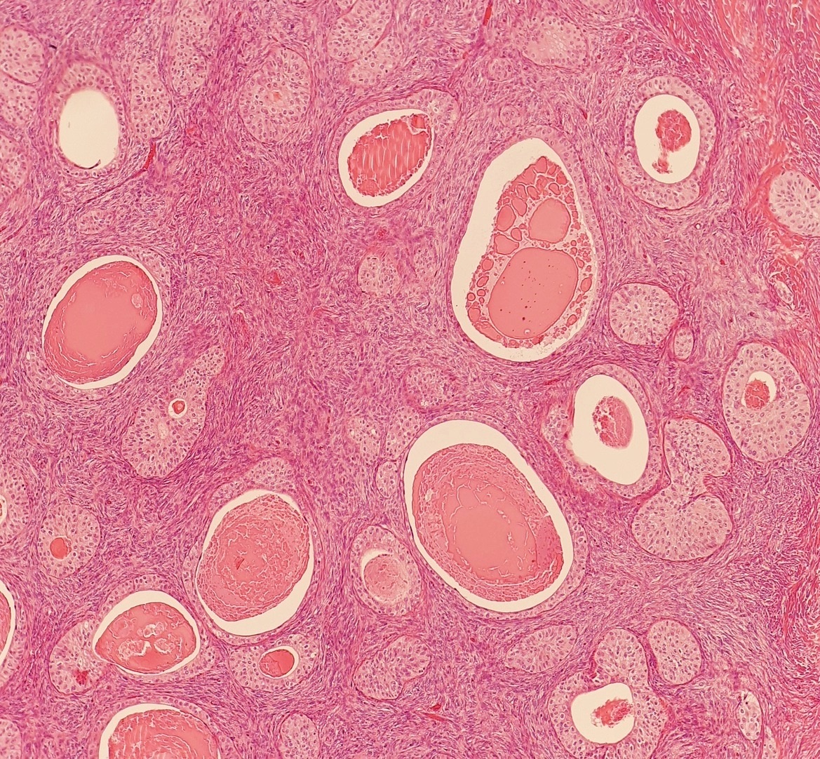
Benign Brenner tumor of the ovary: rounded nests of urothelial-type (transitional) cells with pale cytoplasm and oval, grooved ('coffee bean') nuclei in a dense fibromatous stroma; several nests have central microcysts filled with eosinophilic secretion, resembling cystitis cystica. H&E, low power (magnification not stated).Image: Mikael Häggström, M.D., Wikimedia Commons, CC0.
Ovarian Sex Cord-Stromal Tumors, Small Cell Carcinoma of Hypercalcemic Type and Wolffian and STK11 Adnexal Tumors
Definition. Ovarian sex cord-stromal tumors comprise pure stromal tumors (fibroma and thecoma), pure sex cord tumors (adult and juvenile granulosa cell tumors) and mixed sex cord-stromal tumors (Sertoli-Leydig cell tumors) (Renz et al. 2025). Adult granulosa cell tumors account for 90% of malignant ovarian sex cord-stromal tumors and 2–5% of all ovarian cancers (Pilsworth et al. 2021). Small cell carcinoma of the ovary, hypercalcemic type (SCCOHT) is classified among the miscellaneous ovarian tumors in the WHO 5th edition (McCluggage 2025). Its main differential diagnosis is juvenile granulosa cell tumor. The female adnexal tumor of probable Wolffian origin (FATWO) and the STK11 adnexal tumor arise mostly in para-adnexal soft tissue and share the sex cord-stromal differential (Bennett et al. 2020; Bennett et al. 2021).
Molecular classification of ovarian sex cord-stromal and related adnexal tumors
Recurrent molecular events now define several tumor types. Immunohistochemistry confirms sex cord-stromal lineage but has minimal value in separating the individual tumor types, and the molecular abnormality assists classification in problematic cases (McCluggage 2025).
Tumor
Recurrent alteration
Frequency in the defining study
Associated syndrome
Source
Adult granulosa cell tumor
FOXL2 c.402C>G (p.C134W)
86 of 89 (97%)
None
Shah et al. 2009; McCluggage 2025
Adult granulosa cell tumor, progression
TERT promoter C228T
22% of primary and 41% of recurrent tumors
None
Pilsworth et al. 2018
Juvenile granulosa cell tumor
AKT1 in-frame duplications; GNAS
AKT1 in >60% of tumors in girls under 15 years; GNAS in 30%
Ollier disease, Maffucci syndrome
Bessière et al. 2015; McCluggage 2025
Sertoli-Leydig cell tumor, moderately or poorly differentiated
DICER1 RNase IIIb hotspot
30 of 30
DICER1 syndrome
de Kock et al. 2017
Sertoli-Leydig cell tumor, well differentiated
None (DICER1 wild type)
0 of 18 DICER1 mutated
None
McCluggage et al. 2023
Sclerosing stromal tumor
FHL2::GLI2 fusion
17 of 26 (65%); other GLI2 rearrangements in 15%
None
Kim et al. 2020
Microcystic stromal tumor
CTNNB1 exon 3; less often APC
8 of 14 (57%) CTNNB1
Familial adenomatous polyposis
Irving et al. 2015; McCluggage et al. 2018
Sex cord tumor with annular tubules
STK11 in syndromic cases
Not applicable
Peutz–Jeghers syndrome
McCluggage 2025
Small cell carcinoma, hypercalcemic type
SMARCA4 inactivation
30 of 32 sequenced cases
Familial SCCOHT
Witkowski et al. 2014
STK11 adnexal tumor
STK11 alteration
21 of 21 sequenced
Peutz–Jeghers syndrome
Bennett et al. 2021
Immunohistochemistry of ovarian sex cord-stromal tumors
Inhibin and calretinin. In 215 ovarian tumors, calretinin stained 90% of fibrous tumors and inhibin stained 2% of epithelial tumors. Calretinin had 97% sensitivity and 85% specificity for sex cord-stromal and fibrous tumors (Movahedi-Lankarani and Kurman 2002).
SF-1. In 127 sex cord-stromal tumors of five types, inhibin and calretinin stained 56–100% and 36–100% of cases depending on type (Zhao et al. 2009). SF-1 stained all 27 Sertoli cell tumors and none of 60 endometrioid tumors, including sertoliform carcinomas, or 24 carcinoids (Zhao et al. 2008).
FOXL2. FOXL2 stained >95% of adult and juvenile granulosa cell tumors, fibromas and sclerosing stromal tumors, 1 of 11 steroid cell tumors and none of 368 non-sex cord-stromal tumors (Al-Agha et al. 2011).
Marker
Sex cord-stromal tumors positive
Main pitfall
Source
Inhibin
92%
Negative in many fibromas (22% positive)
Movahedi-Lankarani and Kurman 2002
Calretinin
100%
Stains 22% of epithelial tumors and mesothelium
Movahedi-Lankarani and Kurman 2002
SF-1
100% across five tumor types
Negative in endometrioid tumors and carcinoids
Zhao et al. 2009; Zhao et al. 2008
WT1
All types except steroid cell tumor
Stains 17% of endometrioid tumors
Zhao et al. 2009; Zhao et al. 2008
FOXL2
80%
Only 50% of Sertoli-Leydig cell tumors; positive in FATWO
Al-Agha et al. 2011
Fibroma, cellular fibroma and thecoma of the ovary
Cellular and mitotically active cellular fibroma. Cellular fibroma has bland nuclei and ≤3 mitotic figures per 10 high-power fields. In 75 cellular fibromatous tumors, 40 had ≥4 mitoses per 10 high-power fields with bland cytology (mean 6.7, range 4–19). All were unilateral, and follow-up of 18 of these 40 patients was uneventful. Such tumors are classified as mitotically active cellular fibroma. Fibrosarcoma usually shows moderate to severe atypia with an elevated mitotic rate (Irving et al. 2006).
Meigs syndrome. Meigs syndrome is the triad of a benign ovarian tumor (fibroma, Brenner tumor or occasionally granulosa cell tumor), ascites and hydrothorax that resolves after removal of the tumor. Serum CA125 was elevated in 66 of 580 fibroma or fibrothecoma patients (11.3%), associated with tumor diameter ≥10 cm and ascites (Shen et al. 2018).
Luteinized thecoma associated with sclerosing peritonitis
Clinical features. In 27 cases, sclerosing peritonitis was documented in 25. Patients presented with abdominal distension, pain and ascites, sometimes with bowel obstruction (Staats et al. 2008).
Pathology. The ovarian lesions were clinically bilateral in 24 cases, measured 2–31 cm and often had a cerebriform cut surface. Mitotically active spindle cells, weakly luteinized cells, edema and entrapped follicles were typical. Spindle cells were inhibin negative. Luteinized cells stained for inhibin, calretinin or CD56 (Staats et al. 2008).
Outcome. No ovarian lesion spread during a mean follow-up of 5.9 years in 20 cases, but 3 patients died of sclerosing peritonitis (Staats et al. 2008).
Sclerosing stromal tumor and microcystic stromal tumor of the ovary
Sclerosing stromal tumor.FHL2::GLI2 fusions and other GLI2 rearrangements were absent from 48 other sex cord-stromal tumors. The fusion activates the Sonic Hedgehog pathway (Kim et al. 2020).
Microcystic stromal tumor, morphology. In the original 16 cases, all tumors were unilateral, with a mean size of 8.7 cm, and none had spread. The tumors combined microcysts, solid cellular regions and fibrous stroma with hyaline plaques. Bizarre nuclei were present in 10 cases, and mitoses numbered 0–2 per 10 high-power fields (Irving and Young 2009).
Microcystic stromal tumor, immunophenotype. CD10 and vimentin stained all 16 tumors, whereas inhibin and calretinin were each positive in only 1 of 16 (Irving and Young 2009). Diffuse nuclear FOXL2, WT1, cyclin D1 and β-catenin staining was present in all tumors tested, and SF-1 stained 12 of 15 (Irving et al. 2015).
Microcystic stromal tumor, molecular findings. All 14 tumors tested had wild-type FOXL2 (Irving et al. 2015). Microcystic stromal tumors carry alterations in the β-catenin–APC pathway and may be an extracolonic manifestation of familial adenomatous polyposis (McCluggage et al. 2018).
Signet-ring stromal tumor. This rare benign stromal tumor contains bland signet-ring cells, each with a single large empty vacuole devoid of mucin or lipid, in a cellular fibromatous stroma (Ramzy 1976; Tchrakian et al. 2022). Of 10 tumors diagnosed as signet-ring stromal tumor, 3 were reclassified as microcystic stromal tumor with signet-ring cells because of microcysts, hyalinized stroma, nuclear β-catenin and cyclin D1, or a CTNNB1 mutation, and a CTNNB1 deletion has been reported in a typical example (Kopczynski et al. 2016). The remaining tumors may be related to fibroma (Tchrakian et al. 2022). The tumor cells resemble those of Krukenberg tumor (Ramzy 1976), which is described in Ovarian, Tubal and Peritoneal Cancer Staging and Reporting, Peritoneal Lesions and Metastases to the Ovary.
Adult granulosa cell tumor
Clinical features. Adult granulosa cell tumors occur in perimenopausal women, with a median age at diagnosis of 50–54 years, and secrete estrogen and inhibins (Pilsworth et al. 2021). In a Dutch population cohort of 1031 granulosa cell tumors, concurrent endometrial carcinoma was present in 5.9% and endometrial hyperplasia in 25.5% (van Meurs et al. 2013).
Morphology. In 60 granulosa cell tumors, diffuse sheets were the most frequent pattern (47%). Call-Exner bodies were absent in 22.2%, and the authors concluded that they are not required for the diagnosis. Nuclear grooves were present in 72.9% and may indicate granulosa cell origin (Guleria et al. 2020). Grooves were also a consistent feature of metastatic tumor in aspirates (Saini et al. 2023). Juvenile granulosa cell tumor nuclei lack grooves (Young et al. 1984).
Molecular definition. The FOXL2 c.402C>G (p.C134W) mutation was present in 97% of adult granulosa cell tumors, 21% of thecomas and 10% of juvenile granulosa cell tumors. It was absent from 49 other sex cord-stromal tumors and 329 unrelated ovarian and breast tumors (Shah et al. 2009).
Misdiagnosis.FOXL2 testing of 336 tumors diagnosed as adult granulosa cell tumor reclassified 63 as other tumor types. The misdiagnosed cases accounted for 71.9% of disease-specific deaths within 5 years (McConechy et al. 2016).
Recurrence. Among 164 molecularly defined tumors (92% stage I), 32% recurred. The median time to first relapse was 7.4 years, 75% of relapses occurred within 10 years, and tumor rupture was the only independent predictor of recurrence. Active follow-up for 10–15 years is advised (Bryk et al. 2016). In 176 patients from Taiwan, recurrence was 21%, and residual tumor and tumor size >13.5 cm predicted recurrence (Sun et al. 2012).
TERT promoter.TERT promoter mutations were associated with shorter overall survival (Pilsworth et al. 2018).
High-grade transformation. A p53 immunostain is performed on granulosa cell tumors with high-grade transformation, and TP53 mutations accompany that transformation (CAP 2024).
Juvenile granulosa cell tumor
Clinical features. In 125 cases, the average age was 13 years, and 44% of patients were 10 years or younger. Isosexual pseudoprecocity was the presentation in 82% of prepubertal patients. Two patients had Ollier disease and two had Maffucci syndrome (Young et al. 1984).
Pathology. Diffuse and follicular patterns were present. The follicles varied in size and shape and contained basophilic or eosinophilic secretion, often mucin positive. Granulosa cells were luteinized to varying degrees and had dark round nuclei without grooves. Mitoses ranged from <1 to 32 per 10 high-power fields (average 7) (Young et al. 1984).
Outcome. Of 95 patients with follow-up, 92% were alive and free of disease.
Ollier disease and Maffucci syndrome. In a 4-year-old with Ollier disease, the same IDH1 p.R132C mutation was present in the juvenile granulosa cell tumor and an enchondroma (Zhang et al. 2023). The syndromes are caused by somatic mosaic IDH1 or IDH2 mutations (Amary et al. 2011; Pansuriya et al. 2011).
Sertoli-Leydig cell tumor
Clinical features. In 207 cases, patients averaged 25 years, and 75% were 30 years or younger. One-third presented with androgen excess. At operation, 97.5% were stage I and 1.5% were bilateral. Tumors averaged 13.5 cm, and 15% were ruptured (Young and Scully 1985).
Differentiation and outcome. Eleven percent were well differentiated, 54% moderately differentiated and 13% poorly differentiated, and 22% contained heterologous elements. A retiform pattern was prominent in 15%. All well-differentiated tumors were benign, whereas 11% of moderately differentiated, 59% of poorly differentiated and 19% of heterologous tumors were clinically malignant (Young and Scully 1985).
CAP grading. Grade is required for Sertoli-Leydig cell tumors in the CAP ovary protocol. They are graded with the 3-tier system of the WHO 5th edition (CAP 2024).
CAP grade (WHO 5th edition)
Sertoli cell component
Leydig cells
Stroma
Source
G1, well differentiated
Hollow or solid tubules; no significant atypia or mitotic activity
Readily identified clusters, cords and single cells
Not specified
CAP 2024
G2, moderately differentiated
Nests, tubules or cords; mild, moderate or rarely bizarre atypia; modest mitoses
Minimal, at the periphery
Lobular pattern
CAP 2024
G3, poorly differentiated
Minor component of moderately differentiated tumor
DICER1 mutation.DICER1 RNase IIIb mutations were present in 26 of 43 Sertoli-Leydig cell tumors (60%) in the first series, 4 with additional germline mutations (Heravi-Moussavi et al. 2012). After expert review, all 30 moderately and poorly differentiated tumors carried DICER1 mutations and none of 4 well-differentiated tumors did (de Kock et al. 2017). All 18 well-differentiated tumors in a later series were DICER1 wild type and lacked FOXL2 C134W, and the authors proposed that well-differentiated Sertoli-Leydig cell tumor is a distinct tumor type (McCluggage et al. 2023).
Molecular subtype
Patient age (median)
Differentiation
Retiform or heterologous elements
Source
DICER1 mutant
24.5 years
Moderate or poor
All 10 tumors with retiform or heterologous elements were DICER1 mutant (10 of 18 mutant tumors)
Karnezis et al. 2019
FOXL2 c.402C>G mutant
79.5 years
Moderate or poor
Absent
Karnezis et al. 2019
DICER1 and FOXL2 wild type
51 years
Includes well differentiated
Absent
Karnezis et al. 2019
Molecular subtypes. In 42 tumors, DICER1 mutations (44%) and FOXL2 mutations (19%) were mutually exclusive. All tumors were FOXL2 positive by immunohistochemistry (Karnezis et al. 2019). FOXL2-mutant tumors may represent true Sertoli-Leydig cell tumors or adult granulosa cell tumors with Leydig-like luteinized stromal cells (CAP 2024).
DICER1 syndrome. Among 713 carriers of a germline DICER1 pathogenic or likely pathogenic variant, 103 developed an ovarian Sertoli-Leydig cell tumor at a median age of 14 years. Thirteen percent were diagnosed before 8 years of age, and the registry now recommends pelvic ultrasound from the time the germline variant is detected (Schultz et al. 2024). When a moderately or poorly differentiated Sertoli-Leydig cell tumor is diagnosed, the pathologist raises the possibility of a germline DICER1 variant and recommends genetic referral (McCluggage 2025).
Sertoli cell tumor, gynandroblastoma and sex cord tumor with annular tubules
Sertoli cell tumor. In 54 cases, patients were 2–76 years old (mean 30), and 6 had Peutz–Jeghers syndrome. All tumors were unilateral. A tubular pattern was present in every tumor, at least focally.
Gynandroblastoma. Gynandroblastoma contains both male (Sertoli or Leydig cell) and female (granulosa cell) components. In 16 cases, 3 carried DICER1 hotspot mutations in both components, all with moderately or poorly differentiated Sertoli-Leydig and juvenile granulosa cell components. None had FOXL2 C134W or AKT1 mutations, and all expressed FOXL2 in both components (Wang et al. 2018).
Sex cord tumor with annular tubules. Of 74 cases, 27 were associated with Peutz–Jeghers syndrome. These were benign, multifocal, bilateral, small or microscopic and calcified, and 4 of the 27 patients had a cervical adenocarcinoma, which caused death in 2. The 47 tumors without the syndrome were unilateral and usually large, and 7 were malignant (Young et al. 1982).
Steroid cell tumors of the ovary
Leydig cell (hilus cell) tumor. In 21 hilus cell tumors (12 with Reinke crystals), 62% presented with androgenic manifestations. Testosterone was high (200–2,400 ng/dL) despite a mean diameter of 2.1 cm. All tumors were centered in the hilus. None of the 10 patients with follow-up died of tumor (Paraskevas and Scully 1989).
Steroid cell tumor, not otherwise specified. In 63 cases, virilization was the most common presentation (41%). Four patients had Cushing syndrome. Six percent of tumors were bilateral. Of 50 patients with follow-up, 18 tumors were clinically malignant (Hayes and Scully 1987).
Feature in steroid cell tumor, NOS
Proportion of cases with the feature that were malignant
Source
≥2 mitotic figures per 10 high-power fields
92%
Hayes and Scully 1987
Necrosis
86%
Hayes and Scully 1987
Diameter ≥7 cm
78%
Hayes and Scully 1987
Hemorrhage
77%
Hayes and Scully 1987
Grade 2 or 3 nuclear atypia
64%
Hayes and Scully 1987
Small cell carcinoma of the ovary, hypercalcemic type
Clinical features. In 150 cases, patients were 9–43 years old (average 23.9). Serum calcium was elevated in 62% of the patients in whom it was measured. Tumors were unilateral in 99% and averaged 15.3 cm, and about half had extraovarian spread (Young et al. 1994). SCCOHT is the most common undifferentiated ovarian malignancy in women under 40 years (Witkowski et al. 2014). Long-term survival is about 30% in early-stage cases (Tischkowitz et al. 2020).
Morphology. Diffuse sheets of small cells with follicle-like spaces were the most common pattern. Follicle-like spaces were present in 80% of cases and usually contained eosinophilic fluid (Young et al. 1994).
Genetics. SMARCA4 protein was lost in 82% of SCCOHT and in only 0.4% of other primary ovarian tumors (Ramos et al. 2014). All patients with SCCOHT are referred for germline SMARCA4 testing (McCluggage 2025).
BRG1 immunohistochemistry. Fifty-four of 56 SCCOHT showed complete loss of SMARCA4 expression, and the 2 with retained expression had SMARCA4 alterations. All 217 interpretable mimics retained expression, although 13% showed heterogeneous weak staining. Loss had 96.55% sensitivity and 100% specificity, and only complete loss is interpreted as loss. SMARCB1 (INI1) was retained in all 37 SCCOHT tested (Clarke et al. 2016).
Dual loss of BRG1 and BRM. All SMARCA4-negative SCCOHT also lacked SMARCA2 (BRM). Among ovarian tumors, SMARCA4 loss otherwise occurred only in 4% of clear cell carcinomas, and the dual-loss phenotype was specific for SCCOHT (Karnezis et al. 2016).
Female adnexal tumor of probable Wolffian origin and STK11 adnexal tumor
FATWO. In 15 tumors, patients were 32–69 years old and tumors measured 1.8–30 cm. All but one arose in para-adnexal soft tissue. A tubular pattern was present in all, with solid (73%) and sieve-like (47%) patterns. Tumors were keratin positive and TTF-1 negative. Pathogenic mutations were rare (STK11 in 3, APC in 1, MBD4 in 1) (Bennett et al. 2020).
STK11 adnexal tumor, definition. The STK11 adnexal tumor was first reported in 2021 (Bennett et al. 2021) and is not listed in the WHO 5th edition of 2020 (McCluggage et al. 2022). Its diagnosis currently requires molecular confirmation of STK11 inactivation or alteration (McCluggage 2025).
STK11 adnexal tumor, clinical features. In 22 cases, patients were 17–66 years old (median 39), and nearly half had Peutz–Jeghers syndrome. Eighteen tumors were paratubal. Metastases were present in 11 of 22 patients (50%), and recurrences occurred in 12 of 15 with follow-up (80%) (Bennett et al. 2021).
STK11 adnexal tumor, morphology and immunophenotype. The tumors consist of interanastomosing cords and trabeculae of atypical epithelioid cells in myxoid to edematous stroma, with transitions to tubular, cystic, cribriform and microacinar formations. Tumors express epithelial, sex cord (calretinin, inhibin, WT1) and mesothelial markers and hormone receptors. Claudin-4, FOXL2 and TTF-1 are consistently negative (Bennett et al. 2021).
STK11 immunohistochemistry. All 17 STK11 adnexal tumors showed complete loss of cytoplasmic STK11 staining. Of 105 mimics, all retained staining except one endometrioid carcinoma with mucinous differentiation, which showed complete loss, and one high-grade serous carcinoma with subclonal loss (Dehghani et al. 2024).
Reporting. Many tumors previously reported as malignant FATWO likely represent STK11 adnexal tumors. The report raises the possibility of Peutz–Jeghers syndrome if the patient is not known to have it (McCluggage 2025).
Feature
FATWO
STK11 adnexal tumor
Source
Cytology
Mostly low-grade atypia; mitoses 0–17 per 10 high-power fields
Uniformly atypical; prominent nucleoli; median 9 mitoses per 10 high-power fields
Bennett et al. 2020; Bennett et al. 2021
FOXL2
Positive in 3 of 3
Negative
Al-Agha et al. 2011; Bennett et al. 2021
STK11 immunostain
Retained (10 of 10)
Complete cytoplasmic loss (17 of 17)
Dehghani et al. 2024
Behavior
5 of 6 with follow-up alive and well
Metastases in 11 of 22; recurrence in 12 of 15 with follow-up
Bennett et al. 2020; Bennett et al. 2021
Hereditary syndromes associated with ovarian sex cord-stromal and related tumors
Syndrome
Gene
Ovarian or adnexal tumor
Source
DICER1 syndrome
DICER1
Moderately or poorly differentiated Sertoli-Leydig cell tumor; gynandroblastoma
Schultz et al. 2024; Wang et al. 2018
Peutz–Jeghers syndrome
STK11
Sex cord tumor with annular tubules; STK11 adnexal tumor; Sertoli cell tumor
Young et al. 1982; Bennett et al. 2021; Oliva et al. 2005
Familial SCCOHT
SMARCA4
Small cell carcinoma, hypercalcemic type
Witkowski et al. 2014
Familial adenomatous polyposis
APC
Microcystic stromal tumor
McCluggage et al. 2018
Ollier disease and Maffucci syndrome
IDH1 or IDH2 (somatic mosaic; tumor-level mutations)
Juvenile granulosa cell tumor
Young et al. 1984; Pansuriya et al. 2011; Amary et al. 2011; Zhang et al. 2023
Nevoid basal cell carcinoma (Gorlin) syndrome
PTCH1 (germline)
Ovarian fibroma or calcification (24%)
Evans et al. 1993; Hahn et al. 1996; Johnson et al. 1996
Differential diagnosis of ovarian sex cord-stromal tumors and their mimics
Tumor
Main mimic
Distinguishing finding
Source
Juvenile granulosa cell tumor
SCCOHT
Inhibin positivity; SCCOHT shows complete BRG1 loss
Movahedi-Lankarani and Kurman 2002; Clarke et al. 2016
Sertoli cell tumor
Sertoliform endometrioid carcinoma; carcinoid
SF-1 positive in Sertoli cell tumor and negative in both mimics
Zhao et al. 2008
STK11 adnexal tumor
FATWO; sex cord-stromal tumor
Loss of STK11 staining; FOXL2 negative
Dehghani et al. 2024; Bennett et al. 2021
Staging and CAP reporting of malignant sex cord-stromal tumors
Protocol scope. The CAP ovary protocol version 1.5.0.0 applies to malignant sex cord-stromal tumors. Its histologic type list includes adult and juvenile granulosa cell tumor, steroid cell tumor (NOS and malignant) and Sertoli-Leydig cell tumor (CAP 2024).
Staging. The CAP protocol applies the same pTNM and FIGO 2014 categories to malignant sex cord-stromal tumors as to carcinomas (CAP 2024). Stage at diagnosis is the most important prognostic factor for granulosa cell tumors, and guidelines support fertility-sparing surgery for them (Renz et al. 2025).
Ovarian Germ Cell Tumors: Teratomas, Dysgerminoma, Yolk Sac Tumor and Other Malignant Germ Cell Tumors
Definition. Ovarian germ cell tumors derive from the primitive germ cells of the embryonic gonad (Renz et al. 2025). They range from the benign mature cystic teratoma to malignant tumors: immature teratoma, dysgerminoma, yolk sac tumor, embryonal carcinoma, non-gestational choriocarcinoma and mixed malignant germ cell tumor. The CAP ovary protocol, version 1.5.0.0, applies to the malignant types, to teratoma with malignant transformation and to malignant struma ovarii (CAP 2024).
Epidemiology. Germ cell tumors predominate among ovarian malignancies in women under 20 years and are the most common ovarian tumors in the second and third decades (Renz et al. 2025). In 1,262 malignant germ cell tumors of the female genital tract in the SEER registries (1973–2002), 35.6% were immature teratomas, 32.8% dysgerminomas, 28.7% mixed germ cell tumors and 2.9% mature teratomas with malignant degeneration. The age-adjusted incidence was 0.338 per 100,000 women-years, the highest rate was at 15–19 years, and 5-year relative survival was 83.9% (Smith et al. 2006).
Origin of ovarian teratomas and malignant germ cell tumors
Parthenogenetic origin. Benign ovarian teratomas were homozygous at centromeric chromosome polymorphisms for which the patients' normal tissues were heterozygous. They arise from a single germ cell after the first meiotic division (Linder et al. 1975).
Teratoma in the ovary and testis. Ovarian and prepubertal testicular teratomas derive from benign germ cells. Immaturity is an important finding in ovarian teratomas at any age, although its significance in children has been challenged (Ulbright 2005).
Isochromosome 12p. Five pure mature and three pure immature ovarian teratomas showed no i(12p) or other 12p amplification. In 6 mixed germ cell tumors, i(12p) was present in the non-teratomatous components in 5 and in the teratomatous component in 4. Teratoma in mixed ovarian germ cell tumors therefore has a different pathogenesis from pure ovarian teratoma (Poulos et al. 2006). Dysgerminoma, yolk sac tumor and embryonal carcinoma are associated with chromosome 12 abnormalities (CAP 2024).
Immature teratoma genomics. Ten immature teratomas had 2N near-diploid genomes with extensive loss of heterozygosity and no recurrent somatic mutations. All components within one tumor shared a clonal loss-of-heterozygosity pattern, whereas bilateral tumors had independent clonal origins (Heskett et al. 2020).
Two pathways in dysgerminoma.KIT exon 17 mutations were present in 5 of 13 unilateral dysgerminomas and in none of 4 bilateral dysgerminomas or 22 other germ cell tumors. Five phenotypic females with combined dysgerminoma and gonadoblastoma had Y-chromosome material (Hoei-Hansen et al. 2007).
Mature cystic teratoma and its complications
Clinical and gross features. In 517 patients with 573 mature cystic teratomas, the median age was 30 years and 60% were asymptomatic. Mean tumor size was 6.4 cm. The tumors were bilateral in 10.8%, torsion occurred in 3.5%, and malignant transformation in 0.17% (Comerci et al. 1994).
Anti-NMDA receptor encephalitis. Of 98 patients with anti-NMDA receptor encephalitis and oncologic assessment, 58 (59%) had a tumor, most commonly ovarian teratoma. Early tumor treatment was associated with better outcome and fewer relapses (Dalmau et al. 2008).
Squamous cell carcinoma. Squamous cell carcinoma accounts for 80% of malignant transformations of ovarian teratomas. In a systematic review of 277 patients, it occurred mainly in women older than 50 years with tumors larger than 100 mm and elevated squamous cell carcinoma antigen and CA125. Stage IA tumors had better survival (Hackethal et al. 2008).
Other transformations. In a single institution, 11 of 753 mature cystic teratomas (1.46%) underwent malignant transformation: 4 squamous cell carcinomas and 4 mucinous carcinomas (Oranratanaphan and Khemapech 2013). Somatic malignancy in an ovarian mature cystic teratoma is a de novo transformation (Ulbright 2005).
Reporting. The CAP histologic type list includes "teratoma with malignant transformation", with the type specified (CAP 2024).
Monodermal teratomas: struma ovarii, strumal carcinoid and carcinoid
Definition. Struma ovarii is an ovarian teratoma composed predominantly of thyroid tissue (Cao et al. 2026). Malignant struma ovarii has been defined as a teratoma containing at least 50% thyroid tissue that can metastasize (Lager et al. 2018). Thyroid tissue formed more than 50% of all but 2 of 27 biologically malignant struma ovarii (Shaco-Levy et al. 2010).
Malignant struma ovarii, predictors. Among 88 patients with histologically malignant or proliferative struma ovarii, adhesions, peritoneal fluid ≥1 L or an ovarian serosal rent occurred in 74% of the 27 biologically malignant tumors and in 10% of the 61 clinically benign tumors. A strumal component ≤6 cm recurred in 7%. Except for a papillary pattern or poorly differentiated carcinoma, no microscopic feature reliably predicted outcome. Survival was 89% at 10 years and 84% at 25 years (Robboy et al. 2009).
Biologically malignant struma ovarii. In 27 tumors with extraovarian spread, the thyroid tissue was follicular adenoma in 63% and papillary carcinoma in 26%. Recurrence occurred up to 29 years after surgery, and 37% of patients died of disease 1.5–32 years after diagnosis (Shaco-Levy et al. 2010).
Strumosis and reporting. Peritoneal implants of benign-appearing thyroid follicles, formerly called "strumosis", are regarded as metastases from well-differentiated carcinoma. Thyroid carcinoma arising in struma ovarii is staged with the AJCC or FIGO system, and its subtype follows the WHO 5th edition of thyroid tumors (CAP 2024).
Strumal carcinoid. In 50 cases, strumal carcinoid was an intimate mixture of thyroid tissue and carcinoid. All tumors were unilateral, and three-fifths arose in dermoid cysts or mature solid teratomas. No patient had the carcinoid syndrome, and only one died of tumor (Robboy and Scully 1980).
Primary insular carcinoid. In 48 cases, 33% had the carcinoid syndrome. Pure carcinoids or components 4–7 cm in diameter were associated with the syndrome in half of cases, and no patient had it when the carcinoid formed a small portion of a teratoma. Carcinoid metastatic to the ovary is nearly always bilateral, usually accompanied by peritoneal metastases and has a poor prognosis (Robboy et al. 1975).
Immature teratoma: diagnosis and grading
Diagnosis. Immaturity is usually recognized by embryonic-appearing neuroepithelium with mitotic activity and apoptosis. It is rarely based on cellular, mitotically active glial tissue. Fetal-type tissues alone are not sufficient for the diagnosis (Ulbright 2005).
Grading principle. Immature teratomas are the only malignant germ cell tumors that are graded. The grade is based on the number of low-power (40×) fields containing aggregates of immature neuroepithelium in any one slide. Immature elements other than neuroepithelium are not counted. Grade is a required CAP element, and either the 3-tier or the 2-tier system may be used (CAP 2024).
Origin of the grading system. Norris and colleagues graded 58 pure immature teratomas from 0 to 3. Grade of the primary tumor best determined extraovarian spread, and survival was 81%, 60% and 30% for grades 1, 2 and 3. All seven patients with grade 3 metastases died of tumor (Norris et al. 1976).
Reproducibility and the 2-tier system. In 244 immature teratomas, 16 of 91 patients (18%) with high-grade stage I tumors died with metastases, compared with 3 of 52 (6%) with grade 1 tumors that progressed, all 3 of which were inadequately sampled. Interobserver agreement was moderate with the 3-tier scale (κ=0.54) and better with a 2-tier scale (κ=0.66). Small foci (≤2 mm) of other germ cell elements did not adversely affect prognosis (O'Connor and Norris 1994).
3-tier grade (O'Connor and Norris; CAP)
Immature neuroepithelium in any one slide
2-tier grade
Source
Grade 1
Less than 1 low-power (40×) field
Low grade
CAP 2024; O'Connor and Norris 1994
Grade 2
1 to 3 low-power (40×) fields
High grade
CAP 2024; O'Connor and Norris 1994
Grade 3
4 or more low-power (40×) fields
High grade
CAP 2024; O'Connor and Norris 1994
Behavior by grade. Grade 1 tumors are low grade and curable with resection, whereas grades 2 and 3 are high grade (CAP 2024). In a pediatric intergroup trial, 44 children and adolescents with completely resected immature teratoma were managed with surgery alone, and the authors concluded that resection alone is effective treatment in this age group. Serum AFP was elevated in 34% of patients with pure immature teratoma and in 83% of those with microscopic foci of yolk sac tumor (Cushing et al. 1999).
Gliomatosis peritonei and growing teratoma syndrome
Gliomatosis peritonei. Gliomatosis peritonei consists of mature glial tissue in the peritoneum. In 21 patients aged 5–42 years, the primary tumors were immature teratoma (14), mixed germ cell tumor (6) and mature teratoma with carcinoid (1). Eleven patients also had metastatic mature or immature teratoma, and one developed glioma arising from gliomatosis. Gliomatosis expressed SOX2 and lacked OCT4 and NANOG, and the prognosis was favorable (Liang et al. 2015).
Implant assessment. Implants associated with immature teratoma are assessed for immature elements. Implants may consist entirely of mature glial tissue (CAP 2024).
Origin of gliomatosis. Microsatellite studies found glial implants heterozygous like normal tissue, whereas the associated teratomas were homozygous. These studies concluded that gliomatosis arises from non-teratomatous host cells (Ferguson et al. 2001; Kwan et al. 2004). A later multiregion exome study found that all disseminated components, including gliomatosis, shared the clonal loss-of-heterozygosity pattern of a primary ovarian tumor (Heskett et al. 2020).
Growing teratoma syndrome. Among 103 patients with pure or mixed immature teratoma, 12 (12%) developed growing teratoma syndrome at a median of 9 months. The peritoneum was the first site in 83%. Treatment is complete surgical resection, and late recurrences up to 14 years require prolonged follow-up (Zagamé et al. 2006). Residual mature teratoma can continue to grow and rarely undergoes malignant transformation (Renz et al. 2025).
Dysgerminoma and gonadoblastoma
Pathology. In 140 pure dysgerminomas (mean age 24.1 years), tumors were bilateral in 4%, with a mean diameter of 13 cm. A lymphocytic infiltrate was present in all, fibrovascular septa produced an alveolar pattern in 51%, and granulomatous infiltrates occurred in 46%. Variant patterns (cords, solid tubules, microspaces and follicle-like spaces) can mimic other tumors (Warnnissorn et al. 2021). KIT mutations were found only in unilateral tumors (Hoei-Hansen et al. 2007).
Outcome. In 158 dysgerminomas, 5-year and 10-year survival for pure tumors was 83% and 74%. Unilateral adnexectomy alone for stage IA pure dysgerminoma gave 95% 5-year survival, independent of tumor size (Gordon et al. 1981).
Management. Dysgerminoma is extremely sensitive to chemotherapy. Patients with stage IA disease may be observed after surgery, and disease beyond the ovary is treated with adjuvant chemotherapy (Renz et al. 2025).
Immunohistochemistry. OCT4 showed strong nuclear staining in >90% of cells in all 33 dysgerminomas and in the gonadoblastomas. Non-dysgerminomatous tumors were negative except 4 of 14 clear cell carcinomas, which showed focal staining (Cheng et al. 2004). OCT3/4 also stained 4 of 9 adult granulosa cell tumors (Chang et al. 2009). SALL4 stained >90% of tumor cells in all dysgerminomas (Cao et al. 2009). Dysgerminoma is NANOG positive, D2-40 positive and SOX2 negative (Chang et al. 2009). Dysgerminoma expresses CD117 (Renz et al. 2025).
Serum LDH. Serum lactate dehydrogenase was elevated at diagnosis in three patients with dysgerminoma, normalized after therapy and rose again at progression in one (Levato et al. 1995). LDH, AFP and hCG are part of follow-up of stage I germ cell malignancies (Renz et al. 2025).
Gonadoblastoma morphology. Classical gonadoblastoma consists of rounded islands of germ cells, sex cord elements and hyaline basement membrane material in a variably cellular stroma that may contain steroid cells. About 40% are bilateral. It arises almost entirely in dysgenetic gonads of patients with a Y chromosome or part of one, and TSPY1 is the putative gene. It is a non-invasive (in situ) precursor of germinoma and, indirectly, of other germ cell tumors (Roth and Cheng 2018; Roth and Cheng 2020).
Gonadoblastoma risk in disorders of sex development. Germ cell tumor risk guides the management of patients with disorders of sex development. The undifferentiated gonadal tissue pattern carries a high risk of gonadoblastoma, and TSPY and OCT3/4 are markers of early germ cell neoplasia (Cools et al. 2006).
Gonadoblastoma immunohistochemistry. Gonadoblastomas were positive for OCT4 (Cheng et al. 2004) and showed SALL4 staining in >90% of germ cells (Cao et al. 2009).
Yolk sac tumor
Historical behavior. In 71 endodermal sinus (yolk sac) tumors studied before modern combination chemotherapy, AFP was present in hyaline droplets, cytoplasm and intercellular spaces of all 15 tumors stained. Actuarial survival was 13% at 3 years, and 84% of stage I patients had subclinical metastasis (Kurman and Norris 1976a). Yolk sac tumor and choriocarcinoma are the most aggressive germ cell tumors, and they are highly curable with combination chemotherapy (Renz et al. 2025).
Patterns in 150 tumors. All were unilateral, and 70% measured ≥15 cm. The reticular (microcystic) pattern was conspicuous in 68% and present at least focally in all tumors, usually merging with cysts. Other patterns were polyvesicular (25%), labyrinthine (one-third), glandular (22%), solid (one-third) and festoon (6%). Schiller-Duval bodies (papillae with a central blood vessel) were present in one-third but numerous in only 5%. Hyaline bodies were present in most tumors. The solid pattern can resemble dysgerminoma but lacks its septa and lymphocytic infiltrate (Young et al. 2022).
Patterns and markers. Yolk sac tumors can differentiate into hepatic, intestinal and pulmonary tissues. AFP and glypican-3 are their characteristic immunohistochemical markers. SALL4, Lin28 and IMP-3 aid differential diagnosis, and CDX2, TTF-1 and HepPar1 help recognize unusual variants (Nogales et al. 2012).
Yolk sac tumor versus clear cell carcinoma. SALL4 stained >90% of cells in all 29 yolk sac tumors and only 3 of 45 clear cell carcinomas, focally (<15% of cells). AFP and glypican-3 stained 83% and 69% of yolk sac tumors and 35% and 28% of clear cell carcinomas (Cao et al. 2009). In another series, glypican-3 stained 97% of yolk sac tumors and no teratomas or embryonal carcinomas (Esheba et al. 2008).
Somatically derived yolk sac tumor. In 18 yolk sac tumors of the female genital tract in patients aged 40 years or older, 11 were associated with an epithelial neoplasm, most often high-grade serous carcinoma. Most showed glandular morphology closely mimicking adenocarcinoma, and some expressed epithelial markers (McNamee et al. 2016).
Embryonal carcinoma and non-gestational choriocarcinoma
Embryonal carcinoma, first description. Fifteen ovarian tumors resembling embryonal carcinoma of the adult testis were separated from endodermal sinus tumor. hCG was present in syncytiotrophoblast-like cells in all 10 tumors stained and AFP in mononuclear embryonal cells in 7. Sixty percent of patients had hormonal manifestations such as precocious puberty, irregular bleeding or hirsutism (Kurman and Norris 1976b).
Embryonal carcinoma in mixed tumors. In 55 tumors with an embryonal carcinoma component (median age 20.5 years), every case was part of a malignant mixed germ cell tumor, most often with yolk sac tumor (89%) and immature teratoma (62%). Glandular (45%) and solid (35%) patterns predominated. Many purported pure examples are likely undersampled mixed tumors or mimics such as dysgerminoma, SCCOHT or juvenile granulosa cell tumor (Zanfagnin et al. 2026).
Embryonal carcinoma immunophenotype. Embryonal carcinoma is SOX2, NANOG and OCT3/4 positive and D2-40 negative (Chang et al. 2009). SALL4 stained >90% of cells in all embryonal carcinomas (Cao et al. 2009). CD30 was expressed by 8 of 10 embryonal carcinomas and by the embryonal elements of all 4 mixed germ cell tumors, but by none of 11 other germ cell tumors or 92 non-hematopoietic tumors, in a series not restricted to the ovary (Pallesen and Hamilton-Dutoit 1988).
Non-gestational choriocarcinoma. Short tandem repeat genotyping of 22 choriocarcinomas identified 19 gestational tumors, which contained a paternal genome, and 3 non-gestational tumors, which lacked one: 2 ovarian and 1 pelvic. Ovarian and pelvic choriocarcinomas can be non-gestational and are not assumed to be metastases from an occult gestational tumor (Savage et al. 2017).
Mixed malignant germ cell tumor
Composition and prognosis. In 30 mixed germ cell tumors, dysgerminoma was the most common component (80%), followed by endodermal sinus tumor (70%), teratoma (53%), choriocarcinoma (20%) and embryonal carcinoma (16%). In stage I tumors, more than one-third endodermal sinus tumor, choriocarcinoma or grade 3 teratoma predicted a poor prognosis. All patients with tumors smaller than 10 cm survived. Serum hCG was elevated in 38% of non-pregnant patients and reflected choriocarcinoma or syncytiotrophoblastic giant cells (Kurman and Norris 1976c).
Reporting. The CAP protocol requires the component types and their percentages for mixed malignant germ cell tumors. Quantitation of tumor types within malignant germ cell tumors may be prognostically important (CAP 2024).
Immunohistochemistry and serum markers of ovarian germ cell tumors
Tumor
OCT3/4
SALL4
NANOG
SOX2
D2-40
Other markers
Serum marker
Source
Dysgerminoma
Positive
Positive
Positive
Negative
Positive
CD117
LDH
Cheng et al. 2004; Cao et al. 2009; Chang et al. 2009; Renz et al. 2025; Levato et al. 1995
Embryonal carcinoma
Positive
Positive
Positive
Positive
Negative
CD30
hCG (tissue: syncytiotrophoblast-like cells in 10 of 10); AFP (tissue: mononuclear cells in 7 of 10)
Chang et al. 2009; Cao et al. 2009; Pallesen and Hamilton-Dutoit 1988; Kurman and Norris 1976b
Yolk sac tumor
Negative
Positive
Negative
Negative
Negative
AFP; glypican-3
AFP
Chang et al. 2009; Cao et al. 2009; Esheba et al. 2008; Kurman and Norris 1976a
Choriocarcinoma
Negative
Not reported
Negative
Negative
Negative
β-hCG; glypican-3 in syncytiotrophoblast
hCG
Chang et al. 2009; Esheba et al. 2008; Renz et al. 2025; Kurman and Norris 1976c
Immature teratoma
Scattered epithelial cells only
Variable (11 of 15)
Negative
Positive in neural tissue
Negative
None specific
AFP in 34% of pure pediatric tumors
Chang et al. 2009; Cao et al. 2009; Cushing et al. 1999
Gonadoblastoma
Positive
Positive
Not reported
Not reported
Not reported
None specific
Not applicable
Cheng et al. 2004; Cao et al. 2009
Differential diagnosis of ovarian germ cell tumors
Tumor
Main mimic
Distinguishing finding
Source
Yolk sac tumor
Clear cell carcinoma
SALL4 diffusely positive; focal in only 3 of 45 clear cell carcinomas
Cao et al. 2009
Somatically derived yolk sac tumor
Adenocarcinoma
Associated epithelial neoplasm in an older patient; glandular yolk sac morphology
McNamee et al. 2016
Dysgerminoma
Embryonal carcinoma
D2-40 positive and SOX2 negative in dysgerminoma
Chang et al. 2009
Immature teratoma
Mature teratoma with differentiated neural tissue
Embryonic neuroepithelium with mitoses and apoptosis
Ulbright 2005
Gliomatosis peritonei
Metastatic immature teratoma
Mature glia only, SOX2 positive, OCT4 and NANOG negative
Liang et al. 2015; CAP 2024
Primary insular carcinoid
Metastatic carcinoid
Unilateral, often with teratoma; metastatic carcinoid is nearly always bilateral
Robboy et al. 1975
Non-gestational choriocarcinoma
Gestational choriocarcinoma
No paternal genome on genotyping
Savage et al. 2017
Staging and CAP reporting of ovarian germ cell tumors
Staging. Ovarian germ cell tumors are staged with the same FIGO system as epithelial ovarian cancer (Renz et al. 2025).
Surgery. Conservative surgery is standard for all stages of germ cell tumors, and fertility-sparing surgery is the standard of care for young patients with early-stage disease (Renz et al. 2025).
Peritoneal fluid. Teratomas may shed benign-appearing cells into fluids, and these may be classified as atypia of undetermined significance (CAP 2024).
Ovarian, Tubal and Peritoneal Cancer Staging and Reporting, Peritoneal Lesions and Metastases to the Ovary
Definition. Ovarian, fallopian tube and primary peritoneal cancers share one FIGO staging system, introduced in 2014 (Prat 2014). The FIGO Cancer Reports of 2021 and 2025 restate the 2014 system unchanged (Berek et al. 2021; Renz et al. 2025). The AJCC 8th edition pTNM categories correspond to it. The CAP protocol for primary tumors of the ovary, fallopian tube or peritoneum, version 1.5.0.0 (posted June 2024; required for accreditation from March 2025), uses AJCC-UICC 8 and the FIGO Cancer Report 2021 as its standards. It covers borderline tumors, carcinomas including carcinosarcoma, malignant germ cell and sex cord-stromal tumors and ovarian sarcomas, and it excludes peritoneal mesothelioma and lymphoma (CAP 2024).
Grossing and sampling of adnexal and staging specimens
Specimen part
CAP sampling recommendation
Source
Ovarian or adnexal tumor
One section per centimeter of greatest dimension, adjusted for heterogeneity and diagnostic difficulty
CAP 2024
Serous borderline tumor (especially micropapillary/cribriform) and mucinous borderline tumor
Two sections per centimeter, excluding smooth-walled cystic areas
CAP 2024
Ovarian surface
Area most closely approached by tumor; adhesions and rupture sites labeled
CAP 2024
Risk-reducing salpingo-oophorectomy or known BRCA1/BRCA2 carrier
Ovaries and tubes entirely, with SEE-FIM of the fimbriae
CAP 2024
Fallopian tubes with high-grade serous carcinoma and no gross tubal lesion
SEE-FIM of both fimbriated ends
CAP 2024
Grossly normal omentum (borderline tumor, serous carcinoma, immature teratoma)
5–10 sections
CAP 2024; Skala and Hagemann 2015
Omentum after neoadjuvant chemotherapy
4–6 sections of the most abnormal areas for the chemotherapy response score
CAP 2024
Grossly negative lymph nodes
Entirely submitted; largest metastatic deposit measured
CAP 2024
Staging biopsies
Entirely processed unless grossly positive
CAP 2024
Omental sampling model. In 44 gynecologic cancers with a grossly negative but microscopically positive omentum, a simulation estimated 82% sensitivity for 5 blocks and 95% for 10 blocks (Skala and Hagemann 2015).
Purpose. Frozen section determines the extent of surgery: cystectomy for benign tumors, oophorectomy or limited staging for borderline tumors in younger patients who wish to preserve fertility, and full staging for carcinoma (Buza 2019).
Accuracy. In a Cochrane review of 38 studies with 11,181 participants, frozen section had 90.0% sensitivity and 99.5% specificity for invasive cancer. With borderline and invasive tumors together as positive, sensitivity was 96.5% and specificity 89.5% (Ratnavelu et al. 2016).
Borderline diagnoses. A frozen section diagnosis of benign or invasive cancer was unchanged on paraffin sections in 94% and 99% of cases. A frozen section diagnosis of borderline tumor became invasive cancer in 21% of cases (Ratnavelu et al. 2016).
Capsule rupture, surface involvement and specimen integrity
Prognosis of rupture. In 1545 stage I invasive epithelial ovarian carcinomas, degree of differentiation was the strongest prognostic factor. Rupture before surgery (hazard ratio 2.65) and rupture during surgery (hazard ratio 1.64) were independent adverse factors (Vergote et al. 2001).
Intraoperative rupture. In 161 comprehensively staged stage I cancers, intraoperative capsule rupture (hazard ratio 4.2) and positive washings (hazard ratio 6.4) independently predicted worse disease-free survival (Bakkum-Gamez et al. 2009).
CAP reporting rule. Ovarian and tubal surface involvement and specimen integrity are reported for each ovary (capsule intact, capsule ruptured with preoperative, intraoperative or unknown timing, or fragmented). An ovary removed intact into a laparoscopic bag and opened in the bag without spillage into the peritoneal cavity is recorded as capsule intact, with an explanatory comment (CAP 2024).
FIGO 2014 staging of ovarian, fallopian tube and peritoneal cancer with AJCC 8th edition pTNM
FIGO 2014 stage
Definition
AJCC 8th edition
Source
IA
Tumor limited to one ovary (capsule intact) or fallopian tube; no surface tumor; no malignant cells in ascites or washings
pT1a N0 M0
Prat 2015; CAP 2024
IB
Tumor limited to both ovaries (capsules intact) or fallopian tubes; no surface tumor; no malignant cells in ascites or washings
pT1b N0 M0
Prat 2015; CAP 2024
IC1
Surgical spill
pT1c1
Prat 2015; CAP 2024
IC2
Capsule ruptured before surgery or tumor on ovarian or fallopian tube surface
pT1c2
Prat 2015; CAP 2024
IC3
Malignant cells in ascites or peritoneal washings
pT1c3
Prat 2015; CAP 2024
IIA
Extension or implants on the uterus, fallopian tubes or ovaries
pT2a
Prat 2015; CAP 2024
IIB
Extension to other pelvic intraperitoneal tissues
pT2b
Prat 2015; CAP 2024
IIIA1(i)
Positive retroperitoneal lymph nodes only; metastasis ≤10 mm
pT1 or pT2, pN1a
Prat 2015; CAP 2024
IIIA1(ii)
Positive retroperitoneal lymph nodes only; metastasis >10 mm
pT1 or pT2, pN1b
Prat 2015; CAP 2024
IIIA2
Microscopic extrapelvic peritoneal involvement, with or without positive retroperitoneal nodes
pT3a
Prat 2015; CAP 2024
IIIB
Macroscopic extrapelvic peritoneal metastasis ≤2 cm, with or without positive retroperitoneal nodes
pT3b
Prat 2015; CAP 2024
IIIC
Macroscopic extrapelvic peritoneal metastasis >2 cm, with or without positive retroperitoneal nodes; includes extension to the liver or spleen capsule without parenchymal involvement
pT3c
Prat 2015; CAP 2024
IVA
Pleural effusion with positive cytology
pM1a
Prat 2015; CAP 2024
IVB
Parenchymal metastases and metastases to extra-abdominal organs, including inguinal nodes and nodes outside the abdominal cavity
pM1b
Prat 2015; CAP 2024
Primary site and histotype. The primary site (ovary, fallopian tube or peritoneum) is designated where possible, and the histologic type is recorded (Prat 2015). FIGO lists cases whose primary cannot be delineated as "undesignated" (Prat 2015; Renz et al. 2025), whereas the CAP protocol says "undesignated" should be avoided and uses "tubo-ovarian" for small biopsies, prior incomplete salpingo-oophorectomy or some treated cases (CAP 2024). Primary peritoneal cancer has no stage I and begins at stage II (Prat 2015; Renz et al. 2025).
Lymph node measurement. The IIIA1 subdivision refers to the size of the metastatic deposit in the node, and the node itself is not measured (CAP 2024). FIGO introduced the 10-mm cutoff without retrospective data supporting it (Prat 2015).
Liver and spleen. Extension of tumor from the omentum to the spleen or liver is IIIC, whereas isolated parenchymal splenic or liver metastasis is IVB (Prat 2015).
Changes from the FIGO 1988 system (pre-2014 rules)
Topic
Pre-2014 FIGO rule
FIGO 2014 rule
Source
Scope
Ovarian, fallopian tube and peritoneal cancer not incorporated in one system
One system for ovary, fallopian tube and peritoneum, with the primary site designated
Prat 2015; Renz et al. 2025
Stage IC
Single stage IC for rupture, surface tumor or malignant ascites or washings
IC1 surgical spill; IC2 rupture before surgery or surface tumor; IC3 malignant ascites or washings
Prat 2015; Renz et al. 2025; Paik et al. 2015
Stage IIC
IIA or IIB with surface tumor, ruptured capsule, ascites or positive washings
Eliminated as redundant
Prat 2015
Retroperitoneal nodes only
Stage IIIC
Stage IIIA1, divided into (i) ≤10 mm and (ii) >10 mm
Onda et al. 1998; Renz et al. 2025
Microscopic extrapelvic peritoneum
Stage IIIA
Stage IIIA2, with or without positive retroperitoneal nodes
Renz et al. 2025
Stage IIIB
Macroscopic extrapelvic disease ≤2 cm
Same threshold; wording now includes nodal status
Renz et al. 2025; Rosendahl et al. 2016
Stage IV
Single stage IV
IVA pleural effusion with positive cytology; IVB parenchymal and extra-abdominal metastases, now including inguinal nodes
Renz et al. 2025; Rosendahl et al. 2016; Paik et al. 2015
Number of substages
10
14
Rosendahl et al. 2016
Evidence for IIIA1. Fewer than 10% of ovarian carcinomas extend beyond the pelvis with exclusively retroperitoneal nodal involvement, and these patients have a better prognosis than those with abdominal peritoneal spread (Prat 2015). In one series, 5-year survival was 84% for patients upstaged to stage III by nodes alone and 26% for patients with intraperitoneal tumor beyond the pelvis (Onda et al. 1998).
Restaging data. Of 4036 Danish patients, 1532 were assigned new stages. IA and IC1 had similar 5-year survival (87%), and IB, IC2 and IC3 had 75–80%. Sixteen stage IIIC cases with nodal disease alone became IIIA1, and 63 became IIIB (Rosendahl et al. 2016).
IIIA1(i) and IIIA1(ii). Among 46 stage IIIA1 patients, 5-year overall survival was 83.1% for IIIA1(i) and 80.2% for IIIA1(ii) (p=0.44) (Matsuoka et al. 2022).
Stage distribution. Most high-grade serous carcinomas present at stage III, mostly IIIC, and 12–21% present at stage IV (Prat 2015).
AJCC 8th edition and CAP rules for pN, pM and special situations
Regional nodes. Pelvic nodes (listed in Endometrial Carcinoma Staging, CAP Reporting and Biomarker Testing), para-aortic and retroperitoneal nodes are regional. Intra-omental and peri-intestinal nodes are not regional, and their involvement counts as T3 intraperitoneal disease. Other involved nodes are pM1 (CAP 2024).
Distant metastasis. Diaphragmatic surface involvement is pT3. Involvement of diaphragmatic skeletal muscle or abdominal wall tissue beyond the peritoneum is pM1. Transmural involvement of the intestine into the mucosa is pM1b, stage IVB (CAP 2024).
Ascites. Ascites does not affect stage unless malignant cells are present (CAP 2024).
Peritoneal washings and ascites. Malignant cells in fluid define IC3 when tumor is confined to the ovaries or tubes. Reporting categories and the CAP rule for borderline tumor cells in fluids are given in Pelvic washing cytology and ovarian cyst FNA (Prat 2015; CAP 2024).
Final stage. The pathologist reports pT, pN and pM from the information available, and the managing physician assigns the final pathologic stage (CAP 2024).
CAP 1.5.0.0 reporting elements for ovarian, tubal and peritoneal tumors
Element
Rule
Linked detail
Source
Histologic grade
Required for endometrioid and mucinous carcinomas, immature teratomas and Sertoli-Leydig cell tumors
ICCR dataset. The second edition of the ICCR dataset for ovarian, tubal and primary peritoneal carcinoma replaces the 2015 first edition and reflects the WHO 5th edition (Gilks et al. 2022).
Nodal serous borderline tumor, endosalpingiosis and endocervicosis
Nodal endosalpingiosis. Nodal endosalpingiosis was present in 58% of cases with nodal involvement and 35% without (McKenney et al. 2006).
Endosalpingiosis and cancer. Among 838 women with endosalpingiosis in a surgical cohort, concurrent endometriosis, uterine cancer and ovarian cancer were more frequent. Endosalpingiosis was associated with serous borderline (odds ratio 10.2), clear cell and invasive mucinous tumors (Esselen et al. 2016).
Endocervicosis. Endocervicosis of the urinary bladder forms a 2–5 cm mass of benign or mildly atypical endocervical-type glands in the bladder wall of women of reproductive age. Three of six cases were initially diagnosed as adenocarcinoma (Clement and Young 1992).
Mesothelial lesions of the female peritoneum
Reactive mesothelial hyperplasia. In 44 cases of florid peritoneal mesothelial hyperplasia, usually on the ovarian surface, endometriosis was the most common associated lesion. Small bland tubules, nests and cords in fibrous tissue were typical, and true or apparent lymphovascular invasion occurred. Calretinin positivity and Ber-EP4 negativity identified mesothelial cells (Oparka et al. 2011).
Multilocular peritoneal inclusion cyst. In 25 cases, all but 4 had prior surgery, endometriosis or pelvic inflammatory disease. Cysts were lined by flat to cuboidal mesothelial cells, sometimes with mural proliferation simulating mesothelioma. Twelve recurred locally, and the lesion is a non-neoplastic reactive mesothelial proliferation (Ross et al. 1989).
Well-differentiated papillary mesothelial tumor. The WHO 5th edition of thoracic tumors (2021) renamed well-differentiated papillary mesothelioma as well-differentiated papillary mesothelial tumor because of its indolent behavior (Sauter et al. 2022). In 26 women aged 23–75 years, lesions were 0.1–2 cm and usually incidental, and 22 of 25 with follow-up were free of disease (Malpica et al. 2012). All 10 peritoneal tumors sequenced carried mutually exclusive TRAF7 or CDC42 mutations, lacked BAP1, NF2 and CDKN2A alterations and retained BAP1 expression (Stevers et al. 2019).
Peritoneal mesothelioma, BAP1. BAP1 loss occurred in 1 of 395 serous carcinomas (0.3%) and in 6 of 9 abdominal mesotheliomas (67%) (Andrici et al. 2016). In another series, 16 of 25 peritoneal mesotheliomas lost BAP1, whereas all benign mesothelium and ovarian serous tumors retained it (Chapel et al. 2017).
Peritoneal mesothelioma, PAX8. PAX8 stained 99% of high-grade serous carcinomas, no pleural mesotheliomas and 2 of 23 peritoneal mesotheliomas (Laury et al. 2010). PAX8 staining of benign mesothelium and peritoneal mesothelioma is given in Mesothelial vs Carcinoma Immunohistochemistry in Effusions (Chapel et al. 2017).
Peritoneal mesothelioma, MTAP. MTAP loss is less frequent in peritoneal than in pleural mesothelioma (11 of 53 versus 35 of 60) (Davidson et al. 2024). In 39 peritoneal mesotheliomas, CDKN2A homozygous deletion was present in 27, and MTAP immunohistochemistry agreed poorly with FISH (κ=0.091), so MTAP is not a surrogate for CDKN2A FISH at this site (Quaranta et al. 2025). Retained MTAP therefore does not exclude mesothelioma. MTAP loss was absent from reactive peritoneal mesothelial proliferations in these series, and it may still serve as an independent adjunct marker of malignancy in a proven mesothelial proliferation (Quaranta et al. 2025; Quaranta et al. 2026). MTAP does not separate mesothelioma from serous tumors: loss occurred in 3 of 18 peritoneal mesotheliomas and 1 of 25 serous borderline tumors (Devins et al. 2023).
Peritoneal mesothelioma, claudin-4. Membranous claudin-4 stained 44 of 45 serous carcinomas (98%) and none of 40 peritoneal epithelioid mesotheliomas (Ordóñez 2013). It was positive in all 49 low-grade serous carcinomas and serous borderline tumors and negative in all 18 peritoneal mesotheliomas (Devins et al. 2023).
WT1 and other lineage markers. Calretinin, WT1, CK5/6 and D2-40 and the Müllerian markers PAX8 and SOX17 are each expressed by a subset of both mesothelial and Müllerian tumors (Churg et al. 2026). WT1 stained 72% of primary ovarian carcinomas in one series (Espinosa et al. 2015).
Peritoneal mesothelioma, ALK. ALK immunohistochemistry was positive in 11 of 88 peritoneal mesotheliomas. The 8 with focal weak staining lacked rearrangement, whereas the 3 with diffuse strong staining had ALK rearrangements (partners ATG16L1, STRN and TPM1). Patients with the rearrangement were women and younger (median 36 versus 62 years), had no asbestos fibers and lacked BAP1, SETD2 and NF2 alterations. All 205 pleural mesotheliomas were ALK negative (Hung et al. 2018).
Lesion
Architecture
Key immunohistochemistry
Molecular finding
Source
Reactive mesothelial hyperplasia
Bland tubules, nests and cords in fibrous tissue
Calretinin positive; Ber-EP4 negative
Not applicable (non-neoplastic)
Oparka et al. 2011
Multilocular peritoneal inclusion cyst
Cysts lined by flat to cuboidal mesothelium
L1CAM negative
Not applicable (non-neoplastic)
Ross et al. 1989; Goode et al. 2018
Adenomatoid tumor
Not applicable
L1CAM positive
TRAF7
Goode et al. 2018
Well-differentiated papillary mesothelial tumor
Papillae with bland mesothelial cells; rare mitoses
BAP1 retained; L1CAM positive
TRAF7 or CDC42
Malpica et al. 2012; Stevers et al. 2019
Peritoneal mesothelioma
Not applicable
BAP1 lost in 64–67%; PAX8 positive in up to 18%; claudin-4 negative
CDKN2A deletion in 27 of 39; MTAP loss less frequent than in pleura; poor surrogate for CDKN2A FISH
Andrici et al. 2016; Chapel et al. 2017; Quaranta et al. 2025; Davidson et al. 2024; Ordóñez 2013
Serous carcinoma
Not applicable
PAX8 positive in 99%; BAP1 retained in 99.7%; claudin-4 positive (98%)
Laury et al. 2010; Andrici et al. 2016; Ordóñez 2013; CAP 2024
Desmoplastic small round cell tumor
Clinical features. In 109 cases, 90 patients were male and 19 female, aged 6–49 years (mean 22). Tumor involved the abdominal cavity in 103 (Gerald et al. 1998).
Immunophenotype. Tumor cells were usually positive for keratin (86%), epithelial membrane antigen (93%), vimentin (97%), desmin (90%) and neuron-specific enolase (81%), and for the EWS-WT1 chimeric protein (93%). Muscle-specific actin, myogenin and chromogranin were usually negative (Gerald et al. 1998).
Molecular finding. The t(11;22)(p13;q12) translocation fuses EWSR1 and WT1 (Ladanyi and Gerald 1994). A functional fusion was found in 25 of 26 cases tested (Gerald et al. 1998).
Reactive peritoneal lesions
Keratin granulomas. Peritoneal keratin granulomas accompany endometrioid carcinoma with squamous differentiation of the endometrium or ovary and atypical polypoid adenomyoma. In 22 cases they had no prognostic significance and are distinguished from viable tumor implants (Kim and Scully 1990).
Deciduosis. Pregnancy-related peritoneal deciduosis forms small subperitoneal nodules of large polygonal eosinophilic cells that express vimentin and progesterone receptor and lack keratin, calretinin and HMB-45 staining. It regresses after delivery (Bolat et al. 2012).
Splenosis. Splenosis is autotransplantation of splenic tissue after splenic trauma or splenectomy. Peritoneal and pelvic nodules can mimic peritoneal metastases at staging laparotomy or endometriosis. A history of splenic injury is the clue, and frozen section or a Tc-99m heat-damaged red cell scan confirms splenic tissue (Braun et al. 2021; Abdul Jawad et al. 2025).
Pseudomyxoma peritonei and ovarian mucinous tumors
All 30 women with pseudomyxoma peritonei and ovarian mucinous tumors had an associated appendiceal or intestinal mucinous tumor (Ronnett et al. 1995). Identical KRAS mutations were present in the appendiceal and ovarian tumors of all 16 cases studied, supporting a single, most likely appendiceal, origin (Szych et al. 1999). The PSOGI classification of pseudomyxoma peritonei is given in Appendiceal Neoplasms: Mucinous Neoplasms, Pseudomyxoma Peritonei, Goblet Cell Adenocarcinoma and Neuroendocrine Tumors (Carr et al. 2016). Extensive poorly cellular mucin dissecting the ovarian stroma (pseudomyxoma ovarii) suggests an appendiceal mucinous neoplasm (CAP 2024).
Metastases to the ovary and Krukenberg tumor
General principles. Metastatic tumors are often cystic in the ovary even when the primary tumor is not, and many show well-differentiated areas in the ovary that mimic benign or borderline tumors. Surface implants suggest metastasis (Young 2006). Tumor mainly on the ovarian surface or centered on the hilum is more likely metastatic (CAP 2024).
Krukenberg tumor. In 120 cases, the average age was 45 years, and 63% were documented as bilateral. Signet-ring cells by definition occupied at least 10% of the tumor. Two-thirds of the primaries were gastric, followed by appendix, colon, breast, small intestine, rectum, gallbladder and urinary bladder. Two-thirds of the primaries were detected with or after the ovarian tumor (Kiyokawa et al. 2006).
Colorectal carcinoma. In 86 cases, involvement was bilateral in 49, and 20 unilateral tumors measured ≥10 cm. Garland pattern and dirty necrosis were typical, and benign-appearing or borderline-like foci occurred in 23. CK20 stained all 29 tumors tested and CK7 was focal in 17% (Lewis et al. 2006).
Pancreaticobiliary carcinoma. In 35 cases, 89% were bilateral, 63% nodular and 40% surface involved. Borderline-like and cystadenomatous patterns occurred in 69%. DPC4 (SMAD4) expression was lost in 61% and gave the most useful immunohistochemical evidence of a pancreaticobiliary source (Meriden et al. 2011).
Endocervical adenocarcinoma. In 29 cases, ovarian metastases were unilateral in 65.5% and often simulated borderline tumors or well-differentiated carcinomas. Identical HPV types in paired cervical and ovarian tumors established metastasis, even from microinvasive or in situ cervical tumors (Ronnett et al. 2008).
Breast carcinoma. Seven of 9 breast metastases to the ovary were lobular. All metastases were GATA3 positive and PAX8 negative, and only 1 expressed WT1. Independent primary ovarian carcinomas were PAX8 positive in 88% and GATA3 negative in all (Espinosa et al. 2015).
Primary ovarian mucinous tumors are SATB2 and CDX2 negative
CAP 2024; Ronnett et al. 1995
Pancreas and biliary tract
Bilateral, nodular, surface involvement; borderline-like areas
DPC4 loss in 61%
Meriden et al. 2011
Endocervix (HPV-associated)
Borderline-like or villoglandular patterns; often unilateral
Same HPV type as the cervical tumor
Ronnett et al. 2008
Breast
Lobular pattern; low nuclear grade
GATA3 positive; PAX8 negative
Espinosa et al. 2015
References
Abdul Jawad K, Denny K, Osumah TS, et al. Splenosis Masquerading as Endometriosis: A Rare Pelvic Post-Trauma Presentation. Am J Case Rep. 2025;26:e946307. doi:10.12659/AJCR.946307. PMID 40055891
Al-Agha OM, Huwait HF, Chow C, et al. FOXL2 is a sensitive and specific marker for sex cord-stromal tumors of the ovary. Am J Surg Pathol. 2011;35(4):484–494. doi:10.1097/PAS.0b013e31820a406c. PMID 21378549
Aljassim F, Rigby CH, Drury JA, et al. Hydrosalpinx perturbs the tubal and endometrial immune environment. Reprod Fertil. 2026;7(2). doi:10.1530/RAF-25-0204. PMID 42284130 (PMC13317832)
Amary MF, Damato S, Halai D, et al. Ollier disease and Maffucci syndrome are caused by somatic mosaic mutations of IDH1 and IDH2. Nat Genet. 2011;43(12):1262–1265. doi:10.1038/ng.994. PMID 22057236
American College of Obstetricians and Gynecologists Committee on Clinical Consensus–Gynecology. ACOG Clinical Practice Update: Salpingectomy for the prevention of epithelial ovarian cancer. Obstet Gynecol. 2026;148(4):e275–e280. doi:10.1097/AOG.0000000000006400. PMID 42623672 (cited as ACOG 2026)
American College of Obstetricians and Gynecologists. Adnexal torsion in adolescents: ACOG Committee Opinion No. 783. Obstet Gynecol. 2019;134(2):e56–e63. doi:10.1097/AOG.0000000000003373. PMID 31348225 (cited as ACOG 2019)
American College of Obstetricians and Gynecologists' Committee on Practice Bulletins–Gynecology. ACOG Practice Bulletin No. 193: Tubal ectopic pregnancy. Obstet Gynecol. 2018;131(3):e91–e103. doi:10.1097/AOG.0000000000002560. PMID 29470343 (cited as ACOG 2018)
Andrici J, Jung J, Sheen A, et al. Loss of BAP1 expression is very rare in peritoneal and gynecologic serous adenocarcinomas and can be useful in the differential diagnosis with abdominal mesothelioma. Hum Pathol. 2016;51:9–15. doi:10.1016/j.humpath.2015.12.012. PMID 27067777
Anglesio MS, Papadopoulos N, Ayhan A, et al. Cancer-Associated Mutations in Endometriosis without Cancer. N Engl J Med. 2017;376(19):1835–1848. doi:10.1056/NEJMoa1614814. PMID 28489996 (PMC5555376)
Assem H, Rambau PF, Lee S, et al. High-grade Endometrioid Carcinoma of the Ovary: A Clinicopathologic Study of 30 Cases. Am J Surg Pathol. 2018;42(4):534–544. doi:10.1097/PAS.0000000000001016. PMID 29309296
Atileh LIA, Dahbour D, Hammo H, et al. Laparoscopic Removal of a 40-cm Paratubal Cyst in a Morbidly Obese Patient. Gynecol Minim Invasive Ther. 2020;9(1):39–41. doi:10.4103/GMIT.GMIT_110_18. PMID 32090013 (PMC7008651)
Bagué S, Rodríguez IM, Prat J. Sarcoma-like mural nodules in mucinous cystic tumors of the ovary revisited: a clinicopathologic analysis of 10 additional cases. Am J Surg Pathol. 2002;26(11):1467–1476. doi:10.1097/00000478-200211000-00009. PMID 12409723
Bakkum-Gamez JN, Richardson DL, Seamon LG, et al. Influence of intraoperative capsule rupture on outcomes in stage I epithelial ovarian cancer. Obstet Gynecol. 2009;113(1):11–17. doi:10.1097/AOG.0b013e3181917a0c. PMID 19104354
Banet N, Kurman RJ. Two types of ovarian cortical inclusion cysts: proposed origin and possible role in ovarian serous carcinogenesis. Int J Gynecol Pathol. 2015;34(1):3–8. doi:10.1097/PGP.0000000000000120. PMID 25473746
Bell KA, Kurman RJ. A clinicopathologic analysis of atypical proliferative (borderline) tumors and well-differentiated endometrioid adenocarcinomas of the ovary. Am J Surg Pathol. 2000;24(11):1465–1479. doi:10.1097/00000478-200011000-00002. PMID 11075848
Bennett JA, Pesci A, Morales-Oyarvide V, et al. Incidence of Mismatch Repair Protein Deficiency and Associated Clinicopathologic Features in a Cohort of 104 Ovarian Endometrioid Carcinomas. Am J Surg Pathol. 2019;43(2):235–243. doi:10.1097/PAS.0000000000001165. PMID 30256257
Bennett JA, Ritterhouse LL, Furtado LV, et al. Female adnexal tumors of probable Wolffian origin: morphological, immunohistochemical, and molecular analysis of 15 cases. Mod Pathol. 2020;33(4):734–747. doi:10.1038/s41379-019-0375-9. PMID 31591497 (cited as Bennett et al. 2020)
Bennett JA, Young RH, Howitt BE, et al. A Distinctive Adnexal (Usually Paratubal) Neoplasm Often Associated With Peutz-Jeghers Syndrome and Characterized by STK11 Alterations (STK11 Adnexal Tumor): A Report of 22 Cases. Am J Surg Pathol. 2021;45(8):1061–1074. doi:10.1097/PAS.0000000000001677. PMID 33534223 (cited as Bennett et al. 2021)
Berek JS, Renz M, Kehoe S, et al. Cancer of the ovary, fallopian tube, and peritoneum: 2021 update. Int J Gynaecol Obstet. 2021;155 Suppl 1:61–85. doi:10.1002/ijgo.13878. PMID 34669199
Bessière L, Todeschini AL, Auguste A, et al. A Hot-spot of In-frame Duplications Activates the Oncoprotein AKT1 in Juvenile Granulosa Cell Tumors. EBioMedicine. 2015;2(5):421–431. doi:10.1016/j.ebiom.2015.03.002. PMID 26137586
Bogaerts JMA, Steenbeek MP, van Bommel MHD, et al. Recommendations for diagnosing STIC: a systematic review and meta-analysis. Virchows Arch. 2022;480(4):725–737. doi:10.1007/s00428-021-03244-w. PMID 34850262 (PMC9023413)
Bogaerts JMA, van Bommel MHD, Hermens RPMG, et al. Consensus based recommendations for the diagnosis of serous tubal intraepithelial carcinoma: an international Delphi study. Histopathology. 2023;83(1):67–79. doi:10.1111/his.14902. PMID 36939551
Böhm S, Faruqi A, Said I, et al. Chemotherapy Response Score: Development and Validation of a System to Quantify Histopathologic Response to Neoadjuvant Chemotherapy in Tubo-Ovarian High-Grade Serous Carcinoma. J Clin Oncol. 2015;33(22):2457–2463. doi:10.1200/JCO.2014.60.5212. PMID 26124480
Bolat F, Canpolat T, Tarim E. Pregnancy-related peritoneal ectopic decidua (deciduosis): morphological and clinical evaluation. Turk Patoloji Derg. 2012;28(1):56–60. doi:10.5146/tjpath.2012.01098. PMID 22207433
Bonadona V, Bonaïti B, Olschwang S, et al. Cancer risks associated with germline mutations in MLH1, MSH2, and MSH6 genes in Lynch syndrome. JAMA. 2011;305(22):2304–2310. doi:10.1001/jama.2011.743. PMID 21642682
Boudaouara O, Sellami A, Roger P. Ovarian stromal hyperthecosis: An underestimated virilizing disorder in postmenopausal women. Ann Endocrinol (Paris). 2025;86(5):101800. doi:10.1016/j.ando.2025.101800. PMID 40513985
Braun T, De Gregorio A, Baumann L, et al. Coincidence of Intra-Abdominal Splenosis in a Patient with Advanced Ovarian Cancer: Case Report and Review of the Literature. Surg J (N Y). 2021;7(2):e116–e120. doi:10.1055/s-0041-1731426. PMID 34179460
Bryk S, Färkkilä A, Bützow R, et al. Characteristics and outcome of recurrence in molecularly defined adult-type ovarian granulosa cell tumors. Gynecol Oncol. 2016;143(3):571–577. doi:10.1016/j.ygyno.2016.10.002. PMID 27729108
Burks RT, Sherman ME, Kurman RJ. Micropapillary serous carcinoma of the ovary. A distinctive low-grade carcinoma related to serous borderline tumors. Am J Surg Pathol. 1996;20(11):1319–1330. doi:10.1097/00000478-199611000-00003. PMID 8898836
Busca A, Nofech-Mozes S, Olkhov-Mitsel E, et al. Histological grading of ovarian mucinous carcinoma - an outcome-based analysis of traditional and novel systems. Histopathology. 2020;77(1):26–34. doi:10.1111/his.14039. PMID 31782197
Buza N. Frozen Section Diagnosis of Ovarian Epithelial Tumors: Diagnostic Pearls and Pitfalls. Arch Pathol Lab Med. 2019;143(1):47–64. doi:10.5858/arpa.2018-0289-RA. PMID 30785337
Callahan MJ, Crum CP, Medeiros F, et al. Primary fallopian tube malignancies in BRCA-positive women undergoing surgery for ovarian cancer risk reduction. J Clin Oncol. 2007;25(25):3985–3990. doi:10.1200/JCO.2007.12.2622. PMID 17761984
Cancer Genome Atlas Research Network. Integrated genomic analyses of ovarian carcinoma. Nature. 2011;474(7353):609–615. doi:10.1038/nature10166. PMID 21720365 (PMC3163504)
Cao D, Guo S, Allan RW, et al. SALL4 is a novel sensitive and specific marker of ovarian primitive germ cell tumors and is particularly useful in distinguishing yolk sac tumor from clear cell carcinoma. Am J Surg Pathol. 2009;33(6):894–904. doi:10.1097/PAS.0b013e318198177d. PMID 19295406 (cited as Cao et al. 2009)
Cao J, Meng Z, Li C. Sonographic and Clinicopathological Characterization of Struma Ovarii: A Retrospective Analysis for Enhanced Preoperative Diagnosis. Curr Med Imaging. 2026;22:e15734056393689. doi:10.2174/0115734056393689250929185251. PMID 41088975 (cited as Cao et al. 2026)
Cao Y, Wu W, Wang W, et al. Three cases of ovarian actinomycosis with literature review. Int J Clin Exp Pathol. 2023;16(7):158–163. PMID 37559683 (PMC10408434)
Carr NJ, Cecil TD, Mohamed F, et al. A Consensus for Classification and Pathologic Reporting of Pseudomyxoma Peritonei and Associated Appendiceal Neoplasia: The Results of the Peritoneal Surface Oncology Group International (PSOGI) Modified Delphi Process. Am J Surg Pathol. 2016;40(1):14–26. doi:10.1097/PAS.0000000000000535. PMID 26492181
Chae HS, Rheu CH. Precocious pseudopuberty due to an autonomous ovarian follicular cyst: case report with a review of literatures. BMC Res Notes. 2013;6:319. doi:10.1186/1756-0500-6-319. PMID 23937919 (PMC3766658)
Chang MC, Vargas SO, Hornick JL, et al. Embryonic stem cell transcription factors and D2-40 (podoplanin) as diagnostic immunohistochemical markers in ovarian germ cell tumors. Int J Gynecol Pathol. 2009;28(4):347–355. doi:10.1097/PGP.0b013e318195da86. PMID 19483629
Chapel DB, Husain AN, Krausz T, et al. PAX8 Expression in a Subset of Malignant Peritoneal Mesotheliomas and Benign Mesothelium has Diagnostic Implications in the Differential Diagnosis of Ovarian Serous Carcinoma. Am J Surg Pathol. 2017;41(12):1675–1682. doi:10.1097/PAS.0000000000000935. PMID 28877056
Cheasley D, Wakefield MJ, Ryland GL, et al. The molecular origin and taxonomy of mucinous ovarian carcinoma. Nat Commun. 2019;10(1):3935. doi:10.1038/s41467-019-11862-x. PMID 31477716 (PMC6718426)
Cheng L, Thomas A, Roth LM, et al. OCT4: a novel biomarker for dysgerminoma of the ovary. Am J Surg Pathol. 2004;28(10):1341–1346. doi:10.1097/01.pas.0000135528.03942.1f. PMID 15371950
Churg A, Malpica A, Hung YP. Mesothelial Proliferations Involving the Gynecologic Tract. Adv Anat Pathol. 2026. doi:10.1097/PAP.0000000000000560. PMID 43000614
Clarke BA, Witkowski L, Ton Nu TN, et al. Loss of SMARCA4 (BRG1) protein expression as determined by immunohistochemistry in small-cell carcinoma of the ovary, hypercalcaemic type distinguishes these tumours from their mimics. Histopathology. 2016;69(5):727–738. doi:10.1111/his.12988. PMID 27100627
Clement PB, Young RH. Endocervicosis of the urinary bladder. A report of six cases of a benign müllerian lesion that may mimic adenocarcinoma. Am J Surg Pathol. 1992;16(6):533–542. doi:10.1097/00000478-199206000-00001. PMID 1318002
Clement PB. The pathology of endometriosis: a survey of the many faces of a common disease emphasizing diagnostic pitfalls and unusual and newly appreciated aspects. Adv Anat Pathol. 2007;14(4):241–260. doi:10.1097/PAP.0b013e3180ca7d7b. PMID 17592255
Clement PB. Tumor-like lesions of the ovary associated with pregnancy. Int J Gynecol Pathol. 1993;12(2):108–115. doi:10.1097/00004347-199304000-00004. PMID 8463033
Coatham M, Li X, Karnezis AN, et al. Concurrent ARID1A and ARID1B inactivation in endometrial and ovarian dedifferentiated carcinomas. Mod Pathol. 2016;29(12):1586–1593. doi:10.1038/modpathol.2016.156. PMID 27562491
Cohen PA, Powell A, Böhm S, et al. Corrigendum to "Pathological chemotherapy response score is prognostic in tubo-ovarian high-grade serous carcinoma: A systematic review and meta-analysis of individual patient data" Gynecol. Oncol. 154 (2019) 441-448. Gynecol Oncol. 2021;161(1):328–329. doi:10.1016/j.ygyno.2021.01.032. PMID 33551198 (cited as Cohen et al. 2021)
Cohen PA, Powell A, Böhm S, et al. Pathological chemotherapy response score is prognostic in tubo-ovarian high-grade serous carcinoma: A systematic review and meta-analysis of individual patient data. Gynecol Oncol. 2019;154(2):441–448. doi:10.1016/j.ygyno.2019.04.679. PMID 31118141
Comerci JT, Licciardi F, Bergh PA, et al. Mature cystic teratoma: a clinicopathologic evaluation of 517 cases and review of the literature. Obstet Gynecol. 1994;84(1):22–28. PMID 8008317
Cools M, Drop SL, Wolffenbuttel KP, et al. Germ cell tumors in the intersex gonad: old paths, new directions, moving frontiers. Endocr Rev. 2006;27(5):468–484. doi:10.1210/er.2006-0005. PMID 16735607
Cushing B, Giller R, Ablin A, et al. Surgical resection alone is effective treatment for ovarian immature teratoma in children and adolescents: a report of the pediatric oncology group and the children's cancer group. Am J Obstet Gynecol. 1999;181(2):353–358. doi:10.1016/s0002-9378(99)70561-2. PMID 10454682
Dalmau J, Gleichman AJ, Hughes EG, et al. Anti-NMDA-receptor encephalitis: case series and analysis of the effects of antibodies. Lancet Neurol. 2008;7(12):1091–1098. doi:10.1016/S1474-4422(08)70224-2. PMID 18851928
Davidson B, Holth A, Hummel C, et al. Different Frequency and Clinical Role for MTAP Loss in Pleural and Peritoneal Mesothelioma. Appl Immunohistochem Mol Morphol. 2024;32(6):280–284. doi:10.1097/PAI.0000000000001206. PMID 38812360
de Kock L, Terzic T, McCluggage WG, et al. DICER1 Mutations Are Consistently Present in Moderately and Poorly Differentiated Sertoli-Leydig Cell Tumors. Am J Surg Pathol. 2017;41(9):1178–1187. doi:10.1097/PAS.0000000000000895. PMID 28654427
Dehghani A, Sharma AE, Siegmund SE, et al. STK11 (LKB1) immunohistochemistry is a sensitive and specific marker for STK11 adnexal tumours. Histopathology. 2024;85(5):769–782. doi:10.1111/his.15303. PMID 39169716
Devins KM, Zukerberg L, Watkins JC, et al. BAP1 and Claudin-4, But Not MTAP, Reliably Distinguish Borderline and Low-grade Serous Ovarian Tumors From Peritoneal Mesothelioma. Int J Gynecol Pathol. 2023;42(2):159–166. doi:10.1097/PGP.0000000000000877. PMID 35512220
Esheba GE, Longacre TA, Atkins KA, et al. Expression of the urothelial differentiation markers GATA3 and placental S100 (S100P) in female genital tract transitional cell proliferations. Am J Surg Pathol. 2009;33(3):347–353. doi:10.1097/PAS.0b013e3181908e24. PMID 19092634
Esheba GE, Pate LL, Longacre TA. Oncofetal protein glypican-3 distinguishes yolk sac tumor from clear cell carcinoma of the ovary. Am J Surg Pathol. 2008;32(4):600–607. doi:10.1097/PAS.0b013e31815a565a. PMID 18277882
Espinosa I, Gallardo A, D'Angelo E, et al. Simultaneous carcinomas of the breast and ovary: utility of Pax-8, WT-1, and GATA3 for distinguishing independent primary tumors from metastases. Int J Gynecol Pathol. 2015;34(3):257–265. doi:10.1097/PGP.0000000000000155. PMID 25844549
Esselen KM, Terry KL, Samuel A, et al. Endosalpingiosis: More than just an incidental finding at the time of gynecologic surgery? Gynecol Oncol. 2016;142(2):255–260. doi:10.1016/j.ygyno.2016.05.036. PMID 27261327
Evans DG, Ladusans EJ, Rimmer S, et al. Complications of the naevoid basal cell carcinoma syndrome: results of a population based study. J Med Genet. 1993;30(6):460–464. doi:10.1136/jmg.30.6.460. PMID 8326488
Ferguson AW, Katabuchi H, Ronnett BM, et al. Glial implants in gliomatosis peritonei arise from normal tissue, not from the associated teratoma. Am J Pathol. 2001;159(1):51–55. doi:10.1016/S0002-9440(10)61672-0. PMID 11438453
Folkins AK, Jarboe EA, Saleemuddin A, et al. A candidate precursor to pelvic serous cancer (p53 signature) and its prevalence in ovaries and fallopian tubes from women with BRCA mutations. Gynecol Oncol. 2008;109(2):168–173. doi:10.1016/j.ygyno.2008.01.012. PMID 18342932 (PMC2746001)
Fukunaga M, Nomura K, Ishikawa E, et al. Ovarian atypical endometriosis: its close association with malignant epithelial tumours. Histopathology. 1997;30(3):249–255. doi:10.1046/j.1365-2559.1997.d01-592.x. PMID 9088954
Garg K, Karnezis AN, Rabban JT. Uncommon hereditary gynaecological tumour syndromes: pathological features in tumours that may predict risk for a germline mutation. Pathology. 2018;50(2):238–256. doi:10.1016/j.pathol.2017.10.009. PMID 29373116
Genestie C, Auguste A, Al Battal M, et al. Histological classification of mucinous ovarian tumors: inter-observer reproducibility, clinical relevance, and role of genetic biomarkers. Virchows Arch. 2021;478(5):885–891. doi:10.1007/s00428-020-02939-w. PMID 33009577
Gerald WL, Ladanyi M, de Alava E, et al. Clinical, pathologic, and molecular spectrum of tumors associated with t(11;22)(p13;q12): desmoplastic small round-cell tumor and its variants. J Clin Oncol. 1998;16(9):3028–3036. doi:10.1200/JCO.1998.16.9.3028. PMID 9738572
Gershenson DM, Miller A, Brady WE, et al. Trametinib versus standard of care in patients with recurrent low-grade serous ovarian cancer (GOG 281/LOGS): an international, randomised, open-label, multicentre, phase 2/3 trial. Lancet. 2022;399(10324):541–553. doi:10.1016/S0140-6736(21)02175-9. PMID 35123694 (PMC8819271)
Gilks CB, Selinger CI, Davidson B, et al. Data Set for the Reporting of Ovarian, Fallopian Tube and Primary Peritoneal Carcinoma: Recommendations From the International Collaboration on Cancer Reporting (ICCR). Int J Gynecol Pathol. 2022;41(Suppl 1):S119–S142. doi:10.1097/PGP.0000000000000908. PMID 36305537
González-Martín A, Pothuri B, Vergote I, et al. Niraparib in Patients with Newly Diagnosed Advanced Ovarian Cancer. N Engl J Med. 2019;381(25):2391–2402. doi:10.1056/NEJMoa1910962. PMID 31562799
Goode B, Joseph NM, Stevers M, et al. Adenomatoid tumors of the male and female genital tract are defined by TRAF7 mutations that drive aberrant NF-kB pathway activation. Mod Pathol. 2018;31(4):660–673. doi:10.1038/modpathol.2017.153. PMID 29148537
Gordon A, Lipton D, Woodruff JD. Dysgerminoma: a review of 158 cases from the Emil Novak Ovarian Tumor Registry. Obstet Gynecol. 1981;58(4):497–504. PMID 7279343
Grisham RN, Slomovitz B, Podder V, et al. Low-grade serous ovarian cancer: 2026 expert consensus report. Gynecol Oncol. 2026;214:144–163. doi:10.1016/j.ygyno.2026.10.011. PMID 42913278 (cited as Grisham et al. 2026b)
Grisham RN, Van Nieuwenhuysen E, Santin AD, et al. Long-term efficacy and safety of avutometinib + defactinib in recurrent low-grade serous ovarian cancer: Results of a 2-year follow-up of ENGOT-OV60/GOG-3052/RAMP 201. Gynecol Oncol. 2026;213:59–67. doi:10.1016/j.ygyno.2026.09.003. PMID 42748602 (cited as Grisham et al. 2026a)
Guleria P, Kumar L, Kumar S, et al. A clinicopathological study of granulosa cell tumors of the ovary: Can morphology predict prognosis? Indian J Pathol Microbiol. 2020;63(1):53–59. doi:10.4103/IJPM.IJPM_403_19. PMID 32031123
Hackethal A, Brueggmann D, Bohlmann MK, et al. Squamous-cell carcinoma in mature cystic teratoma of the ovary: systematic review and analysis of published data. Lancet Oncol. 2008;9(12):1173–1180. doi:10.1016/S1470-2045(08)70306-1. PMID 19038764
Hahn H, Wicking C, Zaphiropoulous PG, et al. Mutations of the human homolog of Drosophila patched in the nevoid basal cell carcinoma syndrome. Cell. 1996;85(6):841–851. doi:10.1016/s0092-8674(00)81268-4. PMID 8681379
Hayes MC, Scully RE. Ovarian steroid cell tumors (not otherwise specified). A clinicopathological analysis of 63 cases. Am J Surg Pathol. 1987;11(11):835–845. doi:10.1097/00000478-198711000-00002. PMID 2823622
Heravi-Moussavi A, Anglesio MS, Cheng SW, et al. Recurrent somatic DICER1 mutations in nonepithelial ovarian cancers. N Engl J Med. 2012;366(3):234–242. doi:10.1056/NEJMoa1102903. PMID 22187960
Heskett MB, Sanborn JZ, Boniface C, et al. Multiregion exome sequencing of ovarian immature teratomas reveals 2N near-diploid genomes, paucity of somatic mutations, and extensive allelic imbalances shared across mature, immature, and disseminated components. Mod Pathol. 2020;33(6):1193–1206. doi:10.1038/s41379-019-0446-y. PMID 31911616
Hoei-Hansen CE, Kraggerud SM, Abeler VM, et al. Ovarian dysgerminomas are characterised by frequent KIT mutations and abundant expression of pluripotency markers. Mol Cancer. 2007;6:12. doi:10.1186/1476-4598-6-12. PMID 17274819
Hollis RL, Thomson JP, Stanley B, et al. Molecular stratification of endometrioid ovarian carcinoma predicts clinical outcome. Nat Commun. 2020;11(1):4995. doi:10.1038/s41467-020-18819-5. PMID 33020491 (PMC7536188)
Hughesdon PE. Morphology and morphogenesis of the Stein-Leventhal ovary and of so-called "hyperthecosis". Obstet Gynecol Surv. 1982;37(2):59–77. doi:10.1097/00006254-198202000-00001. PMID 7033852
Hung YP, Dong F, Watkins JC, et al. Identification of ALK Rearrangements in Malignant Peritoneal Mesothelioma. JAMA Oncol. 2018;4(2):235–238. doi:10.1001/jamaoncol.2017.2918. PMID 28910456
Iannone V, Martinelli C, Maiorano P, et al. Laparoscopic treatment of a rare primary peritoneal serous borderline tumor: Case report and narrative review of the literature. Gynecol Oncol Rep. 2025;59:101742. doi:10.1016/j.gore.2025.101742. PMID 40276631 (PMC12018051)
Irving JA, Alkushi A, Young RH, et al. Cellular fibromas of the ovary: a study of 75 cases including 40 mitotically active tumors emphasizing their distinction from fibrosarcoma. Am J Surg Pathol. 2006;30(8):929–938. doi:10.1097/00000478-200608000-00001. PMID 16861962 (cited as Irving et al. 2006)
Irving JA, Lee CH, Yip S, et al. Microcystic Stromal Tumor: A Distinctive Ovarian Sex Cord-Stromal Neoplasm Characterized by FOXL2, SF-1, WT-1, Cyclin D1, and β-catenin Nuclear Expression and CTNNB1 Mutations. Am J Surg Pathol. 2015;39(10):1420–1426. doi:10.1097/PAS.0000000000000482. PMID 26200099 (cited as Irving et al. 2015)
Irving JA, Young RH. Microcystic stromal tumor of the ovary: report of 16 cases of a hitherto uncharacterized distinctive ovarian neoplasm. Am J Surg Pathol. 2009;33(3):367–375. doi:10.1097/PAS.0b013e31818479c3. PMID 18971779
Jarboe E, Folkins A, Nucci MR, et al. Serous carcinogenesis in the fallopian tube: a descriptive classification. Int J Gynecol Pathol. 2008;27(1):1–9. doi:10.1097/pgp.0b013e31814b191f. PMID 18156967
Jenkins CS, Williams SR, Schmidt GE. Salpingitis isthmica nodosa: a review of the literature, discussion of clinical significance, and consideration of patient management. Fertil Steril. 1993;60(4):599–607. doi:10.1016/s0015-0282(16)56207-4. PMID 8405510
Johnson RL, Rothman AL, Xie J, et al. Human homolog of patched, a candidate gene for the basal cell nevus syndrome. Science. 1996;272(5268):1668–1671. doi:10.1126/science.272.5268.1668. PMID 8658145
Karnezis AN, Wang Y, Keul J, et al. DICER1 and FOXL2 Mutation Status Correlates With Clinicopathologic Features in Ovarian Sertoli-Leydig Cell Tumors. Am J Surg Pathol. 2019;43(5):628–638. doi:10.1097/PAS.0000000000001232. PMID 30986800 (cited as Karnezis et al. 2019)
Karnezis AN, Wang Y, Ramos P, et al. Dual loss of the SWI/SNF complex ATPases SMARCA4/BRG1 and SMARCA2/BRM is highly sensitive and specific for small cell carcinoma of the ovary, hypercalcaemic type. J Pathol. 2016;238(3):389–400. doi:10.1002/path.4633. PMID 26356327 (cited as Karnezis et al. 2016)
Kato N, Sasou S, Motoyama T. Expression of hepatocyte nuclear factor-1beta (HNF-1beta) in clear cell tumors and endometriosis of the ovary. Mod Pathol. 2006;19(1):83–89. doi:10.1038/modpathol.3800492. PMID 16258507
Kim HS, Ahn JH, Chung HH, et al. Impact of intraoperative rupture of the ovarian capsule on prognosis in patients with early-stage epithelial ovarian cancer: a meta-analysis. Eur J Surg Oncol. 2013;39(3):279–289. doi:10.1016/j.ejso.2012.12.003. PMID 23273873
Kim KR, Scully RE. Peritoneal keratin granulomas with carcinomas of endometrium and ovary and atypical polypoid adenomyoma of endometrium. A clinicopathological analysis of 22 cases. Am J Surg Pathol. 1990;14(10):925–932. doi:10.1097/00000478-199010000-00005. PMID 1698340
Kim SH, Da Cruz Paula A, Basili T, et al. Identification of recurrent FHL2-GLI2 oncogenic fusion in sclerosing stromal tumors of the ovary. Nat Commun. 2020;11(1):44. doi:10.1038/s41467-019-13806-x. PMID 31896750
Kindelberger DW, Lee Y, Miron A, et al. Intraepithelial carcinoma of the fimbria and pelvic serous carcinoma: Evidence for a causal relationship. Am J Surg Pathol. 2007;31(2):161–169. doi:10.1097/01.pas.0000213335.40358.47. PMID 17255760
Kiyokawa T, Young RH, Scully RE. Krukenberg tumors of the ovary: a clinicopathologic analysis of 120 cases with emphasis on their variable pathologic manifestations. Am J Surg Pathol. 2006;30(3):277–299. doi:10.1097/01.pas.0000190787.85024.cb. PMID 16538048
Köbel M, Kang EY. The Evolution of Ovarian Carcinoma Subclassification. Cancers (Basel). 2022;14(2). doi:10.3390/cancers14020416. PMID 35053578 (PMC8774015)
Köbel M, Kang EY. The Many Uses of p53 Immunohistochemistry in Gynecological Pathology: Proceedings of the ISGyP Companion Society Session at the 2020 USCAP Annual9 Meeting. Int J Gynecol Pathol. 2021;40(1):32–40. doi:10.1097/PGP.0000000000000725. PMID 33290354
Köbel M, Piskorz AM, Lee S, et al. Optimized p53 immunohistochemistry is an accurate predictor of TP53 mutation in ovarian carcinoma. J Pathol Clin Res. 2016;2(4):247–258. doi:10.1002/cjp2.53. PMID 27840695 (PMC5091634)
Konstantinopoulos PA, Norquist B, Lacchetti C, et al. Germline and Somatic Tumor Testing in Epithelial Ovarian Cancer: ASCO Guideline. J Clin Oncol. 2020;38(11):1222–1245. doi:10.1200/JCO.19.02960. PMID 31986064 (PMC8842911)
Kopczynski J, Kowalik A, Chłopek M, et al. Oncogenic Activation of the Wnt/β-Catenin Signaling Pathway in Signet Ring Stromal Cell Tumor of the Ovary. Appl Immunohistochem Mol Morphol. 2016;24(5):e28–e33. doi:10.1097/PAI.0000000000000271. PMID 26509912
Kostopoulou E, Daponte A, Kallitsaris A, et al. Xanthogranulomatous salpingitis: report of three cases and comparison with a case of pseudoxanthomatous salpingitis. Clin Exp Obstet Gynecol. 2008;35(4):291–294. PMID 19205448
Krämer P, Talhouk A, Brett MA, et al. Endometrial Cancer Molecular Risk Stratification is Equally Prognostic for Endometrioid Ovarian Carcinoma. Clin Cancer Res. 2020;26(20):5400–5410. doi:10.1158/1078-0432.CCR-20-1268. PMID 32737030
Kuchenbaecker KB, Hopper JL, Barnes DR, et al. Risks of Breast, Ovarian, and Contralateral Breast Cancer for BRCA1 and BRCA2 Mutation Carriers. JAMA. 2017;317(23):2402–2416. doi:10.1001/jama.2017.7112. PMID 28632866
Kuhn E, Ayhan A, Shih IeM, et al. Ovarian Brenner tumour: a morphologic and immunohistochemical analysis suggesting an origin from fallopian tube epithelium. Eur J Cancer. 2013;49(18):3839–3849. doi:10.1016/j.ejca.2013.08.011. PMID 24012099
Kuhn E, Kurman RJ, Vang R, et al. TP53 mutations in serous tubal intraepithelial carcinoma and concurrent pelvic high-grade serous carcinoma--evidence supporting the clonal relationship of the two lesions. J Pathol. 2012;226(3):421–426. doi:10.1002/path.3023. PMID 21990067 (PMC4782784)
Kuo KT, Mao TL, Jones S, et al. Frequent activating mutations of PIK3CA in ovarian clear cell carcinoma. Am J Pathol. 2009;174(5):1597–1601. doi:10.2353/ajpath.2009.081000. PMID 19349352 (PMC2671248)
Kurman RJ, Norris HJ. Embryonal carcinoma of the ovary: a clinicopathologic entity distinct from endodermal sinus tumor resembling embryonal carcinoma of the adult testis. Cancer. 1976;38(6):2420–2433. doi:10.1002/1097-0142(197612)38:6<2420::aid-cncr2820380630>3.0.co;2-2. PMID 63319 (cited as Kurman and Norris 1976b)
Kurman RJ, Norris HJ. Malignant mixed germ cell tumors of the ovary. A clinical and pathologic analysis of 30 cases. Obstet Gynecol. 1976;48(5):579–589. PMID 185553 (cited as Kurman and Norris 1976c)
Kurman RJ, Shih IeM. Seromucinous Tumors of the Ovary. What's in a Name?. Int J Gynecol Pathol. 2016;35(1):78–81. doi:10.1097/PGP.0000000000000266. PMID 26598986 (PMC5512580)
Kurman RJ, Shih IeM. The Dualistic Model of Ovarian Carcinogenesis: Revisited, Revised, and Expanded. Am J Pathol. 2016;186(4):733–747. doi:10.1016/j.ajpath.2015.11.011. PMID 27012190 (PMC5808151)
Kwan MY, Kalle W, Lau GT, et al. Is gliomatosis peritonei derived from the associated ovarian teratoma? Hum Pathol. 2004;35(6):685–688. doi:10.1016/j.humpath.2004.01.025. PMID 15188134
Labidi-Galy SI, Papp E, Hallberg D, et al. High grade serous ovarian carcinomas originate in the fallopian tube. Nat Commun. 2017;8(1):1093. doi:10.1038/s41467-017-00962-1. PMID 29061967 (PMC5653668)
Ladanyi M, Gerald W. Fusion of the EWS and WT1 genes in the desmoplastic small round cell tumor. Cancer Res. 1994;54(11):2837–2840. PMID 8187063
Lager CJ, Koenig RJ, Lieberman RW, et al. Rare Clinical Entity: Metastatic malignant struma ovarii diagnosed during pregnancy - Lessons for management. Clin Diabetes Endocrinol. 2018;4:13. doi:10.1186/s40842-018-0064-5. PMID 29946481
Laury AR, Hornick JL, Perets R, et al. PAX8 reliably distinguishes ovarian serous tumors from malignant mesothelioma. Am J Surg Pathol. 2010;34(5):627–635. doi:10.1097/PAS.0b013e3181da7687. PMID 20414098
Lawson BC, Euscher ED, Bassett RL, et al. A 3-Tier Chemotherapy Response Score for Ovarian/Fallopian Tube/Peritoneal High-grade Serous Carcinoma: Is it Clinically Relevant?. Am J Surg Pathol. 2020;44(2):206–213. doi:10.1097/PAS.0000000000001391. PMID 31651523 (PMC6954274)
Lee KR, Scully RE. Mucinous tumors of the ovary: a clinicopathologic study of 196 borderline tumors (of intestinal type) and carcinomas, including an evaluation of 11 cases with 'pseudomyxoma peritonei'. Am J Surg Pathol. 2000;24(11):1447–1464. doi:10.1097/00000478-200011000-00001. PMID 11075847
Lee S, Piskorz AM, Le Page C, et al. Calibration and Optimization of p53, WT1, and Napsin A Immunohistochemistry Ancillary Tests for Histotyping of Ovarian Carcinoma: Canadian Immunohistochemistry Quality Control (CIQC) Experience. Int J Gynecol Pathol. 2016;35(3):209–221. doi:10.1097/PGP.0000000000000251. PMID 26598982
Lee Y, Miron A, Drapkin R, et al. A candidate precursor to serous carcinoma that originates in the distal fallopian tube. J Pathol. 2007;211(1):26–35. doi:10.1002/path.2091. PMID 17117391
Leskela S, Romero I, Rosa-Rosa JM, et al. Molecular Heterogeneity of Endometrioid Ovarian Carcinoma: An Analysis of 166 Cases Using the Endometrial Cancer Subrogate Molecular Classification. Am J Surg Pathol. 2020;44(7):982–990. doi:10.1097/PAS.0000000000001478. PMID 32384322
Levato F, Martinello R, Campobasso C, et al. LDH and LDH isoenzymes in ovarian dysgerminoma. Eur J Gynaecol Oncol. 1995;16(3):212–215. PMID 7664770
Lewis MR, Deavers MT, Silva EG, et al. Ovarian involvement by metastatic colorectal adenocarcinoma: still a diagnostic challenge. Am J Surg Pathol. 2006;30(2):177–184. doi:10.1097/01.pas.0000176436.26821.8a. PMID 16434891
Liang L, Zhang Y, Malpica A, et al. Gliomatosis peritonei: a clinicopathologic and immunohistochemical study of 21 cases. Mod Pathol. 2015;28(12):1613–1620. doi:10.1038/modpathol.2015.116. PMID 26564007
Linder D, McCaw BK, Hecht F. Parthenogenic origin of benign ovarian teratomas. N Engl J Med. 1975;292(2):63–66. doi:10.1056/NEJM197501092920202. PMID 162806
Lomme M, Kostadinov S, Zhang C. Large solitary luteinized follicle cyst of pregnancy and puerperium: report of two cases. Diagn Pathol. 2011;6:3. doi:10.1186/1746-1596-6-3. PMID 21219622 (PMC3022785)
Longacre TA, McKenney JK, Tazelaar HD, et al. Ovarian serous tumors of low malignant potential (borderline tumors): outcome-based study of 276 patients with long-term (> or =5-year) follow-up. Am J Surg Pathol. 2005;29(6):707–723. doi:10.1097/01.pas.0000164030.82810.db. PMID 15897738
Lozoya Araque T, Monfort Ortiz IR, Martín González JE, et al. Ovarian Stromal Hyperplasia: A Rare Cause of Postmenopausal Hyperandrogenism. J Menopausal Med. 2020;26(1):39–43. doi:10.6118/jmm.19012. PMID 32307950 (PMC7160587)
Malakzai HA, Abdul-Ghafar J, Ibrahimkhil AS, et al. A rare ovarian hilus cell tumour accompanying bilateral serous cystadenomas: report of a case. J Ovarian Res. 2021;14(1):81. doi:10.1186/s13048-021-00833-w. PMID 34154625 (PMC8218513)
Malpica A, Deavers MT, Lu K, et al. Grading ovarian serous carcinoma using a two-tier system. Am J Surg Pathol. 2004;28(4):496–504. doi:10.1097/00000478-200404000-00009. PMID 15087669
Malpica A, Deavers MT, Tornos C, et al. Interobserver and intraobserver variability of a two-tier system for grading ovarian serous carcinoma. Am J Surg Pathol. 2007;31(8):1168–1174. doi:10.1097/PAS.0b013e31803199b0. PMID 17667538
Malpica A, Sant'Ambrogio S, Deavers MT, et al. Well-differentiated papillary mesothelioma of the female peritoneum: a clinicopathologic study of 26 cases. Am J Surg Pathol. 2012;36(1):117–127. doi:10.1097/PAS.0b013e3182354a79. PMID 22024662
Matsuoka A, Tate S, Nishikimi K, et al. Validity of the 2014 FIGO Stage IIIA1 Subclassification for Ovarian, Fallopian Tube, and Peritoneal Cancers. In Vivo. 2022;36(5):2453–2460. doi:10.21873/invivo.12980. PMID 36099140
McCluggage WG, Irving JA, Chong AS, et al. Ovarian Microcystic Stromal Tumors Are Characterized by Alterations in the Beta-Catenin-APC Pathway and May be an Extracolonic Manifestation of Familial Adenomatous Polyposis. Am J Surg Pathol. 2018;42(1):137–139. doi:10.1097/PAS.0000000000000981. PMID 29076875
McCluggage WG, Rivera B, Chong AS, et al. Well-differentiated Sertoli-Leydig Cell Tumors (SLCTs) Are Not Associated With DICER1 Pathogenic Variants and Represent a Different Tumor Type to Moderately and Poorly Differentiated SLCTs. Am J Surg Pathol. 2023;47(4):490–496. doi:10.1097/PAS.0000000000002010. PMID 36583307
McCluggage WG, Singh N, Gilks CB. Key changes to the World Health Organization (WHO) classification of female genital tumours introduced in the 5th edition (2020). Histopathology. 2022;80(5):762–778. doi:10.1111/his.14609. PMID 34996131
McCluggage WG. New histopathological and molecular findings in gynecological cancers. Int J Gynaecol Obstet. 2025;171 Suppl 1:109–119. doi:10.1002/ijgo.70283. PMID 40739655 (cited as McCluggage 2025)
McConechy MK, Färkkilä A, Horlings HM, et al. Molecularly Defined Adult Granulosa Cell Tumor of the Ovary: The Clinical Phenotype. J Natl Cancer Inst. 2016;108(11):djw134. doi:10.1093/jnci/djw134. PMID 27297428
McKenney JK, Balzer BL, Longacre TA. Lymph node involvement in ovarian serous tumors of low malignant potential (borderline tumors): pathology, prognosis, and proposed classification. Am J Surg Pathol. 2006;30(5):614–624. doi:10.1097/01.pas.0000194743.33540.e6. PMID 16699316
McKenney JK, Balzer BL, Longacre TA. Patterns of stromal invasion in ovarian serous tumors of low malignant potential (borderline tumors): a reevaluation of the concept of stromal microinvasion. Am J Surg Pathol. 2006;30(10):1209–1221. doi:10.1097/01.pas.0000213299.11649.fa. PMID 17001150
McNamee T, Damato S, McCluggage WG. Yolk sac tumours of the female genital tract in older adults derive commonly from somatic epithelial neoplasms: somatically derived yolk sac tumours. Histopathology. 2016;69(5):739–751. doi:10.1111/his.13021. PMID 27334714
Meagher NS, Wang L, Rambau PF, et al. A combination of the immunohistochemical markers CK7 and SATB2 is highly sensitive and specific for distinguishing primary ovarian mucinous tumors from colorectal and appendiceal metastases. Mod Pathol. 2019;32(12):1834–1846. doi:10.1038/s41379-019-0302-0. PMID 31239549 (PMC8207534)
Medeiros F, Bell DA. Pseudoneoplastic lesions of the female genital tract. Arch Pathol Lab Med. 2010;134(3):393–403. doi:10.5858/134.3.393. PMID 20196667
Medeiros F, Muto MG, Lee Y, et al. The tubal fimbria is a preferred site for early adenocarcinoma in women with familial ovarian cancer syndrome. Am J Surg Pathol. 2006;30(2):230–236. doi:10.1097/01.pas.0000180854.28831.77. PMID 16434898
Medvediev MV, Malvasi A, Gustapane S, et al. Hemorrhagic corpus luteum: Clinical management update. Turk J Obstet Gynecol. 2020;17(4):300–309. doi:10.4274/tjod.galenos.2020.40359. PMID 33343977 (PMC7731611)
Meriden Z, Yemelyanova AV, Vang R, et al. Ovarian metastases of pancreaticobiliary tract adenocarcinomas: analysis of 35 cases, with emphasis on the ability of metastases to simulate primary ovarian mucinous tumors. Am J Surg Pathol. 2011;35(2):276–288. doi:10.1097/PAS.0b013e31820508d0. PMID 21263249
Miller RE, Leary A, Scott CL, et al. ESMO recommendations on predictive biomarker testing for homologous recombination deficiency and PARP inhibitor benefit in ovarian cancer. Ann Oncol. 2020;31(12):1606–1622. doi:10.1016/j.annonc.2020.08.2102. PMID 33004253
Moore K, Colombo N, Scambia G, et al. Maintenance Olaparib in Patients with Newly Diagnosed Advanced Ovarian Cancer. N Engl J Med. 2018;379(26):2495–2505. doi:10.1056/NEJMoa1810858. PMID 30345884
Moore KN, Angelergues A, Konecny GE, et al. Mirvetuximab Soravtansine in FRα-Positive, Platinum-Resistant Ovarian Cancer. N Engl J Med. 2023;389(23):2162–2174. doi:10.1056/NEJMoa2309169. PMID 38055253
Movahedi-Lankarani S, Kurman RJ. Calretinin, a more sensitive but less specific marker than alpha-inhibin for ovarian sex cord-stromal neoplasms: an immunohistochemical study of 215 cases. Am J Surg Pathol. 2002;26(11):1477–1483. doi:10.1097/00000478-200211000-00010. PMID 12409724
Muyldermans K, Moerman P, Amant F, et al. Primary invasive mucinous ovarian carcinoma of the intestinal type: importance of the expansile versus infiltrative type in predicting recurrence and lymph node metastases. Eur J Cancer. 2013;49(7):1600–1608. doi:10.1016/j.ejca.2012.12.004. PMID 23321546
Nogales FF, Preda O, Nicolae A. Yolk sac tumours revisited. A review of their many faces and names. Histopathology. 2012;60(7):1023–1033. doi:10.1111/j.1365-2559.2011.03889.x. PMID 22008025
Norquist BM, Harrell MI, Brady MF, et al. Inherited Mutations in Women With Ovarian Carcinoma. JAMA Oncol. 2016;2(4):482–490. doi:10.1001/jamaoncol.2015.5495. PMID 26720728 (PMC4845939)
O'Connor DM, Norris HJ. The influence of grade on the outcome of stage I ovarian immature (malignant) teratomas and the reproducibility of grading. Int J Gynecol Pathol. 1994;13(4):283–289. doi:10.1097/00004347-199410000-00001. PMID 7814189
Oliva E, Alvarez T, Young RH. Sertoli cell tumors of the ovary: a clinicopathologic and immunohistochemical study of 54 cases. Am J Surg Pathol. 2005;29(2):143–156. doi:10.1097/01.pas.0000149692.21205.9c. PMID 15644771
Onda T, Yoshikawa H, Yasugi T, et al. Patients with ovarian carcinoma upstaged to stage III after systematic lymphadenctomy have similar survival to Stage I/II patients and superior survival to other Stage III patients. Cancer. 1998;83(8):1555–1560. PMID 9781949
Oparka R, McCluggage WG, Herrington CS. Peritoneal mesothelial hyperplasia associated with gynaecological disease: a potential diagnostic pitfall that is commonly associated with endometriosis. J Clin Pathol. 2011;64(4):313–318. doi:10.1136/jcp.2010.086074. PMID 21421699
Oranratanaphan S, Khemapech N. Characteristics and treatment outcomes of patients with malignant transformation arising from mature cystic teratoma of the ovary: experience at a single institution. Asian Pac J Cancer Prev. 2013;14(8):4693–4697. doi:10.7314/apjcp.2013.14.8.4693. PMID 24083728
Ordóñez NG. Value of PAX8, PAX2, claudin-4, and h-caldesmon immunostaining in distinguishing peritoneal epithelioid mesotheliomas from serous carcinomas. Mod Pathol. 2013;26(4):553–562. doi:10.1038/modpathol.2012.200. PMID 23196794
Paik ES, Lee YY, Lee EJ, et al. Survival analysis of revised 2013 FIGO staging classification of epithelial ovarian cancer and comparison with previous FIGO staging classification. Obstet Gynecol Sci. 2015;58(2):124–134. doi:10.5468/ogs.2015.58.2.124. PMID 25798426
Pallesen G, Hamilton-Dutoit SJ. Ki-1 (CD30) antigen is regularly expressed by tumor cells of embryonal carcinoma. Am J Pathol. 1988;133(3):446–450. PMID 2849300
Pansuriya TC, van Eijk R, d'Adamo P, et al. Somatic mosaic IDH1 and IDH2 mutations are associated with enchondroma and spindle cell hemangioma in Ollier disease and Maffucci syndrome. Nat Genet. 2011;43(12):1256–1261. doi:10.1038/ng.1004. PMID 22057234
Paraskevas M, Scully RE. Hilus cell tumor of the ovary. A clinicopathological analysis of 12 Reinke crystal-positive and nine crystal-negative cases. Int J Gynecol Pathol. 1989;8(4):299–310. PMID 2553628
Parker RL, Dadmanesh F, Young RH, et al. Polypoid endometriosis: a clinicopathologic analysis of 24 cases and a review of the literature. Am J Surg Pathol. 2004;28(3):285–297. doi:10.1097/00000478-200403000-00001. PMID 15104291
Patrono MG, Iniesta MD, Malpica A, et al. Clinical outcomes in patients with isolated serous tubal intraepithelial carcinoma (STIC): A comprehensive review. Gynecol Oncol. 2015;139(3):568–572. doi:10.1016/j.ygyno.2015.09.018. PMID 26407480
Pearce CL, Templeman C, Rossing MA, et al. Association between endometriosis and risk of histological subtypes of ovarian cancer: a pooled analysis of case-control studies. Lancet Oncol. 2012;13(4):385–394. doi:10.1016/S1470-2045(11)70404-1. PMID 22361336 (PMC3664011)
Peres LC, Cushing-Haugen KL, Köbel M, et al. Invasive Epithelial Ovarian Cancer Survival by Histotype and Disease Stage. J Natl Cancer Inst. 2019;111(1):60–68. doi:10.1093/jnci/djy071. PMID 29718305 (PMC6335112)
Pilsworth JA, Cochrane DR, Neilson SJ, et al. Adult-type granulosa cell tumor of the ovary: a FOXL2-centric disease. J Pathol Clin Res. 2021;7(3):243–252. doi:10.1002/cjp2.198. PMID 33428330 (cited as Pilsworth et al. 2021)
Pilsworth JA, Cochrane DR, Xia Z, et al. TERT promoter mutation in adult granulosa cell tumor of the ovary. Mod Pathol. 2018;31(7):1107–1115. doi:10.1038/s41379-018-0007-9. PMID 29449679 (cited as Pilsworth et al. 2018)
Poulos C, Cheng L, Zhang S, et al. Analysis of ovarian teratomas for isochromosome 12p: evidence supporting a dual histogenetic pathway for teratomatous elements. Mod Pathol. 2006;19(6):766–771. doi:10.1038/modpathol.3800596. PMID 16547466
Powell CB, Kenley E, Chen LM, et al. Risk-reducing salpingo-oophorectomy in BRCA mutation carriers: role of serial sectioning in the detection of occult malignancy. J Clin Oncol. 2005;23(1):127–132. doi:10.1200/JCO.2005.04.109. PMID 15625367
Prat J; FIGO Committee on Gynecologic Oncology. FIGO's staging classification for cancer of the ovary, fallopian tube, and peritoneum: abridged republication. J Gynecol Oncol. 2015;26(2):87–89. doi:10.3802/jgo.2015.26.2.87. PMID 25872889 (cited as Prat 2015)
Prat J; FIGO Committee on Gynecologic Oncology. Staging classification for cancer of the ovary, fallopian tube, and peritoneum. Int J Gynaecol Obstet. 2014;124(1):1–5. doi:10.1016/j.ijgo.2013.10.001. PMID 24219974 (cited as Prat 2014)
Quaranta A, Mancini D, Caporusso C, et al. Low frequency of MTAP co-deletion with CDKN2A in peritoneal mesothelioma suggests an independent diagnostic role for MTAP. Histopathology. 2026. doi:10.1111/his.70276. PMID 42766860 (cited as Quaranta et al. 2026)
Quaranta A, Marzullo A, Fortarezza F, et al. Loss of MTAP expression is not an accurate surrogate for CDKN2A homozygous deletions in peritoneal mesothelioma. Histopathology. 2025;87(4):526–535. doi:10.1111/his.15502. PMID 40566743 (cited as Quaranta et al. 2025)
Rabban JT, Garg K, Crawford B, et al. Early detection of high-grade tubal serous carcinoma in women at low risk for hereditary breast and ovarian cancer syndrome by systematic examination of fallopian tubes incidentally removed during benign surgery. Am J Surg Pathol. 2014;38(6):729–742. doi:10.1097/PAS.0000000000000199. PMID 24820399
Rafnar T, Gudbjartsson DF, Sulem P, et al. Mutations in BRIP1 confer high risk of ovarian cancer. Nat Genet. 2011;43(11):1104–1107. doi:10.1038/ng.955. PMID 21964575
Rambau PF, McIntyre JB, Taylor J, et al. Morphologic Reproducibility, Genotyping, and Immunohistochemical Profiling Do Not Support a Category of Seromucinous Carcinoma of the Ovary. Am J Surg Pathol. 2017;41(5):685–695. doi:10.1097/PAS.0000000000000812. PMID 28125452
Ramos P, Karnezis AN, Craig DW, et al. Small cell carcinoma of the ovary, hypercalcemic type, displays frequent inactivating germline and somatic mutations in SMARCA4. Nat Genet. 2014;46(5):427–429. doi:10.1038/ng.2928. PMID 24658001
Ratnavelu ND, Brown AP, Mallett S, et al. Intraoperative frozen section analysis for the diagnosis of early stage ovarian cancer in suspicious pelvic masses. Cochrane Database Syst Rev. 2016;3(3):CD010360. doi:10.1002/14651858.CD010360.pub2. PMID 26930463
Ray-Coquard I, Pautier P, Pignata S, et al. Olaparib plus Bevacizumab as First-Line Maintenance in Ovarian Cancer. N Engl J Med. 2019;381(25):2416–2428. doi:10.1056/NEJMoa1911361. PMID 31851799
Renz M, Friedlander M, Berek JS. Cancer of the ovary, fallopian tube, and peritoneum: 2025 update. Int J Gynaecol Obstet. 2025;171 Suppl 1:6–35. doi:10.1002/ijgo.70282. PMID 40908761 (PMC12411830)
Robboy SJ, Shaco-Levy R, Peng RY, et al. Malignant struma ovarii: an analysis of 88 cases, including 27 with extraovarian spread. Int J Gynecol Pathol. 2009;28(5):405–422. doi:10.1097/PGP.0b013e3181a27777. PMID 19696610 (cited as Robboy et al. 2009)
Rodríguez IM, Prat J. Mucinous tumors of the ovary: a clinicopathologic analysis of 75 borderline tumors (of intestinal type) and carcinomas. Am J Surg Pathol. 2002;26(2):139–152. doi:10.1097/00000478-200202000-00001. PMID 11812936
Roma AA, Masand RP. Ovarian Brenner tumors and Walthard nests: a histologic and immunohistochemical study. Hum Pathol. 2014;45(12):2417–2422. doi:10.1016/j.humpath.2014.08.003. PMID 25281026
Romey M, Rodepeter F, Hattesohl A, et al. Systematic Analysis of Homologous Recombination Deficiency Testing in Ovarian Cancer-Development of Recommendations for Optimal Assay Performance. Mod Pathol. 2024;37(4):100445. doi:10.1016/j.modpat.2024.100445. PMID 38341130
Ronnett BM, Kurman RJ, Zahn CM, et al. Pseudomyxoma peritonei in women: a clinicopathologic analysis of 30 cases with emphasis on site of origin, prognosis, and relationship to ovarian mucinous tumors of low malignant potential. Hum Pathol. 1995;26(5):509–524. doi:10.1016/0046-8177(95)90247-3. PMID 7750935 (cited as Ronnett et al. 1995)
Ronnett BM, Yemelyanova AV, Vang R, et al. Endocervical adenocarcinomas with ovarian metastases: analysis of 29 cases with emphasis on minimally invasive cervical tumors and the ability of the metastases to simulate primary ovarian neoplasms. Am J Surg Pathol. 2008;32(12):1835–1853. doi:10.1097/PAS.0b013e3181758831. PMID 18813124 (cited as Ronnett et al. 2008)
Rosendahl M, Høgdall CK, Mosgaard BJ. Restaging and Survival Analysis of 4036 Ovarian Cancer Patients According to the 2013 FIGO Classification for Ovarian, Fallopian Tube, and Primary Peritoneal Cancer. Int J Gynecol Cancer. 2016;26(4):680–687. doi:10.1097/IGC.0000000000000675. PMID 26937751
Roth LM, Cheng L. Classical gonadoblastoma: its relationship to the 'dissecting' variant and undifferentiated gonadal tissue. Histopathology. 2018;72(4):545–555. doi:10.1111/his.13387. PMID 28881049
Rotterdam ESHRE/ASRM-Sponsored PCOS Consensus Workshop Group. Revised 2003 consensus on diagnostic criteria and long-term health risks related to polycystic ovary syndrome. Fertil Steril. 2004;81(1):19–25. doi:10.1016/j.fertnstert.2003.10.004. PMID 14711538
Saini T, Gupta P, Gupta N, et al. Cytomorphologic diagnosis of adult granulosa cell tumors at the metastatic sites with an emphasis on the cytologic mimics. Am J Transl Res. 2023;15(9):5633–5641. PMID 37854236
Sauter JL, Dacic S, Galateau-Salle F, et al. The 2021 WHO Classification of Tumors of the Pleura: Advances Since the 2015 Classification. J Thorac Oncol. 2022;17(5):608–622. doi:10.1016/j.jtho.2021.12.014. PMID 35026477
Savage J, Adams E, Veras E, et al. Choriocarcinoma in Women: Analysis of a Case Series With Genotyping. Am J Surg Pathol. 2017;41(12):1593–1606. doi:10.1097/PAS.0000000000000937. PMID 28877059
Schultz KAP, Nelson AT, Mallinger PHR, et al. DICER1-Related Tumor Predisposition: Identification of At-risk Individuals and Recommended Surveillance Strategies. Clin Cancer Res. 2024;30(24):5681–5692. doi:10.1158/1078-0432.CCR-24-1532. PMID 39400264
Seidman JD, Khedmati F. Exploring the histogenesis of ovarian mucinous and transitional cell (Brenner) neoplasms and their relationship with Walthard cell nests: a study of 120 tumors. Arch Pathol Lab Med. 2008;132(11):1753–1760. doi:10.5858/132.11.1753. PMID 18976011
Seidman JD, Kurman RJ, Ronnett BM. Primary and metastatic mucinous adenocarcinomas in the ovaries: incidence in routine practice with a new approach to improve intraoperative diagnosis. Am J Surg Pathol. 2003;27(7):985–993. doi:10.1097/00000478-200307000-00014. PMID 12826891
Seidman JD, Kurman RJ. Ovarian serous borderline tumors: a critical review of the literature with emphasis on prognostic indicators. Hum Pathol. 2000;31(5):539–557. doi:10.1053/hp.2000.8048. PMID 10836293
Seidman JD, Oberer S, Bitterman P, et al. Pathogenesis of pseudoxanthomatous salpingiosis. Mod Pathol. 1993;6(1):53–55. PMID 8426857
Shaco-Levy R, Bean SM, Bentley RC, et al. Natural history of biologically malignant struma ovarii: analysis of 27 cases with extraovarian spread. Int J Gynecol Pathol. 2010;29(3):212–227. doi:10.1097/PGP.0b013e3181bfb133. PMID 20407319
Shah SP, Köbel M, Senz J, et al. Mutation of FOXL2 in granulosa-cell tumors of the ovary. N Engl J Med. 2009;360(26):2719–2729. doi:10.1056/NEJMoa0902542. PMID 19516027
Shen Y, Liang Y, Cheng X, et al. Ovarian fibroma/fibrothecoma with elevated serum CA125 level: A cohort of 66 cases. Medicine (Baltimore). 2018;97(34):e11926. doi:10.1097/MD.0000000000011926. PMID 30142807
Shimizu Y, Kamoi S, Amada S, et al. Toward the development of a universal grading system for ovarian epithelial carcinoma: testing of a proposed system in a series of 461 patients with uniform treatment and follow-up. Cancer. 1998;82(5):893–901. doi:10.1002/(sici)1097-0142(19980301)82:5<893::aid-cncr14>3.0.co;2-w. PMID 9486579
Silverberg SG. Histopathologic grading of ovarian carcinoma: a review and proposal. Int J Gynecol Pathol. 2000;19(1):7–15. doi:10.1097/00004347-200001000-00003. PMID 10638449
Singer G, Oldt R, Cohen Y, et al. Mutations in BRAF and KRAS characterize the development of low-grade ovarian serous carcinoma. J Natl Cancer Inst. 2003;95(6):484–486. doi:10.1093/jnci/95.6.484. PMID 12644542
Singer G, Stöhr R, Cope L, et al. Patterns of p53 mutations separate ovarian serous borderline tumors and low- and high-grade carcinomas and provide support for a new model of ovarian carcinogenesis: a mutational analysis with immunohistochemical correlation. Am J Surg Pathol. 2005;29(2):218–224. doi:10.1097/01.pas.0000146025.91953.8d. PMID 15644779
Singh N, Gilks CB, Hirschowitz L, et al. Primary site assignment in tubo-ovarian high-grade serous carcinoma: Consensus statement on unifying practice worldwide. Gynecol Oncol. 2016;141(2):195–198. doi:10.1016/j.ygyno.2015.10.022. PMID 26827965
Singh N, Gilks CB, Wilkinson N, et al. Assessment of a new system for primary site assignment in high-grade serous carcinoma of the fallopian tube, ovary, and peritoneum. Histopathology. 2015;67(3):331–337. doi:10.1111/his.12651. PMID 25640750
Singh N, Gilks CB, Wilkinson N, et al. Assignment of primary site in high-grade serous tubal, ovarian and peritoneal carcinoma: a proposal. Histopathology. 2014;65(2):149–154. doi:10.1111/his.12419. PMID 24660659
Skala SL, Hagemann IS. Optimal sampling of grossly normal omentum in staging of gynecologic malignancies. Int J Gynecol Pathol. 2015;34(3):281–287. doi:10.1097/PGP.0000000000000148. PMID 25760907
Skibsted L, Sperling L, Hansen U, et al. Salpingitis isthmica nodosa in female infertility and tubal diseases. Hum Reprod. 1991;6(6):828–831. doi:10.1093/oxfordjournals.humrep.a137436. PMID 1757522
Smith HO, Berwick M, Verschraegen CF, et al. Incidence and survival rates for female malignant germ cell tumors. Obstet Gynecol. 2006;107(5):1075–1085. doi:10.1097/01.AOG.0000216004.22588.ce. PMID 16648414
Soslow RA, Han G, Park KJ, et al. Morphologic patterns associated with BRCA1 and BRCA2 genotype in ovarian carcinoma. Mod Pathol. 2012;25(4):625–636. doi:10.1038/modpathol.2011.183. PMID 22193042
Staats PN, McCluggage WG, Clement PB, et al. Luteinized thecomas (thecomatosis) of the type typically associated with sclerosing peritonitis: a clinical, histopathologic, and immunohistochemical analysis of 27 cases. Am J Surg Pathol. 2008;32(9):1273–1290. doi:10.1097/PAS.0b013e3181666a5f. PMID 18636018
Steenbeek MP, van Bommel MHD, Bulten J, et al. Risk of Peritoneal Carcinomatosis After Risk-Reducing Salpingo-Oophorectomy: A Systematic Review and Individual Patient Data Meta-Analysis. J Clin Oncol. 2022;40(17):1879–1891. doi:10.1200/JCO.21.02016. PMID 35302882 (PMC9851686)
Stevers M, Rabban JT, Garg K, et al. Well-differentiated papillary mesothelioma of the peritoneum is genetically defined by mutually exclusive mutations in TRAF7 and CDC42. Mod Pathol. 2019;32(1):88–99. doi:10.1038/s41379-018-0127-2. PMID 30171198
Stewart I, Twidale E. Ovarian luteoma masses in pregnancy: an uncommon cause of virilisation. BMJ Case Rep. 2024;17(9). doi:10.1136/bcr-2024-261239. PMID 39266026 (PMC11404140)
Stock RJ. Histopathology of fallopian tubes with recurrent tubal pregnancy. Obstet Gynecol. 1990;75(1):9–14. PMID 2296430
Stock RJ. Large intraligamentous cysts. Technique of surgical removal and correlation of surgical observations and histologic findings pertaining to origin. J Reprod Med. 1987;32(5):347–352. PMID 3598983
Sumathi VP, McCluggage WG. CD10 is useful in demonstrating endometrial stroma at ectopic sites and in confirming a diagnosis of endometriosis. J Clin Pathol. 2002;55(5):391–392. doi:10.1136/jcp.55.5.391. PMID 11986349 (PMC1769659)
Sun HD, Lin H, Jao MS, et al. A long-term follow-up study of 176 cases with adult-type ovarian granulosa cell tumors. Gynecol Oncol. 2012;124(2):244–249. doi:10.1016/j.ygyno.2011.10.015. PMID 22019525
Szych C, Staebler A, Connolly DC, et al. Molecular genetic evidence supporting the clonality and appendiceal origin of Pseudomyxoma peritonei in women. Am J Pathol. 1999;154(6):1849–1855. doi:10.1016/S0002-9440(10)65442-9. PMID 10362811
Tavassoli FA, Norris HJ. Peritoneal leiomyomatosis (leiomyomatosis peritonealis disseminata): a clinicopathologic study of 20 cases with ultrastructural observations. Int J Gynecol Pathol. 1982;1(1):59–74. PMID 7184891
Tchrakian N, Oliva E, Chong AS, et al. Ovarian Signet-ring Stromal Tumor: A Morphologic, Immunohistochemical, and Molecular Study of 7 Cases With Discussion of the Differential Diagnosis. Am J Surg Pathol. 2022;46(12):1599–1610. doi:10.1097/PAS.0000000000001954. PMID 36040033
Teede HJ, Tay CT, Laven J, et al. Recommendations from the 2023 International Evidence-based Guideline for the Assessment and Management of Polycystic Ovary Syndrome. Hum Reprod. 2023;38(9):1655–1679. doi:10.1093/humrep/dead156. PMID 37580037 (PMC10477934)
Tischkowitz M, Huang S, Banerjee S, et al. Small-Cell Carcinoma of the Ovary, Hypercalcemic Type-Genetics, New Treatment Targets, and Current Management Guidelines. Clin Cancer Res. 2020;26(15):3908–3917. doi:10.1158/1078-0432.CCR-19-3797. PMID 32156746
Tzelios C, Neuhausser WM, Ryley D, et al. Female Genital Tuberculosis. Open Forum Infect Dis. 2022;9(11):ofac543. doi:10.1093/ofid/ofac543. PMID 36447614 (PMC9697622)
Ulbright TM. Germ cell tumors of the gonads: a selective review emphasizing problems in differential diagnosis, newly appreciated, and controversial issues. Mod Pathol. 2005;18 Suppl 2:S61–S79. doi:10.1038/modpathol.3800310. PMID 15761467
van Meurs HS, Bleeker MC, van der Velden J, et al. The incidence of endometrial hyperplasia and cancer in 1031 patients with a granulosa cell tumor of the ovary: long-term follow-up in a population-based cohort study. Int J Gynecol Cancer. 2013;23(8):1417–1422. doi:10.1097/IGC.0b013e3182a57fb4. PMID 24257556
Vang R, Gown AM, Zhao C, et al. Ovarian mucinous tumors associated with mature cystic teratomas: morphologic and immunohistochemical analysis identifies a subset of potential teratomatous origin that shares features of lower gastrointestinal tract mucinous tumors more commonly encountered as secondary tumors in the ovary. Am J Surg Pathol. 2007;31(6):854–869. doi:10.1097/PAS.0b013e31802efb45. PMID 17527072
Vang R, Levine DA, Soslow RA, et al. Molecular Alterations of TP53 are a Defining Feature of Ovarian High-Grade Serous Carcinoma: A Rereview of Cases Lacking TP53 Mutations in The Cancer Genome Atlas Ovarian Study. Int J Gynecol Pathol. 2016;35(1):48–55. doi:10.1097/PGP.0000000000000207. PMID 26166714 (PMC4696053)
Vang R, Shih IeM, Kurman RJ. Ovarian low-grade and high-grade serous carcinoma: pathogenesis, clinicopathologic and molecular biologic features, and diagnostic problems. Adv Anat Pathol. 2009;16(5):267–282. doi:10.1097/PAP.0b013e3181b4fffa. PMID 19700937 (PMC2745605)
Vang R, Visvanathan K, Gross A, et al. Validation of an algorithm for the diagnosis of serous tubal intraepithelial carcinoma. Int J Gynecol Pathol. 2012;31(3):243–253. doi:10.1097/PGP.0b013e31823b8831. PMID 22498942 (PMC3366037)
Vergote I, De Brabanter J, Fyles A, et al. Prognostic importance of degree of differentiation and cyst rupture in stage I invasive epithelial ovarian carcinoma. Lancet. 2001;357(9251):176–182. doi:10.1016/S0140-6736(00)03590-X. PMID 11213094
Vergote I, González-Martín A, Ray-Coquard I, et al. European experts consensus: BRCA/homologous recombination deficiency testing in first-line ovarian cancer. Ann Oncol. 2022;33(3):276–287. doi:10.1016/j.annonc.2021.11.013. PMID 34861371
Visvanathan K, Vang R, Shaw P, et al. Diagnosis of serous tubal intraepithelial carcinoma based on morphologic and immunohistochemical features: a reproducibility study. Am J Surg Pathol. 2011;35(12):1766–1775. doi:10.1097/PAS.0b013e31822f58bc. PMID 21989347 (PMC4612640)
Wang Y, Karnezis AN, Magrill J, et al. DICER1 hot-spot mutations in ovarian gynandroblastoma. Histopathology. 2018;73(2):306–313. doi:10.1111/his.13630. PMID 29660837
Warnnissorn M, Watkins JC, Young RH. Dysgerminoma of the Ovary: An Analysis of 140 Cases Emphasizing Unusual Microscopic Findings and Resultant Diagnostic Problems. Am J Surg Pathol. 2021;45(8):1009–1027. doi:10.1097/PAS.0000000000001687. PMID 33577182
Watkins JC, Lebok P, Young RH. Hyperreactio Luteinalis (Multiple Luteinized Follicle Cysts): A Report of 10 Cases. Int J Gynecol Pathol. 2021;40(5):427–434. doi:10.1097/PGP.0000000000000746. PMID 33323862
Wiegand KC, Shah SP, Al-Agha OM, et al. ARID1A mutations in endometriosis-associated ovarian carcinomas. N Engl J Med. 2010;363(16):1532–1543. doi:10.1056/NEJMoa1008433. PMID 20942669 (PMC2976679)
Witkowski L, Carrot-Zhang J, Albrecht S, et al. Germline and somatic SMARCA4 mutations characterize small cell carcinoma of the ovary, hypercalcemic type. Nat Genet. 2014;46(5):438–443. doi:10.1038/ng.2931. PMID 24658002
Yamashita Y, Nagasaka T, Naiki-Ito A, et al. Napsin A is a specific marker for ovarian clear cell adenocarcinoma. Mod Pathol. 2015;28(1):111–117. doi:10.1038/modpathol.2014.61. PMID 24721826
Yang X, Leslie G, Doroszuk A, et al. Cancer Risks Associated With Germline PALB2 Pathogenic Variants: An International Study of 524 Families. J Clin Oncol. 2020;38(7):674–685. doi:10.1200/JCO.19.01907. PMID 31841383 (PMC7049229) (cited as Yang et al. 2020b)
Yang X, Song H, Leslie G, et al. Ovarian and Breast Cancer Risks Associated With Pathogenic Variants in RAD51C and RAD51D. J Natl Cancer Inst. 2020;112(12):1242–1250. doi:10.1093/jnci/djaa030. PMID 32107557 (PMC7735771) (cited as Yang et al. 2020a)
Yemelyanova AV, Vang R, Judson K, et al. Distinction of primary and metastatic mucinous tumors involving the ovary: analysis of size and laterality data by primary site with reevaluation of an algorithm for tumor classification. Am J Surg Pathol. 2008;32(1):128–138. doi:10.1097/PAS.0b013e3180690d2d. PMID 18162780
Young RH, Dickersin GR, Scully RE. Juvenile granulosa cell tumor of the ovary. A clinicopathological analysis of 125 cases. Am J Surg Pathol. 1984;8(8):575–596. doi:10.1097/00000478-198408000-00002. PMID 6465418 (cited as Young et al. 1984)
Young RH, Oliva E, Scully RE. Small cell carcinoma of the ovary, hypercalcemic type. A clinicopathological analysis of 150 cases. Am J Surg Pathol. 1994;18(11):1102–1116. doi:10.1097/00000478-199411000-00004. PMID 7943531 (cited as Young et al. 1994)
Young RH, Scully RE. Fibromatosis and massive edema of the ovary, possibly related entities: a report of 14 cases of fibromatosis and 11 cases of massive edema. Int J Gynecol Pathol. 1984;3(2):153–178. doi:10.1097/00004347-198402000-00005. PMID 6490313
Young RH, Scully RE. Ovarian Sertoli-Leydig cell tumors. A clinicopathological analysis of 207 cases. Am J Surg Pathol. 1985;9(8):543–569. doi:10.1097/00000478-198508000-00001. PMID 3911780
Young RH, Welch WR, Dickersin GR, et al. Ovarian sex cord tumor with annular tubules: review of 74 cases including 27 with Peutz-Jeghers syndrome and four with adenoma malignum of the cervix. Cancer. 1982;50(7):1384–1402. doi:10.1002/1097-0142(19821001)50:7<1384::aid-cncr2820500726>3.0.co;2-5. PMID 7104978 (cited as Young et al. 1982)
Young RH, Wong A, Stall JN. Yolk Sac Tumor of the Ovary: A Report of 150 Cases and Review of the Literature. Am J Surg Pathol. 2022;46(3):309–325. doi:10.1097/PAS.0000000000001793. PMID 34469331 (cited as Young et al. 2022)
Young RH. From krukenberg to today: the ever present problems posed by metastatic tumors in the ovary: part I. Historical perspective, general principles, mucinous tumors including the krukenberg tumor. Adv Anat Pathol. 2006;13(5):205–227. doi:10.1097/01.pap.0000213038.85704.e4. PMID 16998315
Zanfagnin V, Oliva E, Young RH. Embryonal Carcinoma Involving the Ovary: A Morphologic Study of 55 Tumors Almost Never Pure, But Common Within Malignant Mixed Germ Cell Tumors. Arch Pathol Lab Med. 2026;150(8):595–602. doi:10.5858/arpa.2025-0482-OA. PMID 41887253
Zeppernick F, Ardighieri L, Hannibal CG, et al. BRAF mutation is associated with a specific cell type with features suggestive of senescence in ovarian serous borderline (atypical proliferative) tumors. Am J Surg Pathol. 2014;38(12):1603–1611. doi:10.1097/PAS.0000000000000313. PMID 25188864 (PMC4314953)
Zhang J, Hua R, Ma L, et al. Ovarian juvenile granulosa cell tumors with Ollier's disease in children with IDH1 gene somatic mutation. Front Endocrinol (Lausanne). 2023;14:1093273. doi:10.3389/fendo.2023.1093273. PMID 37324278
Zhang Y, Chen J, Lu W, et al. Clinical characteristics of persistent ectopic pregnancy after salpingostomy and influence on ongoing pregnancy. J Obstet Gynaecol Res. 2017;43(3):564–570. doi:10.1111/jog.13251. PMID 28127836 (PMC5347973)
Zhao C, Barner R, Vinh TN, et al. SF-1 is a diagnostically useful immunohistochemical marker and comparable to other sex cord-stromal tumor markers for the differential diagnosis of ovarian sertoli cell tumor. Int J Gynecol Pathol. 2008;27(4):507–514. doi:10.1097/PGP.0b013e31817c1b0a. PMID 18753972 (cited as Zhao et al. 2008)
Zhao C, Vinh TN, McManus K, et al. Identification of the most sensitive and robust immunohistochemical markers in different categories of ovarian sex cord-stromal tumors. Am J Surg Pathol. 2009;33(3):354–366. doi:10.1097/PAS.0b013e318188373d. PMID 19033865 (cited as Zhao et al. 2009)
Zhao X, Yuan C, Wang X, et al. Clinical manifestation and pathological changes of serous papillary adenofibroma: A case series. Mol Clin Oncol. 2025;23(5):98. doi:10.3892/mco.2025.2893. PMID 41019454 (PMC12464471)
Image credits
Endometriosis in the wall of the fallopian tube. Image: Ed Uthman, Wikimedia Commons, CC BY 2.0.
Serous borderline tumor of the ovary. Image: Mikael Häggström, M.D., Wikimedia Commons, CC0.
Benign Brenner tumor of the ovary. Image: Mikael Häggström, M.D., Wikimedia Commons, CC0.