Library › Uterine corpus and cervix pathology: endometrium, endometrial carcinoma, uterine mesenchymal tumors and cervical neoplasia
Uterine corpus and cervix pathology: endometrium, endometrial carcinoma, uterine mesenchymal tumors and cervical neoplasia
Last reviewed 2026-11-10 by a board-certified pathologist. 271 references.
Benign Endometrium, Endometrial Polyps and Endometrial Hyperplasia
Definition. Benign endometrial changes include cycle and gestational changes, hormone and drug effects, endometritis, metaplasias, polyps, adenomyosis and atypical polypoid adenomyoma. Endometrial hyperplasia is classified as hyperplasia without atypia or atypical hyperplasia, and endometrioid intraepithelial neoplasia (EIN) is regarded as largely interchangeable with atypical hyperplasia (McCoy et al. 2024; CAP 2024). EIN is the precursor of endometrioid carcinoma and carries a 45-fold elevated cancer risk (Hecht et al. 2005). The WHO 5th edition (2020) is the current published classification (McCluggage et al. 2022); the 6th edition is available only as an online beta (IARC 2026).
Kurman et al. 1985; Writing Group for the PEPI Trial 1996
Atypical hyperplasia/EIN
Crowded glands with altered cytology distinct from background glands
PTEN and PAX2 loss; precursor of endometrioid carcinoma
CAP 2024; Mutter et al. 2000a; Monte et al. 2010
Adenomyosis
Endometrial glands and stroma within myometrium
KRAS mutation in 37.1%
Bergholt et al. 2001; Inoue et al. 2019
Atypical polypoid adenomyoma
Atypical endometrioid glands with morules in smooth muscle stroma
Premenopausal women; infertility
Young et al. 1986; Longacre et al. 1996
Cycle dating and the limits of histologic dating
Cycle day (postovulatory day)
Noyes dating feature
Source
17 (3)
Uniform subnuclear vacuoles give a "piano key" appearance
Montenegro et al. 2021; Li et al. 2020
18 (4)
Smaller vacuoles; some pass the nucleus, and nuclei return toward the base
Montenegro et al. 2021; Li et al. 2020
19 (5)
Intraluminal secretion with few vacuoles; no mitoses or pseudostratification
Montenegro et al. 2021
20 (6)
Peak intraluminal secretion; onset of stromal edema
Montenegro et al. 2021
21 (7)
Stromal edema, maximal in the mid-secretory phase
Li et al. 2020
23 (9)
Prominent spiral arterioles; predecidual reaction begins around vessels
Montenegro et al. 2021; Li et al. 2020
Multiple features. Histologic dating combines several features at once. Subnuclear vacuoles occur on postovulatory day 2 and on postovulatory day 6. Gland mitoses and nuclear pseudostratification are present at the earlier date and absent at the later date, which separates the two (Kliman 2020).
Accuracy in fertile women. In 130 fertile volunteers sampled 1–14 days after the luteinizing hormone surge, no histologic feature or combination of features reliably identified a specific cycle day or a narrow interval of days (Murray et al. 2004).
Fertility status. In 619 timed biopsies, out-of-phase rates overlapped between fertile and infertile women (49.4% and 43.2% in the midluteal phase). Histologic dating is not recommended for the routine evaluation of infertility (Coutifaris et al. 2004).
Gestational endometrium and the Arias-Stella reaction
Definition. The Arias-Stella reaction is a hormone-related atypical change of endometrial glands with cytomegaly, nuclear enlargement and hyperchromasia. The glands may also show cytoplasmic clearing and vacuolization, intraglandular papillae and hobnailing. It is associated with intrauterine or extrauterine pregnancy and with gestational trophoblastic disease (Nili et al. 2023). The change was first described in association with chorionic tissue (Arias-Stella 2002).
Nonpregnant patients. Nine nonpregnant women with the reaction were all perimenopausal or postmenopausal. Eight had received exogenous hormones, seven of them progestational agents. The change did not persist in the four patients who were sampled again, and no patient developed adenocarcinoma (Huettner and Gersell 1994).
Distinction from clear cell carcinoma. In 50 Arias-Stella reactions and 57 clear cell carcinomas, mean patient age was 33.3 and 57.8 years, respectively. AMACR (P504S) expression was significantly higher in clear cell carcinoma, with positive and negative predictive values of 81.1% and 57% (Nili et al. 2023). Misinterpretation as carcinoma is more likely when pregnancy or hormonal drug use is not known to the pathologist (Ip 2018).
Hormone and drug effects on the endometrium
Exposure
Endometrial findings
Source
Unopposed conjugated estrogen (0.625 mg/d)
Over 3 years: simple hyperplasia 27.7%, complex 22.7% and atypical 11.8%, compared with 0.8%, 0.8% and 0% with placebo; estrogen plus progestin regimens matched placebo
Writing Group for the PEPI Trial 1996
Progestins for atypical hyperplasia or low-grade carcinoma
Lower gland-to-stroma ratio, lower glandular cellularity, few or no mitoses, loss of atypia; mucinous, secretory, squamous and eosinophilic metaplasia; progestin-induced cribriform and papillary patterns
Progesterone receptor modulators, including ulipristal acetate
Cystically dilated glands with admixed mitotic and secretory epithelial effects (PAEC); no atypical hyperplasia
Mutter et al. 2008; Whitaker et al. 2017
Tamoxifen and carcinoma risk. In NSABP B-14, the annual hazard of endometrial cancer was 0.2 per 1000 with placebo and 1.6 per 1000 with tamoxifen, a relative risk of 7.5. Of the 24 originally reported cancers, 21 were FIGO stage I, and 18 of 23 gradable cancers were of good to moderate grade (Fisher et al. 1994).
Tamoxifen-associated lesions. In 102 tamoxifen-associated specimens, 18 hyperplasias and one case each of adenofibroma, adenosarcoma, endometrial stromal sarcoma and leiomyosarcoma occurred. No single feature was diagnostic of tamoxifen exposure (Kennedy et al. 1999).
Progesterone receptor modulator-associated endometrial changes (PAEC). A panel of gynecologic pathologists named these cystic changes PAEC in 84 specimens and found no overtly premalignant lesions (Mutter et al. 2008). Ulipristal acetate produced PAEC with low glandular and stromal proliferation (Whitaker et al. 2017).
PAEC in hysterectomies. In 100 hysterectomies after ulipristal acetate, PAEC was present in 59 and was usually focal. Diffuse PAEC occurred only within 4 weeks of stopping treatment (Nogales et al. 2018).
Endometrial findings in abnormal and postmenopausal bleeding: disordered proliferative and atrophic endometrium
Causes of bleeding in the reproductive years. The FIGO PALM-COEIN system, revised in 2018, classifies the causes of abnormal uterine bleeding in the reproductive years (Munro et al. 2018). The structural causes are polyp, adenomyosis, leiomyoma, and malignancy and hyperplasia (PALM). The non-structural causes are coagulopathy, ovulatory dysfunction, endometrial, iatrogenic and not yet classified (COEIN) (Singh et al. 2019).
Anovulation and hyperplasia. EIN is distinguished from the diffuse hormonal changes of hyperplasia seen in anovulation (Mutter 2000b). Endometria exposed to unopposed estrogen contain isolated PTEN-negative glands, while precancers contain contiguous groups of PTEN-negative glands (Mutter et al. 2000a).
Disordered proliferative endometrium. Focal cystically dilated glands are present between tubular proliferative-phase glands, and the pattern results from sustained estrogenic stimulation (Swain and Kulkarni 2021). Glands are architecturally irregular and dilated without significant cytologic atypia. Diagnostic criteria show considerable interobserver variability (Palo and Mangla 2026).
Anovulatory bleeding and breakdown. Anovulatory bleeding follows two patterns. In estrogen breakthrough bleeding, a persistent follicle produces estrogen continuously and the proliferative endometrium outgrows its blood supply. In estrogen withdrawal bleeding, a failed follicle produces subnormal estrogen. The hormonal imbalance pattern comprises disordered proliferative endometrium and non-secretory endometrium with glandular and stromal breakdown (Bhardwaj et al. 2022). In 96 endometrial biopsies from perimenopausal women with abnormal bleeding, the hormonal imbalance pattern was the most common finding (40 cases, 41.7%). It consisted of disordered proliferative endometrium in 32, non-secretory endometrium with glandular and stromal breakdown in 3 and pill effect in 5 (Dhakhwa et al. 2021).
Atrophic endometrium. The atrophic endometrium is thin, with small, widely spaced tubular glands in fibrous stroma. Cystically dilated glands produce cystic (senile cystic) atrophy, which may not be seen in biopsies because fragmentation disrupts the glands. Atrophy is found in 25–48% or more of menopausal women biopsied for bleeding (Swain and Kulkarni 2021).
Postmenopausal bleeding. In a meta-analysis of 40,790 women, 91% of women with endometrial cancer had postmenopausal bleeding. The pooled risk of endometrial cancer among women with postmenopausal bleeding was 9% (Clarke et al. 2018).
Atrophic background of serous carcinoma. The endometrium away from the tumor was inactive or atrophic in 76% of uteri with serous carcinoma and in 29% with endometrioid carcinoma (Ambros et al. 1995).
Acute and chronic endometritis
Chronic endometritis. Chronic endometritis is reported in 3–10% of women biopsied for irregular bleeding. The diagnosis rests on stromal plasma cells. The stroma may be spindled, and the endometrium is often difficult to date (Bayer-Garner and Korourian 2001).
CD138 (syndecan-1). All 18 cases of chronic endometritis showed membranous syndecan-1 staining of plasma cells. None of 25 biopsies for dysfunctional bleeding contained plasma cells on H&E or syndecan-1 stains (Bayer-Garner and Korourian 2001).
Routine CD138 staining. In 47 biopsies for abnormal bleeding, syndecan-1 identified plasma cells in 20, only 7 of which had been diagnosed as chronic endometritis on H&E. Plasma cells were obscured by mononuclear infiltrates, plasmacytoid stromal cells, predecidual or menstrual change and progesterone effect (Bayer-Garner et al. 2004).
Upper genital tract infection. In 69 patients with suspected pelvic inflammatory disease, five or more neutrophils per ×400 field in surface epithelium with one or more plasma cells per ×120 field in stroma predicted infection with salpingitis (sensitivity 92%, specificity 87%) (Kiviat et al. 1990).
Endometrial metaplasias, papillary change and morules
Common metaplasias. Ciliated tubal metaplasia and mucinous metaplasia are the most common types. Intestinal-type mucinous metaplasia is rare, and its presence prompts investigation of the endocervix for an associated lesion. Squamous metaplasia is often linked to chronic irritation and is separated from secondary involvement by an HPV-related cervical lesion (Nicolae et al. 2011).
Mucinous proliferations. Carcinoma followed a curettage diagnosis in 0% of architecturally simple (type A), 64.7% of complex (type B) and 100% of atypical or filiform (type C) mucinous proliferations. Carcinomas after type B lesions were all well differentiated (Nucci et al. 1999).
Papillary proliferation. Papillary proliferations have fibrovascular cores covered by epithelium without atypia and are associated with hormonal drugs and polyps. Complex papillary proliferations carry a high risk of concurrent or subsequent atypical hyperplasia or carcinoma (Ip 2018).
Morules. Morules show diffuse nuclear CDX2 and β-catenin, CD10 and high-molecular-weight keratin, and are usually negative for estrogen receptor and p63. Typical squamous metaplasia is usually positive for estrogen receptor and p63 and lacks nuclear β-catenin (Houghton et al. 2008). SATB2 was positive in 38 of 43 (88%) morules and negative in 13 cases of nonmorular squamous metaplasia (McCluggage and Van de Vijver 2019).
Molecular basis of morules.CTNNB1 mutations were present in 7 of 13 (54%) atypical hyperplasias with morules, without PTEN or KRAS mutations. CTNNB1 mutations occur in 15–20% of endometrioid carcinomas (Brachtel et al. 2005).
Isolated morular metaplasia. Isolated morular metaplasia was found in 0.17% of 32,800 endometrial samples, and endometrioid carcinoma developed in 2 of 37 patients with follow-up (Zanfagnin et al. 2026).
Change
Immunophenotype
Significance
Source
Ciliated tubal metaplasia
PAX2, bcl-2, p16 and LhS28 positive
Complex forms may precede ciliated endometrioid carcinoma
Nicolae et al. 2011
Morular metaplasia
Nuclear β-catenin, CDX2, SATB2, CD10; ER and p63 negative
Marker of complex endometrioid architecture
Houghton et al. 2008; McCluggage and Van de Vijver 2019
Typical squamous metaplasia
ER and p63 positive; no nuclear β-catenin; SATB2 negative
Chronic irritation
Houghton et al. 2008; Nicolae et al. 2011
Papillary syncytial change
p16 positive
Mimic of serous carcinoma
Nicolae et al. 2011
Infarct-associated epithelial change in polyps
p53 wild-type, diffuse p16, low proliferation
Mimic of serous carcinoma
Fadare et al. 2019
Endometrial polyps
Risk of neoplasia. In a meta-analysis of 17 studies, premalignant or malignant polyps occurred in 5.42% of postmenopausal and 1.7% of reproductive-aged women (relative risk 3.86). The rate was 4.15% with bleeding and 2.16% without bleeding. Among postmenopausal women, it was 4.47% with symptoms and 1.51% without symptoms (Lee et al. 2010).
Cytogenetics. Sporadic and tamoxifen-associated polyps share rearrangements of HMGA2 (formerly HMGIC, 12q14–15) and HMGA1 (formerly HMGIY, 6p21), which are associated with benign neoplasia (Dal Cin et al. 1998; NCBI Gene 2026). The four carcinomas in the series lacked these changes.
Infarcted polyps. Infarcted polyps showed pseudopapillary change (39%), grade 2 nuclear pleomorphism (37%) and hobnail cells (27%), with p53 wild-type staining, diffuse p16 and low proliferation (Fadare et al. 2019).
Mimics. Polyps may show features misinterpreted as hyperplasia or as müllerian adenosarcoma (Ip 2018).
Endometrial hyperplasia without atypia and atypical hyperplasia/EIN
Classification of endometrial hyperplasia by edition
WHO 5th edition. The two-tier system of 2014 (hyperplasia without atypia and atypical hyperplasia) was unchanged in 2020 (McCoy et al. 2024). The WHO 5th edition term for EIN is endometrioid intraepithelial neoplasia (CAP 2024).
Previous rule. The 1994 WHO schema had four classes: simple and complex hyperplasia, each with or without atypia (McCoy et al. 2024). It equated atypical hyperplasia with precancerous behavior (American College of Obstetricians and Gynecologists 2015).
Basis in outcome data. In 170 patients followed for a mean of 13.4 years, 1.6% with hyperplasia and 23% with atypical hyperplasia progressed to carcinoma, including 29% with complex atypical hyperplasia (Kurman et al. 1985).
Reproducibility. Interobserver agreement among nine experts was poor for the WHO (κ=0.337) and EIN (κ=0.419) schemes and rose when only two categories were used (Ordi et al. 2014).
Diagnostic criteria for atypical hyperplasia/EIN
EIN concept. The Endometrial Collaborative Group designated monoclonal endometrial precancers as EIN. A stromal volume below about half of the sample volume was proposed as an architectural criterion (Mutter 2000b). The 2000 term was endometrial intraepithelial neoplasia; the WHO 5th edition term is endometrioid intraepithelial neoplasia (CAP 2024).
EIN criteria. The published criteria are gland area greater than stromal area, cytologic change in the focus of altered architecture, lesion size greater than 1 mm, and exclusion of carcinoma and mimics. All eight follow-up cancers in 97 hyperplastic biopsies occurred in patients diagnosed with EIN (Hecht et al. 2005).
CAP definition. Atypical hyperplasia/EIN consists of crowded glands with altered cytology (nuclear enlargement, pleomorphism, rounding, loss of polarity, prominent nucleoli) distinct from adjacent benign glands. Confluent glandular, cribriform, maze-like and solid growth and myoinvasion are absent. Artifacts, metaplasia, basalis glands, polyps and dyssynchronous endometrium are excluded (CAP 2024).
Threshold for carcinoma. Glandular crowding with endometrial stromal exclusion and significant cribriform, confluent glandular, labyrinthine, papillary or villoglandular, or nonsquamous solid architecture distinguishes low-grade endometrioid carcinoma from atypical hyperplasia (Rabban et al. 2019).
Feature
Hyperplasia without atypia
Atypical hyperplasia/EIN
Low-grade endometrioid carcinoma
Source
Gland crowding
Ranges from mild to back-to-back crowding
Gland area exceeds stromal area
Stromal exclusion
Kurman et al. 1985; Hecht et al. 2005; Rabban et al. 2019
Cytology
No cytologic atypia
Altered relative to background glands
Variable
Kurman et al. 1985; Hecht et al. 2005; CAP 2024
Distribution
Diffuse hormonal change
Focus >1 mm
Any size
Mutter 2000b; Hecht et al. 2005
Architecture
No confluence
No cribriform, maze-like, confluent or solid growth
Cribriform, confluent, labyrinthine, villoglandular or nonsquamous solid growth
CAP 2024; Rabban et al. 2019
PTEN immunostain
Isolated PTEN-negative glands under unopposed estrogen
Contiguous groups of PTEN-negative glands in most
Complete PTEN loss in 61%
Mutter et al. 2000a
PTEN and PAX2 immunohistochemistry in hyperplasia
PTEN.PTEN mutations were present in 83% of endometrioid carcinomas and 55% of precancers and in no normal endometria. Complete loss of PTEN protein occurred in 61% of carcinomas (Mutter et al. 2000a).
Pattern of PTEN loss. Cancers and most precancers showed contiguous groups of PTEN-negative glands. Endometria altered by unopposed estrogen showed isolated PTEN-negative glands (Mutter et al. 2000a).
PAX2 and PTEN together. PAX2 loss was present in 36% of normal endometria, 71% of EIN and 77% of carcinomas. In EIN and carcinoma, PAX2 and PTEN loss overlapped in a clonal distribution (Monte et al. 2010).
PAX2 across categories. Complete PAX2 loss occurred in 0% of normal endometria, 17.4% of simple hyperplasias, 59.0% of complex hyperplasias, 74.1% of atypical hyperplasias and 73.3% of grade 1 endometrioid carcinomas. PAX2 did not separate these diagnostic categories (Allison et al. 2012).
Concurrent carcinoma and progression risk in hyperplasia
Concurrent carcinoma. In a Gynecologic Oncology Group study of 289 women with a community diagnosis of atypical endometrial hyperplasia, the panel reclassified 25.6% of biopsies as less than atypical hyperplasia and 29.1% as carcinoma. Carcinoma was present in 42.6% of hysterectomies. Of these carcinomas, 30.9% were myoinvasive and 10.6% involved the outer half of the myometrium (Trimble et al. 2006).
Meta-analysis. The pooled prevalence of concurrent carcinoma in atypical hyperplasia was 32.6%. The annual progression rate was 8.2% for atypical hyperplasia and 2.6% for nonatypical hyperplasia, the latter from a single study (Doherty et al. 2020).
Cumulative progression. Among 7947 women with hyperplasia, cumulative progression risk for nonatypical hyperplasia was 1.2%, 1.9% and 4.6% through 4, 9 and 19 years. For atypical hyperplasia it was 8.2%, 12.4% and 27.5% (Lacey et al. 2010).
Sampling and treatment. Hysteroscopy with directed biopsy is more sensitive than dilation and curettage for uterine lesions. Total hysterectomy provides definitive assessment of a concurrent carcinoma. Progestin therapy is an unproven but commonly used alternative for women who are poor surgical candidates or who wish to retain fertility (American College of Obstetricians and Gynecologists 2015).
Response of atypical hyperplasia to progestin therapy
Outcome. Of 18 women with atypical hyperplasia treated with oral progestins or a progestin-releasing intrauterine device, 12 (67%) had complete resolution, 2 (11%) regressed to hyperplasia without atypia and 4 (22%) had persistent disease. Of 26 women with well-differentiated carcinoma, 11 (42%) had complete resolution (Wheeler et al. 2007).
Predictors of failure. Only persistent architectural abnormalities or cytologic atypia in biopsies taken at 7–9 months predicted treatment failure. Architectural changes resolved later than cytologic changes. Some cribriform and papillary patterns were induced by progestins and mimicked progression (Wheeler et al. 2007).
Adenomyosis, adenomyoma and atypical polypoid adenomyoma
Adenomyosis prevalence. In 549 consecutive hysterectomies, the prevalence of adenomyosis was 10.0–18.2%, depending on the diagnostic criteria. Concurrent endometrial hyperplasia was the only factor significantly associated with adenomyosis (odds ratio 3.0) (Bergholt et al. 2001).
Adenomyosis threshold. A commonly used histologic threshold is endometrial glands and stroma at least 2.5 mm below the endomyometrial junction (Shiwali et al. 2025). Prevalence varies with the threshold used (Bergholt et al. 2001).
Adenomyosis genetics.KRAS mutations were present in 26 of 70 (37.1%) cases of adenomyosis, and multiregional sequencing showed oligoclonality. KRAS mutations were more frequent with coexisting endometriosis, low progesterone receptor expression or dienogest pretreatment (Inoue et al. 2019).
Adenomyoma. Stromal cells of adenomyomas, endometrial polyps and adenosarcomas were usually SATB2 negative. Stromal cells of atypical polypoid adenomyoma usually showed diffuse nuclear SATB2 (McCluggage and Van de Vijver 2019).
Atypical polypoid adenomyoma: clinical features. In 27 cases, patients were 21–53 years old (mean 39.7 years) and most presented with abnormal bleeding. Two had Turner syndrome. Lesions most often involved the lower uterine segment, measured up to 6 cm (mean 1.9 cm) and were polypoid and well circumscribed (Young et al. 1986).
Atypical polypoid adenomyoma: histology. Endometrial glands with varying hyperplasia and atypia are present within smooth muscle, and squamous differentiation is common (Young et al. 1986). In 55 cases, the stroma was actin or desmin positive, and morular or squamous metaplasia was present in all but two cases (Longacre et al. 1996).
Behavior. After polypectomy or curettage with hormonal therapy, persistent or recurrent lesions developed in 45%, and superficial myoinvasion was found in 2 of 12 hysterectomies with a high architectural index (Longacre et al. 1996). In a meta-analysis of 296 cases, relapse occurred in 44%, and endometrial carcinoma was diagnosed concurrently or during follow-up in 16% (Biasioli et al. 2020).
CD10. CD10 was absent from the myofibromatous stroma in 6 of 7 atypical polypoid adenomyomas. All 19 deeply myoinvasive endometrioid carcinomas showed CD10-positive cells immediately around invasive glands (Ohishi et al. 2008).
Differential diagnosis of benign and precursor endometrial lesions
Entity
Main mimic
Distinguishing finding
Source
Arias-Stella reaction
Clear cell carcinoma
Young patient, pregnancy or progestin exposure; lower AMACR expression
Nili et al. 2023; Huettner and Gersell 1994
Chronic endometritis
Proliferative or secretory endometrium
Stromal plasma cells highlighted by CD138
Bayer-Garner et al. 2004
Unopposed estrogen effect
Atypical hyperplasia/EIN
Isolated PTEN-negative glands; no focus of altered cytology
Mutter et al. 2000a; Hecht et al. 2005
Atypical hyperplasia/EIN
Low-grade endometrioid carcinoma
No stromal exclusion, cribriform, confluent or solid growth
Rabban et al. 2019; CAP 2024
Morular metaplasia
Squamous cell carcinoma or solid carcinoma
Bland cells with nuclear β-catenin and CDX2; ER and p63 negative
Houghton et al. 2008
Papillary syncytial change or infarcted polyp
Serous carcinoma
p53 wild-type pattern and low proliferation
Fadare et al. 2019; Nicolae et al. 2011
Progestin-treated hyperplasia
Persistent or progressive hyperplasia
Low gland-to-stroma ratio, absent mitoses and metaplasias
Wheeler et al. 2007
PAEC after ulipristal acetate
Hyperplasia
Cystic glands with mixed mitoses and apoptosis; drug history
Nogales et al. 2018; Mutter et al. 2008
Tamoxifen-associated polyp
Müllerian adenosarcoma
A cambium layer occurs in 72% of tamoxifen polyps and does not separate them. Adenosarcoma shows leaf-like architecture with periglandular cuffing of cellular, atypical stroma and stromal mitoses; tamoxifen polyps are multiple and fibrotic with polarized and staghorn glands and diverse metaplasias
Kennedy et al. 1999; Posite et al. 2026
Atypical polypoid adenomyoma
Myoinvasive endometrioid carcinoma
Smooth muscle stroma without CD10 around glands; SATB2-positive stroma
Ohishi et al. 2008; McCluggage and Van de Vijver 2019
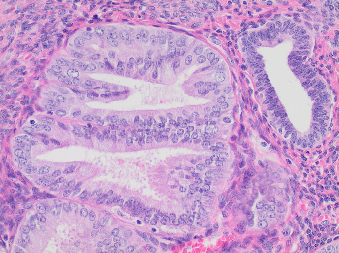
Endometrial intraepithelial neoplasia (atypical hyperplasia): a crowded, complex gland (left and center) lined by cells with enlarged, rounded, vesicular nuclei, visible nucleoli and loss of polarity, which differ clearly from the smaller, darker, elongated nuclei of the background gland at upper right ('altered cytology relative to background'). H&E, high power (magnification not stated).Image: Mikael Häggström, M.D., Wikimedia Commons, CC0.
Endometrial Carcinoma: Histologic Types and Molecular Classification
Definition. Endometrial carcinoma is a malignant epithelial tumor of the uterine corpus. The CAP endometrium protocol (version 5.1.0.0) lists endometrioid, serous, clear cell, dedifferentiated, undifferentiated, mesonephric-like, squamous cell, gastric (gastrointestinal)-type, mixed and small cell and large cell neuroendocrine carcinoma, and carcinosarcoma (CAP 2024). The WHO 5th edition (2020) integrates morphology with key molecular events (McCluggage et al. 2022). It recommends the ProMisE surrogate classifier, which combines POLE mutation testing with p53 and mismatch repair (MMR) immunohistochemistry (CAP 2024). The WHO 5th edition (2020) is the current published classification; the 6th edition is available only as an online beta (IARC 2026).
Glandular, papillary and solid growth with smooth, round luminal outlines; squamous, mucinous, secretory or ciliated differentiation
PTEN, ARID1A or MMR loss in a subset; p53 abnormal in 2–5% of low-grade and about 20% of high-grade tumors
PTEN, CTNNB1, PIK3CA, ARID1A, KRAS mutations
CAP 2024; TCGA 2013
Serous carcinoma
Papillary, glandular or solid growth with slit-like lumens and high-grade nuclei
Abnormal p53; block-like p16; WT1 negative or focal
TP53 mutation; extensive copy-number alterations
CAP 2024; Murali et al. 2019; TCGA 2013
Clear cell carcinoma
Tubulocystic, papillary and solid growth; clear to eosinophilic hobnail cells
Napsin A, AMACR and HNF1B positive; ER and PR negative
All four molecular groups occur
CAP 2024; DeLair et al. 2017
Carcinosarcoma
High-grade carcinoma with homologous or heterologous sarcoma
Epithelial markers absent or limited in the sarcomatous component; TP53 mutation in most
TP53, PTEN, PIK3CA, PPP2R1A, FBXW7, KRAS
CAP 2024; Rabban et al. 2019; CAP 2025; Cherniack et al. 2017
Undifferentiated and dedifferentiated carcinoma
Sheets of monotonous, dyscohesive cells; low-grade endometrioid component in dedifferentiated carcinoma
Absent or focal keratin, PAX8, ER; SWI/SNF protein loss
MMR deficiency common
CAP 2024; Karnezis et al. 2016
Mesonephric-like adenocarcinoma
Ductal, tubular, retiform and papillary growth; eosinophilic luminal secretions
GATA3, TTF-1, luminal CD10; ER and PR absent or focal
KRAS mutation; microsatellite stable
CAP 2024; Euscher et al. 2020; Kolin et al. 2019
Gastric (gastrointestinal)-type carcinoma
Glands of mucin-secreting epithelium with or without goblet cells
CK7, CEA, MUC6; ER positive in 1 of 4
Mutation-type p53 staining in 2 of 4
CAP 2024; Wong et al. 2020
Squamous cell carcinoma
Pure squamous carcinoma; cervical origin excluded
ER and PR usually negative
HPV independent
CAP 2024; Bures et al. 2013
Neuroendocrine carcinoma
Small cell or large cell neuroendocrine morphology
Diffuse expression of at least one neuroendocrine marker
MMR deficiency in a subset
CAP 2024; Pocrnich et al. 2016
Endometrioid carcinoma of the endometrium
Morphology and variants of endometrioid carcinoma
Architecture. Endometrioid carcinoma shows varying proportions of glandular, papillary and solid architecture. Villoglandular, small non-villous papillary, microglandular, sex cord-like, corded and hyalinized, and sertoliform patterns occur (CAP 2024).
Confirmatory endometrioid features. In high-grade tumors, squamous, mucinous, secretory or ciliated differentiation combined with loss of ARID1A, PTEN or MMR proteins favors endometrioid carcinoma over other histotypes (CAP 2024).
Papillary and villoglandular variants. These patterns do not change tumor behavior. Concordance of low-grade architecture with low-grade cytology separates them from serous carcinoma (Rabban et al. 2019).
Corded and hyalinized and spindle cell variants. Cords of low-grade epithelioid or spindle cells are set in hyaline or myxoid stroma and usually blend with conventional glands. The cells express epithelial markers and wild-type p53 and lack severe atypia, high mitotic activity and atypical mitoses. These findings separate the variants from carcinosarcoma. Grade is assigned from the glandular component (Rabban et al. 2019).
Mucinous differentiation. Low-grade endometrioid carcinoma with extensive mucinous differentiation was previously called mucinous carcinoma. It is separated from gastric (gastrointestinal)-type carcinoma (CAP 2024).
Genomics. Most endometrioid tumors have few copy-number alterations or TP53 mutations. They show frequent PTEN, CTNNB1, PIK3CA, ARID1A and KRAS mutations and mutations of ARID5B (TCGA 2013).
FIGO grading of endometrioid carcinoma
Scope. Only endometrioid carcinoma, including its variants, is graded. All non-endometrioid histotypes are considered high grade (CAP 2024).
Nuclear upgrade. Severe cytologic atypia in >50% of tumor cells raises the grade by one. This finding raises suspicion for serous carcinoma or for TP53-mutated or POLE-mutated endometrioid carcinoma (CAP 2024). Upgrading on nuclear features is rarely appropriate. Microacini are not considered glandular for grading (Soslow et al. 2019).
Validation. In 715 women with clinical stage I and occult stage II carcinoma, FIGO nuclear modification moved 44 patients to a higher grade, and their outcome matched the higher grade (Zaino et al. 1995).
Binary grading. ISGyP recommends grouping FIGO grades 1 and 2 as low grade and grade 3 as high grade (Soslow et al. 2019). ISGyP, ICCR and the WHO 5th edition endorse binary grading for its reproducibility. It has not been widely adopted in practice (CAP 2024).
FIGO grade
Non-squamous solid growth
Binary grade (WHO 5th edition, ISGyP, ICCR)
Source
1
≤5%
Low grade
CAP 2024; Soslow et al. 2019
2
6–50%
Low grade
CAP 2024; Soslow et al. 2019
3
>50%
High grade
CAP 2024; Soslow et al. 2019
Raise by one grade
Severe nuclear atypia in >50% of cells
Applied before binary grouping
CAP 2024; Zaino et al. 1995
Serous carcinoma and serous intraepithelial carcinoma
Morphology. Serous carcinoma has papillary, glandular or solid architecture with marked nuclear pleomorphism, prominent nucleoli and brisk mitoses. Slit-like glands with irregular luminal outlines contrast with the round, smooth lumens of endometrioid carcinoma (CAP 2024).
Behavior of small tumors. In 41 serous carcinomas, widespread metastasis, recurrence and death occurred even when myometrial invasion was less than 1 mm or tumor was confined to a polyp. Intraepithelial carcinoma was present in the surface endometrium next to the tumor in 89% of cases. Clear cell foci were present in 32% (Sherman et al. 1992).
Serous intraepithelial carcinoma. Endometrial intraepithelial carcinoma was present in 34 of 38 uteri with serous carcinoma and in 7 of 113 with endometrioid carcinoma. Atypical hyperplasia was absent in uteri with serous carcinoma (Ambros et al. 1995).
Minimal serous carcinoma. Intraepithelial carcinoma and superficial serous carcinoma measuring 1 cm or less were grouped as minimal uterine serous carcinoma. Thirteen of 14 patients with disease confined to the uterus were disease free. All seven patients with extrauterine disease at presentation, even when microscopic, died of disease or recurred (Wheeler et al. 2000).
Routes of spread. Of 87 serous carcinomas, 54 (62%) had extrauterine spread, and 18 of these lacked myometrial invasion. All 16 cases with tumor clusters in the tubal lumen had peritoneal spread (Snyder et al. 2006). Tubal involvement occurred in 32 of 161 serous carcinomas (20%), mimicked serous tubal intraepithelial carcinoma in 17 and was usually WT1 negative or focal (Kommoss et al. 2017).
Immunohistochemistry. Serous carcinoma expresses p16 in 90–100% of cells, compared with 10–90% in grade 3 endometrioid carcinoma. WT1 is negative or focal (up to 30%) in uterine serous carcinoma. ER, p16 and p53 form the most informative three-marker panel against grade 3 endometrioid carcinoma (Murali et al. 2019).
p53 status. Mutant-pattern p53 staining was present in 63 of 63 (100%) serous carcinomas that were MMR proficient and POLE wild type (Singh et al. 2020).
Genomics. Serous tumors and about 25% of high-grade endometrioid tumors have extensive copy-number alterations, few DNA methylation changes, low ER and PR levels and frequent TP53 mutations. Their genomic features resemble ovarian serous and basal-like breast carcinomas (TCGA 2013).
Clear cell carcinoma of the endometrium
Morphology. Clear cell carcinoma admixes tubulocystic, papillary and solid patterns of clear to eosinophilic cuboidal, polygonal, hobnail or flat cells (CAP 2024).
Immunohistochemistry. Napsin A, AMACR (P504S) and HNF1B are expressed, although they may also occur in endometrioid carcinoma. ER and PR are usually absent (CAP 2024). Monoclonal napsin A is recommended because the polyclonal antibody lacks specificity. HNF1B has high sensitivity but lower specificity than in the ovary. A small percentage of prototypical clear cell carcinomas are ER positive (Murali et al. 2019).
Napsin A performance. Napsin A was expressed in 43 of 49 (88%) clear cell carcinomas, 1 of 13 serous carcinomas and 0 of 49 endometrioid carcinomas. Sensitivity was 0.88 and specificity 0.98 (Fadare et al. 2014).
Genomics. In 32 pure clear cell carcinomas, all four molecular subtypes were represented, and TP53 (46%), PIK3CA (36%) and PPP2R1A (36%) mutations were most frequent in POLE wild-type tumors (DeLair et al. 2017).
Carcinosarcoma of the uterus
Histogenesis. Most carcinosarcomas are monoclonal. The carcinomatous element determines behavior, and the sarcomatous component derives from the carcinoma. Uterine carcinosarcomas are therefore regarded as metaplastic carcinomas, and a small proportion are collision tumors (McCluggage 2002). The WHO 5th edition classifies uterine carcinosarcoma as a high-grade carcinoma (Maiorano et al. 2024), and the CAP endometrial carcinoma protocol applies to it (CAP 2024).
Previous rule for carcinosarcoma. Carcinosarcoma (malignant mixed müllerian tumor) was traditionally regarded as a subtype of uterine sarcoma (McCluggage 2002).
Components. The carcinoma is high grade and often serous or endometrioid. The sarcoma is usually high-grade sarcoma not otherwise specified. Heterologous elements include rhabdomyosarcoma, chondrosarcoma and rarely osteosarcoma. Rhabdomyosarcomatous elements predict poor prognosis (CAP 2024).
Minimum sarcoma. ISGyP authors recommend that the sarcomatous component measure at least 1 mm in one dimension. This threshold is arbitrary (Murali et al. 2019).
Genomics. Carcinosarcomas show extensive copy-number alterations and frequent TP53, PTEN, PIK3CA, PPP2R1A, FBXW7 and KRAS mutations. A subset shows a strong epithelial-to-mesenchymal transition signature (Cherniack et al. 2017).
Undifferentiated and dedifferentiated carcinoma of the endometrium
Morphology. Undifferentiated carcinoma consists of sheets of uniform, small to intermediate, non-cohesive cells. Dedifferentiated carcinoma combines undifferentiated carcinoma with a differentiated component, usually FIGO grade 1 or 2 endometrioid carcinoma (CAP 2024).
Immunophenotype. PAX8, ER, E-cadherin and epithelial markers are absent or focal. EMA and CK8/18 may mark rare cells, and a subset shows abnormal p53. Published criteria allow neuroendocrine marker expression in up to 10% of cells (CAP 2024; Murali et al. 2019).
Clinical course. Of 13 undifferentiated carcinomas with known stage, 7 (54%) presented at advanced stage, and 75% of 16 patients died of disease. In the comparison group, 39.4% of patients with grade 3 endometrioid carcinoma died of disease (Altrabulsi et al. 2005). Of 25 low-grade endometrioid carcinomas with undifferentiated carcinoma, 15 patients died of disease within 1–60 months (Silva et al. 2006).
SWI/SNF loss. In 30 dedifferentiated carcinomas, 50% showed SMARCA4 and SMARCA2 loss (37%) or SMARCB1 and SMARCA2 loss (13%) in the undifferentiated component. SMARCA4 and SMARCB1 loss were mutually exclusive. All 31 grade 3 endometrioid carcinomas retained these proteins. Most SMARCA4-deficient tumors arose in an MMR-deficient background (Karnezis et al. 2016). Concurrent ARID1A and ARID1B inactivation occurred in 12 of 24 BRG1- and INI1-intact dedifferentiated carcinomas, with ARID1B loss confined to the undifferentiated component (Coatham et al. 2016).
Distinction from neuroendocrine carcinoma. Among 44 SWI/SNF-deficient undifferentiated or dedifferentiated carcinomas, 75% expressed a neuroendocrine marker, usually focally. Neuroendocrine carcinomas expressed two or more markers in 93%, with diffuse expression of at least one. MMR loss occurred in 38 of 44 undifferentiated carcinomas and in no neuroendocrine carcinoma. Neuroendocrine carcinomas retained SWI/SNF proteins except for one isolated ARID1A loss (Tessier-Cloutier et al. 2022).
Mesonephric-like, gastric-type, squamous, neuroendocrine and mixed endometrial carcinomas
Mesonephric-like adenocarcinoma. In 23 cases, ductal or tubular architecture (21 of 23), nuclei resembling papillary thyroid carcinoma (18 of 23) and eosinophilic luminal secretions (20 of 23) were present. CD10 (10 of 10), GATA3 (15 of 16) and TTF-1 (11 of 16) were positive, and ER was expressed in ≥10% of nuclei in only 6 of 21. KRAS mutation was found in 14 of 17. Stage III or IV disease was present in 48%, and median progression-free survival was 18.2 months (Euscher et al. 2020). Over one-third of mesonephric-like adenocarcinomas show some napsin A positivity, so a broad panel including GATA3 and TTF-1 is used against clear cell carcinoma (Mills et al. 2026).
Previous rule for mesonephric-like adenocarcinoma. Mesonephric-like adenocarcinoma was first included in the 2020 WHO classification of endometrial and ovarian carcinomas (Mills et al. 2026).
Gastric (gastrointestinal)-type carcinoma. The tumor consists of glands lined by mucin-secreting epithelium, with or without goblet cells. Extension from a cervical primary is excluded (CAP 2024). Four adenocarcinomas expressed CK7 and CEA (4 of 4) and MUC6 (3 of 3), and two of four showed mutation-type p53. Two of the four patients died of disease (Wong et al. 2020). This type was formally included in the WHO 5th edition (Nam et al. 2026).
Previous rule for gastric-type carcinoma. Before the 5th edition, endometrial adenocarcinoma with gastric (gastrointestinal) differentiation was not recognized as a distinct entity (Wong et al. 2020).
Squamous cell carcinoma. Primary squamous cell carcinoma of the endometrium is HPV independent and may follow long-standing obstruction with squamous metaplasia (ichthyosis uteri). Extension from the cervix is excluded (CAP 2024). Five tumors were negative for HPV DNA, and most were negative for ER, PR and p16 (Bures et al. 2013).
Neuroendocrine carcinoma. In 25 cases (median age 57 years), 15 were mixed with other histotypes, usually endometrioid. Large cell type was most common. All tumors expressed at least one neuroendocrine marker, PAX8 was positive in 6 of 18 and MMR was abnormal in 8 of 18. The diagnosis was missed in 89% of referral cases, and 28% of patients survived at least 5 years (Pocrnich et al. 2016).
Mixed carcinoma. Mixed carcinoma consists of two distinct histotypes, at least one of which is usually serous or clear cell carcinoma. It is graded as high grade irrespective of the percentages (CAP 2024). Any amount of serous or clear cell carcinoma confidently recognized on H&E qualifies. The term excludes morphologic variants of endometrioid carcinoma, dedifferentiated carcinoma and carcinosarcoma (Rabban et al. 2019).
Somatic transdifferentiation. Carcinomas with yolk sac-like, choriocarcinoma-like, trophoblastic-like or neuroectodermal-like features are regarded as somatic transdifferentiation of carcinoma and behave aggressively (CAP 2024).
Molecular classification of endometrial carcinoma
TCGA molecular groups and the ProMisE surrogate classifier
TCGA. Integrated genomic analysis of 373 endometrial carcinomas defined four groups: POLE ultramutated, microsatellite instability hypermutated, copy-number low and copy-number high (TCGA 2013).
Frequencies and outcome.POLE-ultramutated tumors form about 7%, MSI-high tumors about 28%, copy-number low tumors about 39% and copy-number high tumors about 26% of cases. Copy-number high tumors carry TP53 mutations in 95%. POLE-mutated tumors have an excellent prognosis and copy-number high tumors a poor prognosis (CAP 2024).
Surrogate assays. A clinically applicable classifier replicated the TCGA groups using MMR immunohistochemistry, POLE mutational analysis and p53 immunohistochemistry as a surrogate for copy-number status (Talhouk et al. 2015).
Confirmation. In 319 new cases, the ProMisE decision tree classified all tumors into four prognostic groups. POLE-mutated tumors had the best and p53-abnormal tumors the worst outcome (Talhouk et al. 2017).
Validation. In 452 population-based cases, molecular group agreed between diagnostic and surgical specimens (κ=0.88) (Kommoss et al. 2018).
Biopsy specimens. Molecular group on biopsy agreed with the hysterectomy (κ=0.86). Grade (κ=0.55) and histotype (κ=0.44) agreed less well (Talhouk et al. 2016).
ProMisE group
Defining test result
Frequency
Prognosis and therapy
Source
POLE-mutated
Pathogenic POLE exonuclease domain mutation
9.3%; 12% of high-risk tumors
5-year recurrence-free survival 98% in PORTEC-3
Kommoss et al. 2018; León-Castillo et al. 2020c
MMR-deficient
Loss of one or more MMR proteins
28.1%; 33% of high-risk tumors
Intermediate; 5-year recurrence-free survival 68% with chemoradiation and 76% with radiation alone (not significant)
Kommoss et al. 2018; León-Castillo et al. 2020c
p53-abnormal
Abnormal p53 immunohistochemistry
12.2%; 23% of high-risk tumors
Worst; 5-year recurrence-free survival 59% with chemoradiation and 36% with radiation alone
Kommoss et al. 2018; León-Castillo et al. 2020c
No specific molecular profile
None of the above
50.4%; 32% of high-risk tumors
Intermediate; ER positivity favorable in high-risk tumors
Kommoss et al. 2018; Vermij et al. 2023
Interpretation of POLE mutations
Pathogenic variants. Eleven exonuclease domain mutations are classified as pathogenic: P286R, V411L, S297F, S459F, A456P, F367S, L424I, M295R, P436R, M444K and D368Y. P286R and V411L are the most frequent (León-Castillo et al. 2020a).
Genomic signature. Tumors with known pathogenic mutations show C>A substitutions >20%, T>G substitutions >4%, C>G substitutions <0.6%, indels <5% and tumor mutational burden >100 mutations/Mb. Most mutations outside the exonuclease domain are not pathogenic (León-Castillo et al. 2020a).
Coexisting MMR deficiency. Tumors with a pathogenic exonuclease domain mutation and MMR deficiency had a 5-year recurrence-free survival of 92.3%, like POLE-ultramutated tumors. Tumors with non-pathogenic mutations and MMR deficiency behaved like MMR-deficient tumors (León-Castillo et al. 2020a).
Prognosis.POLE mutations occurred in 6.1% of PORTEC-1 and PORTEC-2 tumors and were associated with high grade. None of 15 POLE-mutant grade 3 tumors recurred, compared with 30.9% of POLE wild-type grade 3 tumors (Church et al. 2015).
p53 immunohistochemistry patterns in endometrial carcinoma
Subclonal abnormal staining. Subclonal abnormal p53 occurs in up to 21% of endometrial carcinomas. It usually indicates a secondary mutation in an MMR-deficient or POLE-mutated tumor and may indicate a mixed carcinoma (CAP 2024).
Accuracy on biopsies. p53 immunohistochemistry agreed with TP53 sequencing in 92.3% of 168 biopsies overall and in 95.1% after excluding MMR-deficient and POLE-mutated tumors. Subclonal mutant staining occurred in 5.1% (Singh et al. 2020).
High-risk tumors. In 408 PORTEC-3 tumors, p53 was abnormal in 32%. The patterns were overexpression (68%), null (9%), cytoplasmic (2%) and subclonal (21%). Subclonal staining was usually associated with POLE mutation or MMR deficiency (22 of 27). Excluding these tumors raised sensitivity to 95.0% and specificity to 94.1% (Vermij et al. 2022).
p53 pattern
Criteria
Interpretation
Source
Wild-type
Nuclear staining of varying intensity admixed with negative nuclei
Normal
CAP 2024
Overexpression
Diffuse, strong nuclear staining in ≥80% of tumor cells
Abnormal
CAP 2024; Köbel et al. 2019
Null
No nuclear or cytoplasmic staining with positive internal control
Abnormal
CAP 2024
Cytoplasmic
Unequivocal cytoplasmic staining with variable nuclear staining
Abnormal
CAP 2024; Köbel et al. 2019
Subclonal abnormal
Any abnormal pattern in a subset of tumor cells
Secondary event in MMR-deficient or POLE-mutated tumors, or a mixed carcinoma
CAP 2024; Vermij et al. 2022
Multiple classifiers and no specific molecular profile
Multiple classifiers. Of 3518 molecularly profiled carcinomas, 107 (3%) had abnormal p53 with another classifier: 64 MMR-deficient, 31 POLE-mutated and 12 with all three. Subclonal p53 overexpression was seen in 60% of MMR-deficient and 46.7% of POLE-mutated cases. Stage I recurrence-free survival was 92.2% and 94.1%, compared with 70.8% for p53-abnormal carcinoma. These tumors are classified as MMR-deficient and POLE-mutated, respectively (León-Castillo et al. 2020b).
Risk integration in early-stage disease. In 947 PORTEC-1 and PORTEC-2 endometrioid carcinomas, the groups were p53-mutant (9%), MSI (26%), POLE-mutant (6%) and NSMP (59%). Substantial lymphovascular space invasion, p53 mutation and L1CAM expression >10% were unfavorable features (Stelloo et al. 2016).
NSMP and ER. In 648 high-risk carcinomas, ER positivity was independently associated with lower recurrence risk only in NSMP tumors (hazard ratio 0.33) (Vermij et al. 2023).
Histotype
Distribution across molecular groups
Source
Low-grade endometrioid
Mostly MSI-hypermutated or copy-number low; abnormal p53 in 2–5%
CAP 2024
High-grade endometrioid
Represented in all four groups; abnormal p53 in about 20%; POLE-mutated tumors may resemble serous carcinoma
CAP 2024
Serous
Most copy-number high tumors are serous; mutant-pattern p53 in all MMR-proficient, POLE wild-type serous carcinomas
CAP 2024; Singh et al. 2020
Clear cell
POLE mutation 7%, MMR abnormal 19%, p53 abnormal 34%; all four groups represented
DeLair et al. 2017
Undifferentiated and dedifferentiated
MMR loss in 38 of 44 SWI/SNF-deficient tumors; abnormal p53 in a subset
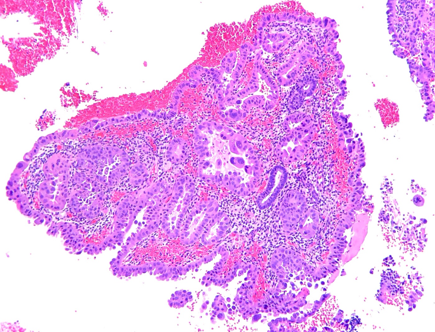
Clear cell carcinoma of the endometrium in an endometrial biopsy: papillae and tubules with hyalinized fibrovascular cores, lined by cells with clear to eosinophilic cytoplasm and enlarged, pleomorphic nuclei, including hobnail cells projecting into the lumina (center); a small benign endometrial gland with dark, uniform nuclei lies in the stroma at center right for comparison. H&E, low to medium power (magnification not stated).Image: Ed Uthman, Wikimedia Commons, CC BY 2.0.
Endometrial Carcinoma Staging, CAP Reporting and Biomarker Testing
Definition. Pathologic staging of endometrial carcinoma records the anatomic extent of tumor in the hysterectomy, adnexa, lymph nodes and other submitted tissues. The CAP endometrium protocol, version 5.1.0.0 (posted December 2024), applies to hysterectomy specimens with carcinoma, including carcinosarcoma. It excludes uterine sarcomas, adenosarcoma, metastatic carcinoma and lymphoma, and it is not required for biopsy or curettage specimens. Its standards are AJCC 8th edition, FIGO 2009 (2018 FIGO Cancer Report) and FIGO 2023. The AJCC pTNM category is a required element, and both FIGO stage fields are optional (CAP 2024). Biomarker results are reported with the CAP Gynecologic Biomarker Reporting Template, version 1.3.0.0 (posted September 2025) (CAP 2025).
Grossing of hysterectomy specimens for endometrial carcinoma
Specimen part
Sampling recommendation
Source
Endometrial tumor
One block per 1 cm of tumor or at least 4 blocks, including the deepest invasion
CAP 2024; Malpica et al. 2019
No gross lesion after a biopsy diagnosis of atypical hyperplasia or carcinoma
Entire endometrium and underlying myometrium
CAP 2024
Lower uterine segment
Longitudinal sections including the endocervical junction; at least 2
Malpica et al. 2019
Ovaries, non-endometrioid carcinoma
Slices at 2–3 mm, submitted entirely
CAP 2024
Fallopian tubes, non-endometrioid carcinoma
Entire tube by SEE-FIM
CAP 2024
Ovaries and tubes, endometrioid carcinoma
At least 2 sections of each ovary; entire fimbriae and representative tube sections
CAP 2024
Omentum. The omentum is sectioned at 5-mm intervals (CAP 2024).
Minimally invasive surgery artifacts. Laparoscopic and robot-assisted hysterectomies may show tumor fragments in vessels and in tubal lumens and myometrial clefts (CAP 2024).
Intraoperative assessment of hysterectomy specimens for endometrial carcinoma
Sections. One random full-thickness section of hysterectomies without a gross lesion showed microscopic tumor in only 3 of 20 cancers (15%) (Malpica et al. 2019).
Sentinel nodes. Routine frozen section of sentinel nodes is not advisable because small tumor foci may be lost in cutting and sensitivity in grossly normal nodes is low (Malpica et al. 2019).
Myometrial invasion by endometrial carcinoma: measurement and patterns
Measurement. Depth is measured from the endomyometrial junction to the deepest point of invasion and related to the myometrial thickness. Exophytic tumor and polyps are excluded from the thickness. Depth is not assessed at the cornu unless tumor involves the entire cornu or serosa. Invasion into a leiomyoma is included, and lymphovascular invasion is excluded (CAP 2024).
Carcinoma in adenomyosis. Carcinoma confined to adenomyosis has a prognosis similar to carcinoma confined to the endometrium and is not counted as myometrial invasion (Singh et al. 2019). When invasion arises from adenomyosis and the deepest point is in the outer half, ICCR and ISGyP recommend staging as pT1b with a comment (CAP 2024).
MELF definition. MELF consists of outpouchings of neoplastic glands that detach, become lined by flattened epithelium and appear microcystic, elongated or fragmented into small clusters and single cells, within a fibromyxoid stromal reaction (Murray et al. 2003).
MELF and nodes. In 464 FIGO grade 1 endometrioid carcinomas, MELF was present in 12.9%. Lymph node metastases occurred in 18.3% of MELF cases and 1.2% of non-MELF cases. All five cases with only micrometastases or isolated tumor cells had MELF invasion (Joehlin-Price et al. 2017).
MELF and LVSI. MELF is associated with lymphovascular invasion and nodal metastasis and is not an independent predictor of overall survival. Nodal deposits may resemble histiocytes, and keratin staining identifies them (CAP 2024).
Invasion pattern
Features
Significance
Source
Adenoma malignum-like
Round bland glands without stromal response
Mimics mesonephric remnants in the cervix
CAP 2024
Adenomyosis-like
Irregular islands without endometrial stroma
Mimics carcinoma in adenomyosis
CAP 2024
MELF
Microcystic, elongated and fragmented glands in fibromyxoid, inflamed stroma
Nodal metastases, often isolated tumor cells
Joehlin-Price et al. 2017; Pelletier et al. 2017
Single-cell infiltration
Individual tumor cells
Extrauterine extension in one study
CAP 2024
Cervical, serosal, lower uterine segment and adnexal involvement by endometrial carcinoma
Cervical stromal invasion. Cervical stromal invasion is pT2 and FIGO stage II. It is recognized by desmoplastic stroma or altered architecture relative to normal endocervical glands. The upper limit of the endocervix is the most proximal endocervical gland. Invasion is present when tumor is located at the level of or distal to non-neoplastic endocervical glands (CAP 2024).
Endocervical glandular involvement. Glandular involvement without stromal invasion is not stage II. It is reported because some oncologists give brachytherapy for it (CAP 2024).
Serosa. The serosa is involved when tumor traverses the myometrium and reaches the submesothelial connective tissue or mesothelium, with or without tumor on the surface (CAP 2024). Tumor within serosal lymphovascular spaces is not serosal involvement (Singh et al. 2019). Serosal involvement carries a higher risk of locoregional recurrence than adnexal involvement (CAP 2024).
Lower uterine segment. Of 1009 women, 35 (3.5%) had carcinoma of the lower uterine segment. Lynch syndrome was confirmed or strongly suspected in 29% of them, compared with 1.8% of endometrial cancer patients overall (Westin et al. 2008).
Adnexa. Tumor invading tubal mucosa or wall is pT3a and stage IIIA in FIGO 2009 and FIGO 2023. Intraluminal tubal fragments alone are disregarded. Lymphovascular invasion in the ovary, tube, cervix or parametrium without stromal invasion does not change stage (CAP 2024).
Lymphovascular space invasion in endometrial carcinoma
Definition. Lymphovascular invasion is a tumor embolus within an endothelium-lined space, often conforming to the lumen and sometimes attached to the endothelium. It is usually seen at the invasive front. Mimics include retraction, artifactually displaced tumor and MELF invasion. The CAP term lymphatic and/or vascular invasion is equivalent to the FIGO term lymphovascular space invasion (CAP 2024).
Prognosis. In 926 PORTEC-1 and PORTEC-2 endometrioid carcinomas, any LVSI was present in 13.9% and substantial LVSI in 4.8%. Substantial LVSI was the strongest independent factor for pelvic regional recurrence (hazard ratio 6.2), distant metastasis (3.6) and overall survival (2.0) (Bosse et al. 2015).
Numeric threshold. In PORTEC patients without external beam radiotherapy, 5-year pelvic nodal recurrence was 3.3%, 6.7% and 26.3% with 0, 1–3 and ≥4 involved vessels (Peters et al. 2022).
CAP reporting. CAP counts LVI foci on the single slide with the most foci. The template responses are "less than or equal to 4 foci", with the number specified, and "greater than or equal to 5 foci". For AJCC and FIGO 2009, CAP recommends ≥4 involved vessels on that slide as substantial, so a count of 4 is entered as ≤4 with the number 4 and is substantial. For FIGO 2023, ≥5 vessels can be used (CAP 2024).
System or study
Threshold for substantial LVSI
How counted
Source
FIGO 2023
≥5 vessels
Not always specified
CAP 2024; Berek et al. 2023
WHO 5th edition
≥5 vessels
Not always specified
CAP 2024
ESGO/ESTRO/ESP 2021
≥5 vessels
Not always specified
CAP 2024
ICCR 2022
≥3 vessels
Not always specified
CAP 2024
ISGyP 2019
≥3 vessels
Distinct, widely separated vessels
CAP 2024; Singh et al. 2019
PORTEC and Danish cohorts
≥4 vessels
At least one H&E slide
Peters et al. 2022
CAP 5.1.0.0
≥4 for AJCC and FIGO 2009; ≥5 usable for FIGO 2023
Single slide with the most foci
CAP 2024
Lymph node staging and sentinel node ultrastaging
Regional nodes. Pelvic (parametrial, obturator, internal iliac, external iliac, common iliac, sacral, presacral) and para-aortic nodes are regional. Other involved nodes are distant metastases (pM1) (CAP 2024).
Size categories. Macrometastases measure >2 mm. Micrometastases measure >0.2 mm to 2 mm or contain >200 cells. Isolated tumor cells measure ≤0.2 mm or form clusters of ≤200 cells and are classified as pN0(i+). Isolated tumor cells are reported only when no micrometastasis or macrometastasis is present. Caution applies to diagnosing isolated tumor cells on a keratin stain without morphologic correlation (CAP 2024).
Reporting. The report states whether ultrastaging was performed and whether metastases were found on routine sections or by ultrastaging (CAP 2024).
Sentinel node accuracy. In the FIRES study, sentinel nodes identified 35 of 36 node-positive patients with a mapped node (sensitivity 97.2%, negative predictive value 99.6%) (Rossi et al. 2017).
Ultrastaging yield. The MSKCC protocol cuts two adjacent 5-μm sections at each of two levels 50 μm apart, one stained with H&E and one with AE1:AE3 keratin. In 508 patients, routine H&E detected nodal metastases in 6.9%, and ultrastaging found another 4.5% (4 micrometastases and 19 isolated tumor cells) (Kim et al. 2013).
MD Anderson protocol. Five H&E levels are cut at 250 μm with two unstained slides per level for keratin immunohistochemistry. No universal ultrastaging protocol exists (Malpica et al. 2019; CAP 2024).
FIGO 2023 and FIGO 2009 staging of endometrial carcinoma
Principles of FIGO 2023. FIGO 2023 adds histotype (aggressive or non-aggressive), grade, LVSI extent and molecular class to anatomic extent. Complete molecular classification is encouraged, and POLE-mutated or p53-abnormal status in stages I and II leads to downstaging or upstaging (Berek et al. 2023).
CAP position. Because the non-anatomic parameters of FIGO 2023 are debated and some are poorly reproducible, CAP restored FIGO 2009 staging and made both FIGO systems optional. Limited availability of POLE testing hinders universal adoption of FIGO 2023 (CAP 2024).
FIGO 2023 stage
FIGO 2023 definition
FIGO 2009 stage and AJCC 8th pT
FIGO 2009 and AJCC definition
Source
IA1
Non-aggressive type limited to a polyp or the endometrium
IA, pT1a
Tumor limited to the endometrium or invading less than half of the myometrium
CAP 2024; Berek et al. 2023
IA2
Non-aggressive type invading <50% of myometrium with no or focal LVSI
IA, pT1a
Tumor limited to the endometrium or invading less than half of the myometrium
CAP 2024; Berek et al. 2023
IA3
Low-grade endometrioid carcinoma limited to the uterus with simultaneous low-grade endometrioid ovarian involvement
IIIA, pT3a
Adnexal involvement
CAP 2024; Berek et al. 2023
IB
Non-aggressive type invading ≥50% of myometrium with no or focal LVSI
IB, pT1b
Invasion of half or more of the myometrium
CAP 2024
IC
Aggressive type limited to a polyp or the endometrium
IA, pT1a
Tumor limited to the endometrium
CAP 2024; Berek et al. 2023
IIA
Cervical stromal invasion by a non-aggressive type
II, pT2
Cervical stromal invasion without extension beyond the uterus
CAP 2024
IIB
Substantial LVSI in a non-aggressive type
IA or IB, pT1a or pT1b
No LVSI criterion; staged by depth of invasion
CAP 2024
IIC
Aggressive type with any myometrial invasion
IA, IB or II; pT1a, pT1b or pT2
Staged by depth of invasion, or II and pT2 if the cervical stroma is invaded
CAP 2024; Berek et al. 2023
IIIA1
Spread to ovary or fallopian tube, except when IA3 criteria are met
IIIA, pT3a
Serosa and/or adnexa
CAP 2024
IIIA2
Involvement of uterine subserosa or spread through the serosa
IIIA, pT3a
Serosa and/or adnexa
CAP 2024
IIIB1
Metastasis or direct spread to the vagina or parametria
IIIB, pT3b
Vaginal or parametrial involvement
CAP 2024
IIIB2
Metastasis to the pelvic peritoneum
IIIA, pT3a
Pelvic serosal structures (cul-de-sac, bladder or sigmoid serosa)
CAP 2024
IIIC1i and IIIC1ii
Pelvic node micrometastasis or macrometastasis
IIIC1, pN1mi or pN1a
Pelvic node metastasis
CAP 2024
IIIC2i and IIIC2ii
Para-aortic node (up to the renal vessels) micrometastasis or macrometastasis
IIIC2, pN2mi or pN2a
Para-aortic node metastasis with or without pelvic nodes
CAP 2024
IVA
Invasion of bladder or bowel mucosa
IVA, pT4
Bladder or bowel mucosa; bullous edema is insufficient
CAP 2024
IVB
Abdominal peritoneal metastasis beyond the pelvis
IVB, pM1
Omentum and abdominal peritoneum are distant metastasis
CAP 2024
IVC
Distant metastasis, including nodes above the renal vessels, lung, liver, brain or bone
IVB, pM1 for distant organs and non-regional nodes such as inguinal nodes; para-aortic nodes above the renal vessels remain IIIC2, pN2
Pelvic and para-aortic nodes are regional; other involved nodes are pM1
CAP 2024
IAmPOLEmut
POLE-mutated carcinoma confined to the corpus or with cervical extension, regardless of LVSI or histotype
No molecular category
Not applicable
CAP 2024; Berek et al. 2023
IICmp53abn
p53-abnormal carcinoma confined to the uterine corpus with any myometrial invasion, with or without cervical invasion, regardless of LVSI or histotype
No molecular category
Not applicable
CAP 2024; Berek et al. 2023
Synchronous endometrial and ovarian carcinoma
Clonality. In 18 synchronous endometrial and ovarian carcinomas, 17 were clonally related, including 10 of 11 cases that met clinicopathologic criteria for independent primaries (Anglesio et al. 2016).
Management and staging rule. The WHO and ESGO/ESTRO/ESP recommend conservative management without adjuvant therapy when the tumor is low-grade endometrioid, with no more than superficial myometrial invasion, no LVSI and no other metastasis. FIGO 2023 stage IA3 adds the requirement of a unilateral ovarian tumor without surface involvement, that is, an ovarian tumor confined to one ovary (ovarian pT1a). Under FIGO 2009 and AJCC 8th edition, the same case is stage IIIA and endometrial pT3a (CAP 2024).
CAP required and optional staging elements (version 5.1.0.0)
Element
Status
Note
Source
pTNM (AJCC 8th edition)
Required
y and r prefixes; (sn) and (f) suffixes
CAP 2024
FIGO 2009 and FIGO 2023 stage
Optional
FIGO 2009 follows the 2018 FIGO Cancer Report
CAP 2024; Amant et al. 2018
Lymphatic and/or vascular invasion
Required
≤4 foci with the number, or ≥5 foci, on the single slide with the most foci; ≥4 is substantial for AJCC and FIGO 2009
CAP 2024
Margins
Required only if the cervix or parametrium is involved
Ectocervical, vaginal cuff, parametrial and paracervical
CAP 2024
Molecular type, ProMisE and TCGA class
Optional
MMR, MSI, p53, TP53 and POLE results
CAP 2024
The full case summary is available on the CAP cancer protocol templates page (CAP 2024).
ESGO-ESTRO-ESP 2025 risk groups for endometrial carcinoma
The 2025 ESGO-ESTRO-ESP update incorporates FIGO 2023 staging, LVSI and molecular classification (Concin et al. 2025). Its risk groups divide NSMP into low-grade, ER-positive tumors and high-grade or ER-negative tumors, and it recommends sentinel node dissection for all patients with early-stage endometrial carcinoma (Abacjew-Chmylko et al. 2026).
FIGO 2023 stage
POLE-mutated
MMR-deficient
NSMP, low grade and ER positive
NSMP, high grade or ER negative
p53-abnormal
Source
IA1 (low grade, endometrium)
Low
Low
Low
Uncertain
Uncertain
Abacjew-Chmylko et al. 2026
IA2 (low grade, <50% invasion)
Low
Low
Low
High
High
Abacjew-Chmylko et al. 2026
IA3 (low grade, ovary)
Low
Low
Low
High
High
Abacjew-Chmylko et al. 2026
IB (low grade, ≥50% invasion)
Low
Intermediate
Intermediate
High
High
Abacjew-Chmylko et al. 2026
IC (high grade, endometrium)
Low
Low
Not applicable
Uncertain
Uncertain
Abacjew-Chmylko et al. 2026
IIA (low grade, cervical stroma)
Low
High-intermediate
Intermediate
High
High
Abacjew-Chmylko et al. 2026
IIB (low grade, substantial LVSI)
Low
High-intermediate
High-intermediate
High
High
Abacjew-Chmylko et al. 2026
IIC (high grade, any invasion)
Low
Intermediate or high-intermediate*
Not applicable
High
High
Abacjew-Chmylko et al. 2026
IIIA–IVA
Uncertain
High
High
High
High
Abacjew-Chmylko et al. 2026
*IIC, MMR-deficient: intermediate if there is myometrial invasion only with no or focal LVSI; high-intermediate if there is cervical stromal invasion or substantial LVSI. "Uncertain" indicates insufficient data. "Not applicable" indicates a FIGO stage that applies to low-grade tumors only (Abacjew-Chmylko et al. 2026).
Biomarker testing in endometrial carcinoma (CAP Gynecologic Biomarker Template 1.3.0.0)
Changes in version 1.3.0.0. All immunohistochemical tests are optional. HER2 in situ hybridization status is required when HER2 is equivocal (2+). MLH1 promoter methylation status is required when MLH1 and PMS2 are lost (CAP 2025).
Mismatch repair immunohistochemistry and Lynch syndrome triage
Frequency. About 30% of endometrial carcinomas are MMR deficient, and universal MMR testing of all endometrial carcinomas, including carcinosarcoma, can be used for Lynch syndrome screening (CAP 2025). About 3–5% of endometrial carcinomas are attributable to Lynch syndrome (CAP 2024).
Universal screening. In 605 consecutive endometrial cancers, 40 were MMR deficient without MLH1 methylation, and only 25% of these patients were younger than 50 years (Mills et al. 2014).
Interpretation. Intact expression is nuclear staining equal to or stronger than internal control cells. Loss is reported only when internal controls stain. Cytoplasmic, nucleolar and punctate nuclear staining is not intact expression. Missense mutations may produce intact staining of a non-functional protein (CAP 2025).
Subclonal loss. Subclonal loss is a discrete area of complete loss of nuclear expression next to tumor with retained expression. It is distinguished from patchy intact staining (CAP 2025). It occurred in 9 of 125 (7.2%) carcinomas, only in endometrioid components, and all subclonal MLH1 losses were associated with epigenetic silencing (Watkins et al. 2016). Subclonal loss may reflect MLH1 methylation, POLE mutation or, rarely, Lynch syndrome and is reported with its likely explanation (Mendoza et al. 2023; CAP 2025).
MSI and immunohistochemistry. MSI and MMR immunohistochemistry agreed in 655 of 696 tumors (κ=0.854). Most discordant cases were explained by MLH1 promoter hypermethylation (Stelloo et al. 2017).
BRAF.BRAF mutations are extremely rare in endometrial cancer (0.1%), and BRAF testing has no role in gynecologic tumors (CAP 2025).
MMR immunohistochemistry result
Interpretation and next step
Source
All four proteins intact
Low probability of MSI-high phenotype
CAP 2025
Loss of MLH1 and PMS2
MLH1 promoter methylation testing; methylation suggests a sporadic tumor, and its absence indicates germline MLH1 testing
CAP 2025
Loss of MSH2 and MSH6, isolated loss of MSH6 or isolated loss of PMS2
High probability of Lynch syndrome; genetic counseling
CAP 2025
Subclonal loss
Report with likely explanation (methylation, POLE mutation); rare in Lynch syndrome
CAP 2025; Watkins et al. 2016
MMR status and immune checkpoint inhibitor therapy
Assay choice. The CAP, AMP and Fight Colorectal Cancer guideline prefers MMR immunohistochemistry over MSI testing to determine eligibility of endometrial cancer patients for immune checkpoint inhibitors (strong recommendation) (CAP 2025; Bartley et al. 2022).
Trial evidence. In RUBY, 23.9% of 494 patients had dMMR, MSI-high tumors, and dostarlimab added to chemotherapy improved their progression-free survival (hazard ratio 0.28) (Mirza et al. 2023).
PD-L1. PD-L1 testing is not required in endometrial cancer, because immunotherapy eligibility depends on MMR or MSI status (CAP 2025).
HER2 testing in serous carcinoma and carcinosarcoma
Frequency and indications. HER2 overexpression or amplification has been reported in 30% of serous carcinomas, 16% of carcinosarcomas and 48% of clear cell carcinomas. NCCN guidelines recommend HER2 testing in serous carcinoma and carcinosarcoma and consideration of testing for all p53-abnormal carcinomas (CAP 2025).
Trastuzumab trial. Adding trastuzumab to carboplatin-paclitaxel prolonged progression-free survival in HER2-positive advanced or recurrent serous carcinoma (hazard ratio 0.44) (Fader et al. 2018).
Staining pattern. HER2 staining in endometrial carcinoma is often lateral or basolateral and spares the apical membrane. Heterogeneity is frequent. Biopsy and hysterectomy agreed on final HER2 status in 84%, and testing of multiple specimens is encouraged (CAP 2025).
In situ hybridization. For trastuzumab eligibility, a HER2/CEP17 ratio ≥2.0, or a ratio <2.0 with an average HER2 copy number ≥6 per nucleus, is positive. At least 20 contiguous tumor cells are counted in the area with the highest protein expression (CAP 2025).
Trastuzumab deruxtecan. In April 2024, the FDA granted tumor-agnostic accelerated approval of trastuzumab deruxtecan for previously treated unresectable or metastatic solid tumors with HER2 3+ immunohistochemistry. Eligibility scoring uses gastric cancer criteria, and in situ hybridization is not indicated for this purpose (CAP 2025).
No staining or membrane staining in <10% of tumor cells
CAP 2025
1+
Faint incomplete staining in any proportion, or weak complete staining in <10%
Faint incomplete staining in ≥10%
CAP 2025
2+ (equivocal)
Intense complete or basolateral/lateral staining in ≤30%, or weak to moderate staining in ≥10%; reflex in situ hybridization
Weak to moderate complete, basolateral or lateral staining in ≥10%
CAP 2025
3+
Intense complete or basolateral/lateral staining in >30%
Strong complete, basolateral or lateral staining in ≥10%
CAP 2025
ER, PR and folate receptor alpha testing in endometrial carcinoma
ER and PR. A tumor is positive when ≥1% of tumor cells show nuclear staining, and the percentage and intensity are reported. Non-neoplastic endometrium, stroma or myometrium serves as internal control (CAP 2025).
Folate receptor alpha. Positivity is defined as ≥75% of viable tumor cells with moderate to strong membrane staining with the FDA-approved VENTANA FOLR1 RxDx assay (CAP 2025).
Differential diagnosis of staging findings in endometrial carcinoma
Finding
Main mimic
Distinguishing feature
Source
Carcinoma confined to adenomyosis
Myometrial invasion
Endometrial stroma around glands; no invasive front
Singh et al. 2019
MELF invasion
Lymphovascular invasion
Microcystic and fragmented glands in fibromyxoid, inflamed stroma
CAP 2024; Murray et al. 2003
Artifactual tumor displacement
Lymphovascular invasion
Fragments in vessels and clefts away from the invasive front after laparoscopic surgery
CAP 2024; Logani et al. 2008
Endocervical glandular involvement
Cervical stromal invasion
No desmoplasia or altered architecture beyond native glands
CAP 2024
Histiocyte-like nodal deposits after MELF invasion
Sinus histiocytes
Keratin staining
CAP 2024
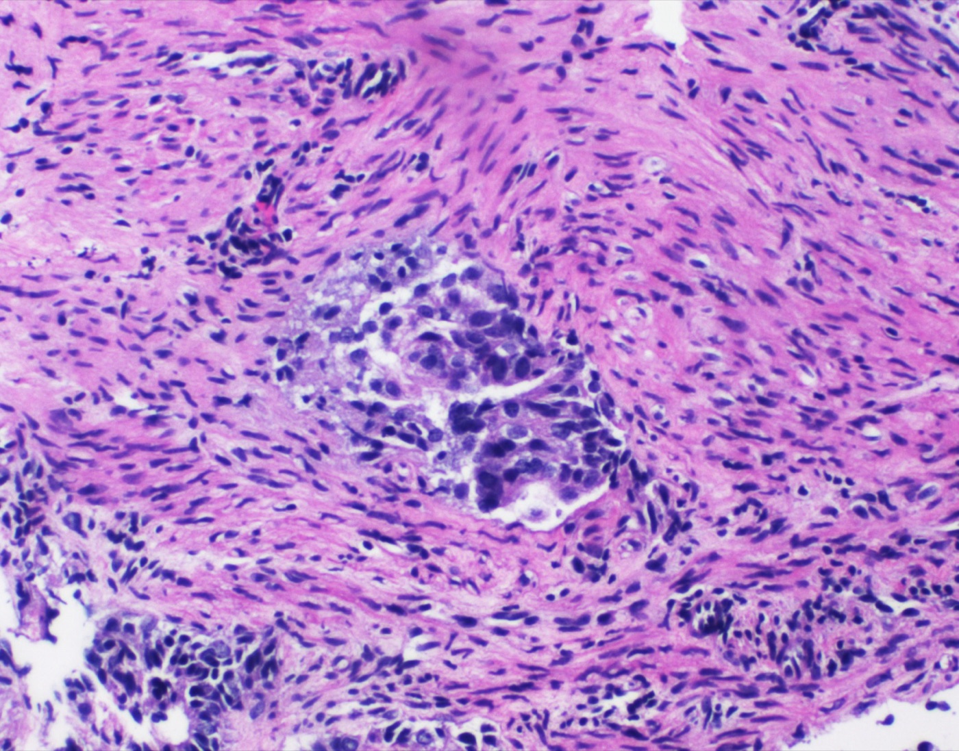
Myometrial invasion by endometrioid carcinoma: an irregular gland of tumor cells with enlarged, crowded nuclei (center) lies deep among smooth muscle bundles of the myometrium, with further tumor at lower left; the depth of invasion relative to myometrial thickness (less than half or half or more) sets FIGO stage IA versus IB. H&E, medium power (magnification not stated).Image: Mikael Häggström, M.D., Wikimedia Commons, CC0.
Uterine Mesenchymal Tumors: Smooth Muscle Tumors, Endometrial Stromal Tumors, Adenosarcoma and Other Sarcomas
Definition. Uterine mesenchymal tumors arise from the myometrium, the endometrial stroma or other mesenchymal elements of the corpus and cervix. Most are smooth muscle or endometrial stromal neoplasms. Molecular studies have added sarcomas defined by BCOR alterations, NTRK rearrangements, COL1A1::PDGFB fusion and SMARCA4 loss (Momeni-Boroujeni and Chiang 2020; Croce et al. 2019). Uterine sarcomas account for about 3–7% of uterine cancers, and leiomyosarcoma is the most common subtype (Mbatani et al. 2018). Carcinosarcoma is classified with the endometrial carcinomas and is excluded from the uterine sarcoma protocol (CAP Uterine Sarcoma 2022).
Classification standard. Entity definitions follow the WHO Classification of Female Genital Tumours, 5th edition (2020). The 6th-edition Female Genital Tumours volume is listed as an online beta version (IARC 2026). The ICCR dataset for uterine malignant and potentially malignant mesenchymal tumors incorporates the WHO 5th-edition updates (Nucci et al. 2022). The WHO Classification of Soft Tissue and Bone Tumours, 6th edition (2026), excludes organ-restricted entities such as uterine sarcomas, which are classified in the organ volumes (Hornick et al. 2026).
Frequency. In 1364 randomly selected women aged 35–49 years, the estimated cumulative incidence of leiomyoma by age 50 was more than 80% in Black women and nearly 70% in White women. Leiomyomas developed at earlier ages in Black women (Baird et al. 2003).
MED12 mutation.MED12 was mutated in 70% (159 of 225) of leiomyomas. All mutations lay in exon 2 (Mäkinen et al. 2011).
HMGA2 and other drivers. About 40% of leiomyomas carry nonrandom cytogenetic rearrangements, most often t(12;14)(q15;q24) with HMGA2 overexpression. MED12 mutation, HMGA2 overexpression and FH inactivation were mutually exclusive. Conventional leiomyomas had MED12 mutation in 56.9% and HMGA2 overexpression in 24.6%. HMGA2 overexpression was most frequent in cellular leiomyomas (32%), and FH deficiency in leiomyomas with bizarre nuclei (33.3%) (Mäkinen et al. 2017). COL4A5::COL4A6 deletions define a further subtype (Mehine et al. 2016).
Histologic variants of leiomyoma
Mitotically active leiomyoma. In 15 leiomyomas with more than 4 mitoses per 10 high-power fields (HPF) and no atypia, counts ranged from 5 to 15 mitoses per 10 HPF. Most patients were in the secretory phase, and no tumor recurred or metastasized (Prayson and Hart 1992).
Leiomyoma with increased mitotic index. Of 89 tumors with 5 to fewer than 20 mitoses per 10 HPF, no coagulative tumor cell necrosis and no more than mild atypia, 88 were clinically benign. The estimated failure rate was about 0.5% (Bell et al. 1994).
Leiomyoma with bizarre nuclei. In 31 tumors, 17 had aberrant FH or S-(2-succino)cysteine (2SC) staining. Staghorn vessels were the only feature that remained significant for an abnormal immunoprofile on multivariate analysis. Tumors with normal FH and 2SC staining more often had TP53 or RB1 alterations (60% vs 14%) (Bennett et al. 2017a).
Lipoleiomyoma. Lipoleiomyoma occurred in 2.1% of patients with leiomyomas. In 50 tumors (median size 2.1 cm, 13% cervical), none had mitoses, atypia or necrosis, and none recurred (Wang et al. 2006). MDM2 FISH was negative in all 14 tumors tested in a second series (Swanson et al. 2023).
Apoplectic (hemorrhagic cellular) leiomyoma. Dense cellularity with hemorrhage and edema occurred mainly in women taking oral contraceptives or pregnant. Atypia was absent or slight, and mitoses were confined to a narrow zone next to the hemorrhage (Norris et al. 1988). The pattern was more frequent with hormonal therapy (23.3% vs 13.2%) (Turner et al. 2022).
Hydropic leiomyoma. Edema with collagen, sometimes perinodular, mimicked intravenous leiomyomatosis or myxoid leiomyosarcoma. Acid mucin was prominent in only 1 of 10 tumors (Clement et al. 1992).
Cotyledonoid dissecting leiomyoma. An exophytic, congested, placenta-like mass extends from a dissecting intramural leiomyoma into the broad ligament (Roth et al. 1996). Among 41 reported cases (mean size 15.4 cm), none recurred, and intravascular growth was present in 20% (Smith et al. 2012).
Diffuse leiomyomatosis. Numerous small, ill-defined nodules of uniform smooth muscle merge with the myometrium and enlarge the uterus symmetrically. Four patients aged 28–35 years were well on follow-up (Mulvany et al. 1995).
Immunohistochemistry of cellular leiomyoma. Desmin and h-caldesmon were positive in almost all nonepithelioid smooth muscle tumors, and pure endometrial stromal tumors were usually desmin-negative. CD10 was expressed in almost all leiomyosarcomas and almost half of highly cellular leiomyomas, so a panel of CD10, h-caldesmon and desmin is used (Oliva et al. 2002). h-caldesmon was the most useful single marker in separating cellular leiomyoma from endometrial stromal sarcoma (Rush et al. 2001).
Fumarate hydratase-deficient leiomyoma and hereditary leiomyomatosis and renal cell cancer syndrome
Syndrome. Hereditary leiomyomatosis and renal cell cancer (HLRCC) is a dominantly inherited predisposition to uterine leiomyomas, cutaneous leiomyomas and papillary renal cell carcinoma. In the index kindred, four kidney cancers occurred in women aged 33–48 years and had metastasized at diagnosis (Launonen et al. 2001).
Gene. The predisposing gene encodes fumarate hydratase, an enzyme of the tricarboxylic acid cycle. Germline mutations predict absent, truncated or altered protein (Tomlinson et al. 2002).
Morphology. HLRCC-associated leiomyomas show eosinophilic cytoplasmic inclusions, prominent eosinophilic nucleoli and perinucleolar halos (Joseph et al. 2015). FH-deficient leiomyomas also show a distinctive hemangiopericytomatous vasculature (Harrison et al. 2016).
Immunohistochemistry. In 194 leiomyomas from women younger than 40 years, FH aberrations were present in 5 (2.6%). All 5 were 2SC-positive. FH staining was lost in only 2, because missense mutations retained FH expression. Positive 2SC staining is therefore sensitive and specific, and FH loss is specific with lower sensitivity (Joseph et al. 2015).
Syndromic and sporadic tumors. One percent (12 of 1152) of unselected leiomyomas and none of 88 leiomyosarcomas were FH-deficient. Somatic FH mutations were found in 6 of 10 informative sporadic tumors. Every patient with HLRCC had at least one FH-deficient leiomyoma (Harrison et al. 2016).
Unusual growth patterns of leiomyoma
Intravenous leiomyomatosis. Benign-appearing smooth muscle grows within myometrial veins and may extend into the broad ligament. Most leiomyoma variants can show this growth. Of 13 patients with follow-up, 11 had no evidence of disease (Clement et al. 1988).
Benign metastasizing leiomyoma. Multiple histologically benign pulmonary smooth muscle tumors occur in women with uterine leiomyomas. The lung nodules are ER- and PR-positive with a very low proliferative index. They are clonally identical to the uterine tumors (Patton et al. 2006).
Disseminated peritoneal leiomyomatosis. Of 20 women, 10 were pregnant or postpartum and 7 were taking oral contraceptives. Nodules contained smooth muscle cells, myofibroblasts and decidual cells. None of 14 women with follow-up had progressive disease (Tavassoli and Norris 1982).
Occult leiomyosarcoma at fibroid surgery. Meta-analyses estimated 1 occult leiomyosarcoma in 8300 (prospective studies) to 1 in 1428 (all studies) surgeries for presumed fibroids, with no outcome difference by morcellation type (Pritts 2018).
Smooth muscle tumor of uncertain malignant potential
Definition. A uterine smooth muscle tumor that cannot be diagnosed histologically as unequivocally benign or malignant is classified as STUMP (Ip et al. 2009).
Outcome. In 16 STUMPs followed for a mean of 80.8 months, 2 recurred at 15 and 51 months. Both had multifocal moderate-to-severe atypia, no tumor cell necrosis and 4–5 mitoses per 10 HPF. Only the recurrent tumors showed diffuse p16 and p53 staining. Long-term surveillance is recommended (Ip et al. 2009).
Diagnostic subcategories. One series applied six subcategories derived from the WHO 5th-edition definition: nuclear atypia with 2–4 mitoses/mm² (6–9 per 10 HPF) and no necrosis; tumor cell necrosis or necrosis of uncertain type without other worrisome features; >6 mitoses/mm² (≥15 per 10 HPF) without atypia or tumor cell necrosis; epithelioid or myxoid tumors below the leiomyosarcoma thresholds; myometrial infiltration without other malignant features; and atypical mitotic figures alone (Liu et al. 2024).
Reproducibility. On blinded review by two gynecologic pathologists, 34 of 44 cases (77.3%) retained the STUMP diagnosis. Frozen section diagnosis agreed with the permanent diagnosis in 3 of 14 cases (21.4%) (Liu et al. 2024).
Outcome after myomectomy or hysterectomy. Of 34 patients (median age 43.5 years) followed for a median of 76 months, 17 underwent myomectomy and 17 hysterectomy. Two (5.9%) recurred, one as STUMP and one as leiomyosarcoma, with one recurrence after each procedure. No patient died (Liu et al. 2024).
Uterine leiomyosarcoma
Diagnostic criteria for leiomyosarcoma by subtype
Subtype
Criteria for leiomyosarcoma
Mitotic threshold
Source
Spindle (conventional)
Two of three: tumor cell necrosis, marked (moderate-to-severe) cytological atypia, mitotic threshold
≥4 mitoses/mm² (≥10 per 10 HPF of 0.55 mm diameter)
CAP Uterine Sarcoma 2022
Epithelioid (>50% round or polygonal cells)
Any one of: moderate-to-severe atypia, tumor cell necrosis, mitotic threshold
≥1.6 mitoses/mm² (≥4 per 10 HPF)
CAP Uterine Sarcoma 2022
Myxoid
Any one of: any degree of atypia, tumor cell necrosis, mitotic threshold
>0.4 mitoses/mm² (>1 per 10 HPF)
CAP Uterine Sarcoma 2022
Stanford study. In 213 problematic smooth muscle tumors, mitotic index, atypia and coagulative tumor cell necrosis predicted behavior. Tumors with diffuse moderate-to-severe atypia, no tumor cell necrosis and at least 10 mitoses per 10 HPF failed in 4 of 10 cases (Bell et al. 1994).
Tumor cell necrosis. Tumor cell necrosis shows an abrupt transition from viable to nonviable tumor cells (CAP Uterine Sarcoma 2022). Ischemic-type necrosis occurs in benign and malignant tumors. Six expert gynecologic pathologists reached only moderate agreement on tumor cell necrosis (κ=0.436), and full agreement in 35% of cases (Lim et al. 2013).
Clinical features. Leiomyosarcoma accounts for about 40–50% of uterine sarcomas, and most patients are older than 50 years (CAP Uterine Sarcoma 2022).
Immunohistochemistry. Leiomyosarcoma expresses h-caldesmon, desmin and SMA, often weakly in myxoid tumors. CD10, EMA and cytokeratin are often positive, especially in epithelioid tumors (CAP Uterine Sarcoma 2022).
Myxoid leiomyosarcoma. Of 25 confirmed tumors, 96% had an infiltrative border. Overall survival at 5 years was 11.1%. Four tumors were reclassified as inflammatory myofibroblastic tumor on the basis of ALK expression (Parra-Herran et al. 2016). PLAG1 rearrangements (TRPS1::PLAG1, RAD51B::PLAG1) underlie about 25% of myxoid leiomyosarcomas (Arias-Stella et al. 2019).
Prognosis. In 419 Norwegian uterine sarcomas, stage was the most important prognostic factor. Tumor size and mitotic index separated leiomyosarcomas confined to the uterus into three risk groups (Abeler et al. 2009).
Genomics. Loss-of-function mutations occur in TP53 (56%), RB1 (51%) and ATRX (31%). Homozygous BRCA2 deletion occurred in 5%, and tumors with somatic BRCA2 alterations responded to PARP inhibitor-containing therapy (Hensley et al. 2020). Of 167 leiomyosarcomas, 94% had an alteration in TP53, RB1, ATRX, PTEN, CDKN2A or MDM2. Abnormal staining for two or more of p53, Rb, PTEN and ATRX was present in 81% of leiomyosarcomas and in no leiomyoma (Momeni-Boroujeni et al. 2023). MED12 mutation was present in 21.6% of leiomyosarcomas (Mäkinen et al. 2017). Chromoplexy or chromothripsis was present in 76% of whole genomes (Choi et al. 2021).
Endometrial stromal tumors
Endometrial stromal nodule and low-grade endometrial stromal sarcoma
Nodule versus sarcoma. Low-grade endometrial stromal sarcoma (LG-ESS) differs from endometrial stromal nodule by myometrial invasion of ≥3 mm, lymphovascular invasion, or ≥3 foci of invasion of any depth. Marginal tongues shorter than 3 mm are allowed in a nodule (CAP Uterine Sarcoma 2022).
Morphology and immunophenotype of LG-ESS. Proliferative-phase stromal-type cells infiltrate the myometrium with or without lymphovascular invasion. Tumors usually express CD10, IFITM1, ER and PR diffusely, with focal cyclin D1 (CAP Uterine Sarcoma 2022).
Intravascular growth in LG-ESS. Intravascular LG-ESS forms cohesive foci attached to the vessel wall and continuous with the main tumor, interpreted as vascular intrusion (CAP Uterine Sarcoma 2022).
Fusions. A recurrent t(7;17)(p15;q21) creates a JAZF1::JJAZ1 (SUZ12) fusion, present in endometrial stromal nodules and sarcomas (Koontz et al. 2001). JAZF1::SUZ12 was present in 50% of nodules and 33% of sarcomas (Chiang et al. 2011). MEAF6::PHF1 is a further fusion (CAP Uterine Sarcoma 2022).
High-grade endometrial stromal sarcoma
YWHAE::NUTM2 (FAM22) fusion. A t(10;17) rearrangement fuses YWHAE with FAM22A or FAM22B (NUTM2). The fusion was absent from 827 other mesenchymal tumors (Lee et al. 2012a).
Morphology. Of 11 primary YWHAE-rearranged tumors, 10 contained nests of round cells with irregular nuclei, a delicate capillary network and more than 10 mitoses per 10 HPF. Seven also contained a bland fibromyxoid spindle cell component. The spindle cells expressed ER, PR and CD10, and the round cells were negative (Lee et al. 2012b).
Cyclin D1. The round cell component of all 12 YWHAE-rearranged tumors showed diffuse (≥70%) nuclear cyclin D1. Diffuse staining was absent from 34 JAZF1-type sarcomas (Lee et al. 2012c).
Stage. Ten of 12 patients with YWHAE-rearranged tumors presented at FIGO stage II–III, compared with 4 of 16 with JAZF1-rearranged tumors (Lee et al. 2012b).
ZC3H7B::BCOR fusion. In 17 tumors (median age 54 years), haphazard fascicles of spindle cells showed myxoid matrix in 82% and collagen plaques in 47%. All expressed CD10. Diffuse cyclin D1 staining was present in 7 of 8 tumors, and diffuse BCOR staining in 7 of 14. Four had been diagnosed as myxoid leiomyosarcoma (Lewis et al. 2018).
BCOR internal tandem duplication.BCOR ITD was found in 3 of 31 uterine sarcomas, in patients with an average age of 24 years. The tumors had diffuse cyclin D1 (Mariño-Enriquez et al. 2018).
CAP diagnostic requirements. High-grade endometrial stromal sarcoma (HG-ESS) requires monomorphic high-grade round or spindle cells, brisk mitotic activity, and cyclin D1 and BCOR positivity with a YWHAE::NUTM2, ZC3H7B::BCOR or BCOR ITD alteration, or a low-grade stromal component (CAP Uterine Sarcoma 2022).
Undifferentiated uterine sarcoma
Definition. Undifferentiated uterine sarcoma is a high-grade sarcoma without specific differentiation, with marked pleomorphism and abundant atypical mitoses. It displaces the myometrium and lacks the infiltrative pattern of LG-ESS. Tumors are usually aneuploid and ER- and PR-negative (CAP Uterine Sarcoma 2022).
Work-up. Immunohistochemistry for BCOR-altered and YWHAE-rearranged HG-ESS and for NTRK sarcomas is required before the diagnosis. Molecular exclusion of other fusion-defined sarcomas is desirable (CAP Uterine Sarcoma 2022).
Genetic and immunophenotypic summary of endometrial stromal tumors and undifferentiated uterine sarcoma
Round cells: cyclin D1 diffuse; CD10, ER and PR negative
Lee et al. 2012b; Lee et al. 2012c
HG-ESS, BCOR type
ZC3H7B::BCOR or BCOR ITD
CD10 positive; cyclin D1 and BCOR diffuse in most
Lewis et al. 2018; Mariño-Enriquez et al. 2018
Undifferentiated uterine sarcoma
No defining fusion; TP53 mutation in pleomorphic tumors
ER and PR usually negative
Kurihara et al. 2008; CAP Uterine Sarcoma 2022
Müllerian adenosarcoma
Clinical and gross features. In 100 uterine adenosarcomas, median age was 58 years. Polypoid masses filled the endometrial cavity (Clement and Scully 1990).
Morphology. Benign or atypical glands lie in a sarcomatous stroma that forms periglandular cuffs of increased cellularity and intraglandular polypoid projections. The stroma was homologous in 78% (Clement and Scully 1990). Stromal atypia and mitoses may be minimal (CAP Uterine Sarcoma 2022).
Separation from adenofibroma. Two or more stromal mitoses per 10 HPF, marked stromal cellularity or significant stromal atypia favored adenosarcoma (Clement and Scully 1990). Under the WHO 5th edition, cervical lesions formerly called adenofibroma are regarded as unusual benign polyps or as adenosarcoma with low-grade stromal morphology (Höhn et al. 2021).
Behavior. Myometrial invasion was the only feature associated with recurrence. Recurrence occurred in 23 of 100 patients, usually in the vagina, pelvis or abdomen (Clement and Scully 1990).
Sarcomatous overgrowth. Overgrowth is pure sarcoma, usually high grade and without epithelium, occupying at least 25% of the tumor (CAP Uterine Sarcoma 2022). Seven of 10 tumors with overgrowth recurred, and 6 patients died of tumor (Clement 1989). Adenosarcoma is the only uterine sarcoma graded in the CAP protocol, and overgrowth is graded as high grade (CAP Uterine Sarcoma 2022).
Immunohistochemistry. The stroma is often CD10-, ER- and PR-positive and p53 wild-type, with a low Ki-67 index. These markers are often lost in sarcomatous overgrowth (CAP Uterine Sarcoma 2022).
DICER1. Somatic DICER1 RNase IIIb hotspot mutations were found in 8 of 19 adenosarcomas (42%), with or without rhabdomyosarcomatous differentiation. Carcinosarcomas with rhabdomyosarcoma lacked DICER1 mutations (Bean et al. 2019).
Other uterine mesenchymal tumors
Perivascular epithelioid cell tumor (PEComa)
Morphology. In 32 uterine PEComas, epithelioid cells were present in all tumors and a spindle component in 37%. Clear to eosinophilic granular cytoplasm was present in 93% and thin, delicate vessels in all. Among the 30 classic tumors, 13% of patients had tuberous sclerosis; two further tumors had a lymphangioleiomyomatosis-like appearance (Bennett et al. 2018).
Immunohistochemistry. All 32 tumors expressed HMB-45, cathepsin K and at least one muscle marker. Melan-A was positive in 77% and MiTF in 79% (Bennett et al. 2018). TFE3-translocation PEComas are diffusely TFE3-positive (CAP Uterine Sarcoma 2022).
Genetics.TSC1 or TSC2 alterations were present in 17 of 19 uterine PEComas (89%), and one had a TFE3 rearrangement. TP53, ATRX or RB1 alterations occurred in 5 of 6 malignant tumors (Bennett et al. 2022).
Risk classification. A PEComa is classified as malignant when three or more of the following are present: size ≥5 cm, high nuclear grade, more than 1 mitosis per 50 HPF, necrosis and vascular invasion. Fewer than three features indicate uncertain malignant potential (CAP Uterine Sarcoma 2022; Bennett et al. 2018).
Previous rule. A four-feature threshold misclassified 4 of 11 aggressive tumors (Bennett et al. 2018). The earlier scheme regarded two or more worrisome features as malignant (Folpe et al. 2005).
Uterine tumor resembling ovarian sex cord tumor (UTROSCT)
Genetics.NCOA1–3 rearrangements were present in 18 of 22 tumors (81.8%). ESR1::NCOA3 was most common (40.9%), followed by GREB1::NCOA1, ESR1::NCOA2 and GREB1::NCOA2 (Goebel et al. 2020).
Behavior. One of 11 patients with follow-up had a recurrence at 66 months, and that tumor carried GREB1::NCOA2 (Goebel et al. 2020). The CAP protocol stages UTROSCT as a uterine sarcoma (CAP Uterine Sarcoma 2022).
Immunohistochemistry. UTROSCTs were h-caldesmon-negative and often keratin-positive. CD99 was positive in 4 of 7 tumors and inhibin in 1 of 7 (Oliva et al. 2002).
Inflammatory myofibroblastic tumor
Immunophenotype and fusions. About 95% of uterine inflammatory myofibroblastic tumors (IMTs) are ALK-positive, with IGFBP5, THBS1 and TIMP3 as common partners (CAP Uterine Sarcoma 2022).
Morphology. Myxoid and compact (fascicular, leiomyoma-like) patterns occur, with a lymphoplasmacytic infiltrate (Parra-Herran et al. 2015; Bennett et al. 2017b).
Aggressive behavior. Size ≥8 cm predicted aggressive behavior (Bennett et al. 2017b). Necrosis, size >7 cm, moderate-to-severe atypia, high mitotic activity and lymphovascular invasion are associated with an aggressive course (CAP Uterine Sarcoma 2022).
NTRK-rearranged and COL1A1::PDGFB sarcomas
Morphology and immunophenotype.NTRK fusion-positive uterine sarcomas resemble fibrosarcoma, with haphazard, storiform or herringbone spindle cells. Median age was 32 years, and most arose in the cervix. They are negative for desmin, ER, PR and SOX10 and retain H3K27me3 (Chiang 2021). The CAP protocol lists S100, CD34, TRK and cyclin D1 as expressed, and CD10, SMA, desmin, BCOR, ER and PR as negative (CAP Uterine Sarcoma 2022). The ISGyP summary describes SMA, CD34 and S100 as variably positive (Chiang 2021), so focal SMA does not exclude the diagnosis.
Fusions. Reported fusions include TPM3::NTRK1, LMNA::NTRK1, TPR::NTRK1 and RBPMS::NTRK3. Pan-Trk or TrkA staining occurred in 6 of 97 leiomyosarcomas without NTRK fusion (Chiang et al. 2018).
Fibrosarcoma-like uterine and vaginal sarcomas. Of 13 uterine and vaginal fibrosarcoma-like sarcomas, 7 had NTRK rearrangements and 3 had COL1A1::PDGFB with diffuse CD34. Three S100-positive tumors had no rearrangement (Croce et al. 2019).
SMARCA4-deficient uterine sarcoma
Clinical features. Patients are young (mean 35.8 years vs 61.2 years for undifferentiated endometrial carcinoma), and disease-specific survival is short (median 9 months) (Kolin et al. 2020).
Morphology. Sheets of large epithelioid cells with rhabdoid features are typical (Kolin et al. 2018). Phyllodiform architecture was present in 36% (Kolin et al. 2020).
Immunophenotype. SMARCA4 is lost. Keratins, EMA and claudin-4 are negative or focal (Kolin et al. 2018). Mismatch repair proteins are intact, and cyclin D1 is often positive (CAP Uterine Sarcoma 2022).
Distinction from undifferentiated carcinoma. A panel of p53, mismatch repair, claudin-4, SMARCA4 and SMARCB1 separated the two tumors with 100% sensitivity and 92% specificity (Kolin et al. 2020).
Cervical embryonal rhabdomyosarcoma
Clinical features. In 14 cervical embryonal rhabdomyosarcomas, mean age was 12.4 years, and 12 presented as a polyp at the os (Dehner et al. 2012).
Morphology. All polyps showed the botryoid pattern, with condensations of primitive and differentiated rhabdomyoblasts beneath the surface epithelium and around glands. Cartilage nodules were present in 43% (Dehner et al. 2012).
Immunohistochemistry. Rhabdomyosarcomas express desmin, muscle-specific actin, myogenin and MyoD1 and are SMA-negative (CAP Uterine Sarcoma 2022).
DICER1.DICER1 alterations were present in 18 of 19 uterine embryonal rhabdomyosarcomas (95%) and 7 of 27 adenosarcomas (26%). Germline alterations were found in 6 of 12 tested patients with rhabdomyosarcoma, so referral to genetics is recommended (de Kock et al. 2020).
Lymphoma in the differential diagnosis of uterine and cervical tumors
Frequency and sites. Primary lymphoma of the female genital tract accounts for 0.2–1.1% of extranodal non-Hodgkin lymphomas. Diffuse large B-cell lymphoma is the most common type. The ovary and cervix are the most frequent sites, and the uterine corpus, vagina and vulva are less often involved (Shi et al. 2024).
Primary versus secondary involvement. The Fox criteria for primary ovarian lymphoma require disease confined to the organ, its regional nodes or adjacent organs at diagnosis, no abnormal cells in peripheral blood or bone marrow, and extragenital disease appearing only months after the genital lesion. The same criteria have been applied to uterine lymphoma (Shi et al. 2024).
Institutional series. Of 13 primary genital tract lymphomas (8 ovarian, 3 uterine, 2 cervical), 12 were diffuse large B-cell lymphoma and 1 was follicular lymphoma. The preoperative diagnoses were ovarian cancer (8), uterine malignancy (3) and cervical cancer (2). All 13 were CD20-positive and negative for keratin AE1/AE3, CD3, CD5 and cyclin D1 (Shi et al. 2024).
Primary cervical lymphoma. In 339 reported cases, the mean age was 48.5 years, and vaginal bleeding was the most common symptom (59.4%). The tumors develop in the cervical stroma beneath the mucosa, which limits the sensitivity of cervical cytology. Of 179 cases staged by the Ann Arbor system, 52% were stage I. The 5-year survival was 86.1% (Kechagias et al. 2025).
Differential diagnosis of uterine mesenchymal tumors
Entity
Main mimic
Distinguishing finding
Source
Highly cellular leiomyoma
LG-ESS
h-caldesmon and desmin positive; CD10 may also be positive
Oliva et al. 2002; Rush et al. 2001
Endometrial stromal nodule
LG-ESS
Invasion <3 mm, fewer than 3 foci, no lymphovascular invasion
CAP Uterine Sarcoma 2022
Leiomyoma with bizarre nuclei
Leiomyosarcoma
No tumor cell necrosis; low mitotic count
CAP Uterine Sarcoma 2022; Bennett et al. 2017a
FH-deficient leiomyoma
Leiomyosarcoma or STUMP
Staghorn vessels, eosinophilic inclusions, 2SC positive, FH loss
HMB-45 and cathepsin K (PEComa); h-caldesmon negative, NCOA fusion (UTROSCT)
Bennett et al. 2018; Oliva et al. 2002; Goebel et al. 2020
Adenosarcoma
Embryonal rhabdomyosarcoma
Rhabdomyoblasts, myogenin; DICER1 in 95%
de Kock et al. 2020
SMARCA4-deficient sarcoma
Undifferentiated endometrial carcinoma
SMARCA4 loss in a young patient; MMR intact; no TP53 mutation
Kolin et al. 2020
Adenomatoid tumor
Leiomyoma or infiltrative carcinoma
Gland-like spaces in hyperplastic myometrium; keratin and HBME-1 positive, calretinin (89.6%) and D2-40 (92.4%) positive, CD31 and CEA negative; somatic TRAF7 mutation
Chen et al. 2017; Goode et al. 2018
NTRK-rearranged sarcoma
Leiomyosarcoma
Desmin, ER and PR negative; pan-Trk, S100, CD34 positive
Chiang et al. 2018; Chiang 2021
Staging and CAP reporting of uterine sarcomas
Protocol. The CAP Uterine Sarcoma protocol, version 4.4.0.0 (posted March 2022), applies to total and supracervical hysterectomy for leiomyosarcoma, adenosarcoma, endometrial stromal sarcoma and undifferentiated sarcoma. Biopsy, myomectomy and fragmented specimens are excluded. Its standards are AJCC-UICC 8th edition and the FIGO Cancer Report 2018 (CAP Uterine Sarcoma 2022).
Required elements. The case summary records procedure, specimen integrity, tumor site and size, histologic type, involvement of other tissues, lymphovascular invasion, margins, regional nodes and pTNM. Grade and myometrial invasion are required only for adenosarcoma (CAP Uterine Sarcoma 2022).
Lymph nodes. Isolated tumor cells ≤0.2 mm are classified as pN0(i+). Involved nodes outside the pelvic and para-aortic groups are pM1 (CAP Uterine Sarcoma 2022).
FIGO 2018 (pT)
Leiomyosarcoma, ESS and undifferentiated sarcoma
Adenosarcoma
Source
IA (pT1a)
Limited to uterus, ≤5 cm
Limited to endometrium or endocervix, no myometrial invasion
CAP Uterine Sarcoma 2022
IB (pT1b)
Limited to uterus, >5 cm
≤50% myometrial invasion
CAP Uterine Sarcoma 2022
IC (pT1c)
Not used
>50% myometrial invasion
CAP Uterine Sarcoma 2022
IIA (pT2a)
Adnexal involvement
Adnexal involvement
CAP Uterine Sarcoma 2022
IIB (pT2b)
Other pelvic tissues
Other pelvic tissues
CAP Uterine Sarcoma 2022
IIIA (pT3a)
Abdominal tissues, one site
Abdominal tissues, one site
CAP Uterine Sarcoma 2022
IIIB (pT3b)
Abdominal tissues, more than one site
Abdominal tissues, more than one site
CAP Uterine Sarcoma 2022
IIIC (pN1)
Pelvic or para-aortic nodes
Pelvic or para-aortic nodes
CAP Uterine Sarcoma 2022
IVA (pT4)
Bladder or rectal mucosa
Bladder or rectal mucosa
CAP Uterine Sarcoma 2022
IVB (pM1)
Distant metastasis
Distant metastasis
CAP Uterine Sarcoma 2022
Abdominal involvement. Stage III requires invasion of abdominal tissues. Protrusion of tumor into the abdominal cavity is insufficient (CAP Uterine Sarcoma 2022).
Cervical Squamous Intraepithelial Lesions, Squamous Cell Carcinoma and Cervical Cancer Staging
Definition. Cervical squamous neoplasia comprises squamous intraepithelial lesions and invasive squamous cell carcinoma (SCC). Almost all cervical carcinomas originate in the transformation zone, between the original and the new squamocolumnar junction (Bhatla et al. 2025). Persistent infection with high-risk human papillomavirus (HPV) is the necessary cause of the disease (Bhatla et al. 2025). GLOBOCAN estimated 662,301 new cases and 348,874 deaths worldwide in 2022. About 85% of cases and 90% of deaths occur in low- and middle-income countries (Bhatla et al. 2025).
Classification standard. WHO 5th edition: cervical SCC is classified as HPV-associated or HPV-independent, and SCC NOS is acceptable when p16 or HPV testing is unavailable (Höhn et al. 2021; CAP Cervix 2023). The 6th-edition Female Genital Tumours volume is listed as an online beta version (IARC 2026). Staging follows FIGO 2018, with its corrigendum, and AJCC-UICC version 9 (CAP Cervix 2023).
Transformation zone and the cell of origin of cervical squamous neoplasia
Transformation zone. The ectocervix is lined by squamous epithelium and the endocervical canal by columnar epithelium.
Squamocolumnar junction cells. A discrete population of junctional cells with a distinct gene-expression profile was identified at the squamocolumnar junction. Their biomarkers were expressed in a high percentage of high-grade lesions and carcinomas associated with carcinogenic HPV. The junctional immunophenotype did not regenerate after excision (Herfs et al. 2012).
Origin of HSIL. High-grade squamous intraepithelial lesion (HSIL) develops in the transformation zone from HPV-infected reserve cells or from CK7-positive cuboidal junctional cells (Stolnicu et al. 2024).
HPV biology and p16 overexpression in cervical neoplasia
Necessary cause. After reanalysis of HPV-negative cases with adequate samples, worldwide HPV prevalence in cervical carcinoma was 99.7% (Walboomers et al. 1999).
High-risk types. A pooled analysis of 1918 squamous carcinomas classified 15 types as high risk: 16, 18, 31, 33, 35, 39, 45, 51, 52, 56, 58, 59, 68, 73 and 82 (Muñoz et al. 2003).
Type attribution. In 10,575 invasive cervical cancers, 85% were HPV DNA-positive. HPV 16 and 18 accounted for 71% of positive cases. HPV 16, 18 and 45 were present in 94% of adenocarcinomas (de Sanjosé et al. 2010).
Mechanism of p16 overexpression. pRB represses p16 transcription, and inactivation of pRB by HPV E7 is reflected by increased p16 expression in dysplastic cells (Klaes et al. 2001). The E6 and E7 oncoproteins are covered in hsil.
p16 in lesions. p16 was overexpressed in all CIN 1 lesions with high-risk HPV, all CIN 2 and CIN 3 lesions, and 58 of 60 carcinomas. Normal epithelium, inflammatory lesions and CIN 1 with low-risk HPV were negative (Klaes et al. 2001).
Terminology. Under the LAST terminology and the WHO 5th edition, CIN 2 and CIN 3 are both classified as HSIL (Darragh et al. 2012; Stolnicu et al. 2024).
CIN qualifier. The 2019 ASCCP guidelines strongly recommend qualifying histologic HSIL as CIN 2 or CIN 3, for example "HSIL (CIN 2)". The qualifier identifies patients for whom observation of CIN 2 is acceptable (Perkins et al. 2020).
Positive pattern. Positivity supporting HPV etiology is diffuse and strong in both nuclei and cytoplasm. Focal strong staining occurs in dysplastic and in benign squamous epithelium (CAP Cervix 2023).
Effect on accuracy. In 500 specimens read by 12 community pathologists, adding p16 increased sensitivity for high-grade CIN by 13% and raised mean κ from 0.566 to 0.749 (Bergeron et al. 2010). In 1455 consecutive biopsies, p16 was 86.7% sensitive and 82.8% specific for CIN 2+. Ki-67 added no accuracy to p16, and HPV L1 staining had no utility (Galgano et al. 2010).
False-negative p16. p16 hypermethylation can cause negative staining, estimated in about 5% of CIN 3. Results depend on the antibody clone, and p16 staining of old or poorly fixed blocks is unreliable (Höhn et al. 2021).
Morphologic interpretation on H&E
p16 recommended
Result and diagnosis
Source
Unequivocal LSIL (CIN 1)
No
LSIL; a positive p16 does not upgrade it
CAP Cervix 2023; Perkins et al. 2020
Unequivocal HSIL (CIN 3)
No
HSIL (CIN 3)
CAP Cervix 2023
CIN 2 morphology
Yes
Block-positive supports HSIL (CIN 2); negative or patchy favors LSIL
CAP Cervix 2023; Perkins et al. 2020
HSIL versus atrophy, immature metaplasia or reactive change
Yes
Block-positive supports HSIL; negative or patchy supports a mimic
CAP Cervix 2023; Stolnicu et al. 2024
Endocervical curettings after recent HSIL
Yes
Interpreted together with morphology
CAP Cervix 2023
Squamous intraepithelial lesions and their mimics
Histologic criteria for HSIL
Criteria. HSIL shows loss of organization, polarity and maturation, with immature cells above the lower third of the epithelium. Nuclei are enlarged, with irregular membranes and dense chromatin, and mitoses are increased and present above the basal third (Stolnicu et al. 2024).
Features raising concern for HSIL in a low-grade lesion. Atypical mitoses, mitoses in the superficial half and koilocytes more than five times larger than classical koilocytes raise concern for HSIL (Stolnicu et al. 2024).
Morphologic variants. Thin, keratinizing, pleomorphic, papillary and eosinophilic HSIL have been described. These terms are not currently used in reports (Stolnicu et al. 2024).
Low-risk HPV lesions. Seborrheic keratosis-like lesions are associated with HPV 42, and papillary immature metaplasia is a further low-risk HPV-associated lesion (Stolnicu et al. 2026).
Reproducibility. In the ALTS trial, κ for punch biopsy interpretation was 0.46 and for LEEP specimens 0.49. Lack of reproducibility was most evident for less severe interpretations (Stoler and Schiffman 2001).
Mimics of HSIL
Benign mimics. Atrophy, atypical immature metaplasia, transitional cell metaplasia, reparative change and tangential sectioning mimic HSIL. Block-type p16, high-risk HPV in situ hybridization and a high Ki-67 index support HSIL (Stolnicu et al. 2024).
Transitional cell metaplasia. In 59 patients (mean age 67.6 years), the epithelium showed spindled nuclei with longitudinal grooves, vertical orientation in the deep layers, horizontal streaming superficially, low nuclear-to-cytoplasmic ratios and absent to rare mitoses (Weir et al. 1997).
Neoplastic mimics. SMILE shows p63 and p40 staining confined to the basal layer or absent. Pagetoid spread of urothelial carcinoma is HPV-negative and positive for CK20 and GATA3 (Stolnicu et al. 2024).
Natural history and management of squamous intraepithelial lesions
CIN 2. In a meta-analysis of 3160 women, untreated CIN 2 regressed in 50%, persisted in 32% and progressed in 18% at 24 months. Regression was 60% in women younger than 30 years (Tainio et al. 2018).
CIN 3. In women with CIN 3 managed only by punch or wedge biopsy, the cumulative incidence of invasive cancer was 31.3% at 30 years. It was 0.7% after adequate treatment (McCredie et al. 2008).
Management. Treatment is recommended for histologic HSIL (CIN 3), and observation is unacceptable outside pregnancy. Observation of HSIL (CIN 2) is acceptable when concerns about future pregnancy outweigh concerns about cancer. Observation is preferred for CIN 1, and excision is preferred to ablation for HSIL in the United States (Perkins et al. 2020).
Margins of excision. Involved excision margins were associated with a relative risk of 6.09 for high-grade post-treatment disease. High-grade disease followed 18% of incomplete and 3% of complete excisions (Ghaem-Maghami et al. 2007).
Invasive squamous cell carcinoma of the cervix
HPV-associated squamous cell carcinoma
Clinical features. The mean age at diagnosis is 51 years, and 30% of patients are younger than 30 years. HSIL precedes invasion by 10–15 years (Stolnicu et al. 2024).
Recognition of stromal invasion. Invasion is recognized by an irregular epithelial-stromal interface, angulated nests and buds, paradoxical maturation with abundant eosinophilic cytoplasm, atypical nuclei, loss of basal palisading and a desmoplastic stromal response (Stolnicu et al. 2024).
Growth patterns. Nonkeratinizing growth is most frequent. Keratinizing (≥1 keratin pearl), basaloid, warty, papillary and lymphoepithelioma-like patterns also occur. The term squamotransitional has been abandoned. Pure verrucous carcinoma is HPV-independent and almost always vulvar (Stolnicu et al. 2024).
Subtype reporting. Growth patterns were neither reproducible nor predictive of survival in 670 SCCs. The WHO 5th edition does not recommend reporting them. A comment in the report is suggested (Stolnicu et al. 2024).
Grading. No grading system for cervical SCC has proven reproducibility or prognostic value. A three-grade scheme (G1–G3) is suggested (CAP Cervix 2023). The WHO 5th edition states that no grading system is established (Höhn et al. 2021).
Immunohistochemistry. HPV-associated SCC is block-positive for p16. p16 may be negative because of methylation, allelic loss, poor fixation or heterogeneous staining, and high-risk HPV in situ hybridization resolves equivocal cases. p53 is almost always wild-type, and the null pattern was found in 1.57% of HPV-associated SCCs (Stolnicu et al. 2024).
HPV testing before classification. p16 testing or molecular HPV typing is recommended before a diagnosis of HPV-associated SCC (CAP Cervix 2023).
HPV-independent squamous cell carcinoma
Frequency. Studies with sensitive HPV assays found 5–7% of cervical SCCs to be HPV-independent (Stolnicu et al. 2024). Confirmed HPV-negative carcinomas were more often adenocarcinomas than SCCs (15.6% vs 2.9%) (Rodríguez-Carunchio et al. 2015).
Clinical features. In 49 tumors that were HPV-negative and p16-negative, median age was 68 years, 77.6% of patients were aged 60 years or older, and 14.2% had uterine prolapse. Tumors were larger and of higher stage, recurred more often and had worse survival than HPV-associated SCC (Stolnicu et al. 2025).
Morphology and p53. HPV-independent SCCs were more often keratinizing and p53-abnormal. p53-abnormal tumors had the highest risk of recurrence and death (Stolnicu et al. 2025). Morphology overlaps, so HPV testing or p16 together with p53 is used, especially in patients older than 60 years (Stolnicu et al. 2025).
Outcome. HPV-negative carcinomas presented at advanced stage more often than HPV-positive carcinomas (91% vs 57%) and had nodal metastases more often (67% vs 36%) (Nicolás et al. 2019).
Precursor lesions. The WHO 5th edition recognized no precursor for HPV-negative SCC (Höhn et al. 2021). HPV-negative precursors have since been described as highly differentiated keratinizing or basaloid intraepithelial proliferations (Regauer et al. 2022). They stratify into p53-abnormal and p53-wild-type groups (Stolnicu et al. 2026).
Measurement of depth, size and lymphovascular invasion
Depth of invasion. Depth is measured from the base of the epithelium of origin, surface or HSIL-involved gland, to the deepest point of invasion. Foci separate from dysplastic epithelium are measured from the base of the nearest dysplastic crypt or surface. Without an obvious origin, depth is measured from the nearest surface epithelium (CAP Cervix 2023).
Tumor thickness. Exophytic carcinomas are measured as thickness from tumor surface to the deepest point. Thickness is also reported, with an explanation, when ulceration prevents measurement of depth (CAP Cervix 2023).
Size. A single set of measurements correlating gross and microscopic findings is reported. When invasion spans three or more consecutive blocks of a cone, the third dimension is calculated as block thickness (usually 2.5–3.0 mm) multiplied by the number of blocks (CAP Cervix 2023).
Horizontal extent. Horizontal extent is optional and is no longer used for staging. Its collection, recorded as ≤7 mm or >7 mm when several blocks are involved, is encouraged (CAP Cervix 2023).
Superficially invasive SCC. The LAST category of superficially invasive squamous cell carcinoma (SISCCA) conforms to FIGO IA1 (CAP Cervix 2023).
Multifocal invasion. Separate foci are invasive foci separated by a block without invasion, foci more than 2.0 mm apart in one block, or foci on different lips. Multifocal tumors are staged by the largest focus (CAP Cervix 2023). Twenty-five multifocal IA1 carcinomas staged in this way had no recurrence or metastasis over a median of 7 years (Day et al. 2016).
Depth in thirds. Depth in thirds of the stroma is a data point in NCCN management (CAP Cervix 2023). In a randomized trial of stage IB carcinoma with two of deep (>1/3) stromal invasion, capillary-lymphatic space involvement and large tumor size, adjuvant pelvic radiotherapy reduced recurrence by 47% (Sedlis et al. 1999).
Lymphovascular space invasion. Lymphovascular space invasion (LVSI) does not change the stage (Bhatla et al. 2025). The CAP protocol allows an "equivocal" category with an explanatory note (CAP Cervix 2023). LVSI is increasingly quantified as focal or extensive, using more than 3 or 5 intravascular emboli as the threshold (Stolnicu et al. 2024).
FIGO 2018 and AJCC version 9 staging of cervical carcinoma
FIGO staging applies to carcinomas whose primary growth is in the cervix (Bhatla et al. 2025). Imaging and pathology may supplement clinical findings at all stages, and pathologic findings supersede imaging and clinical findings (Bhatla et al. 2025).
FIGO 2018 stage
AJCC v9 pT/pN
Definition
Change from FIGO 2009
Source
IA1
pT1a1
Stromal invasion ≤3 mm in depth
Horizontal spread no longer considered; >7 mm was stage IB under FIGO 2009
Bhatla et al. 2025; Nicolai et al. 2023
IA2
pT1a2
Invasion >3 mm and ≤5 mm in depth
Horizontal spread no longer considered; >7 mm was stage IB under FIGO 2009
Bhatla et al. 2025; Wenzel et al. 2024
IB1
pT1b1
Invasion >5 mm and ≤2 cm in greatest dimension
New 2-cm cutoff
Bhatla et al. 2025; Bhatla et al. 2021
IB2
pT1b2
>2 cm and ≤4 cm
IB2 previously meant >4 cm
Bhatla et al. 2025; Bhatla et al. 2021
IB3
pT1b3
>4 cm
Previously stage IB2
Bhatla et al. 2025; Bhatla et al. 2021
IIA1 / IIA2
pT2a1 / pT2a2
Upper two-thirds of vagina, no parametrium; ≤4 cm or >4 cm
Unchanged
Bhatla et al. 2025; CAP Cervix 2023
IIB
pT2b
Parametrial involvement short of the pelvic wall
Unchanged
Bhatla et al. 2025
IIIA
pT3a
Lower third of vagina
Unchanged
Bhatla et al. 2025
IIIB
pT3b
Pelvic wall, hydronephrosis or nonfunctioning kidney
Unchanged
Bhatla et al. 2025
IIIC1
pN1
Pelvic nodal metastasis, including micrometastasis (r or p notation)
New nodal stage
Bhatla et al. 2019; Mohamud et al. 2022
IIIC2
pN2
Para-aortic nodal metastasis (r or p notation)
New nodal stage
Bhatla et al. 2019; Mohamud et al. 2022
IVA
pT4
Biopsy-proven bladder or rectal mucosa; bullous edema insufficient
Imaging may now be used
Bhatla et al. 2025; Mohamud et al. 2022
IVB
pM1
Distant organs
Imaging may now be used
Bhatla et al. 2025; Mohamud et al. 2022
Corrigendum. The original 2019 summary described IB1 as ≥5 mm and <2 cm (Bhatla et al. 2019). The CAP protocol incorporates the 2019 corrigendum, and the CAP protocol and the FIGO Cancer Reports define IB1 as >5 mm deep and ≤2 cm (FIGO corrigendum 2019; CAP Cervix 2023; Bhatla et al. 2025).
Notation. IIIC assigned on imaging is recorded as IIIC1r or IIIC2r, and on pathology as IIIC1p or IIIC2p. When in doubt, the lower stage is assigned (Bhatla et al. 2025).
Stage migration. Restaging 4461 Danish patients moved 41.4% to a different stage, mainly through stage IIIC and the new size criteria (Mohamud et al. 2022).
Nodal risk in the new IA tumors. Among 992 FIGO 2009 IB1 tumors ≤5 mm deep with horizontal spread >7 mm, 4.1% had nodal metastasis. LVSI was the main risk factor (odds ratio 4.26) (Wenzel et al. 2024). In a second series of 389 such tumors, 2.6% had nodal metastasis (Nicolai et al. 2023).
AJCC and FIGO differences. In AJCC version 9, uterine serosa and adnexal involvement is pM1. FIGO does not assign stage IV for serosal or tubal involvement alone, and adnexal involvement of a tumor limited to the cervix is stage I in FIGO (CAP Cervix 2023).
Lymph node assessment and sentinel node ultrastaging in cervical carcinoma
Definitions. Nodal metastases are classified by size as in Endometrial Carcinoma Staging, CAP Reporting and Biomarker Testing. Isolated tumor cells are classified as pN0(i+) (CAP Cervix 2023), leave the FIGO stage unchanged, and are recorded (Bhatla et al. 2025). Micrometastases are included in stage IIIC (Bhatla et al. 2025).
AJCC pN categories. pN1mi and pN1a denote pelvic micrometastasis and macrometastasis. pN2mi and pN2a denote para-aortic involvement. Suffixes (sn) and (f) mark sentinel node and needle biopsy findings (CAP Cervix 2023).
Sentinel node processing. Sentinel nodes are sliced at 2.0-mm intervals and ultrastaged (CAP Cervix 2023). Ultrastaging protocols are covered in Endometrial Carcinoma Staging, CAP Reporting and Biomarker Testing. In SENTIX, 43.2% of node-positive patients had metastases that intraoperative assessment missed and ultrastaging detected. Four levels detected more than 90% of node-positive patients (Kocian et al. 2024).
Prognosis of micrometastasis. Disease-free survival was 69% with micrometastasis, 89% with macrometastasis and 93% in node-negative patients, and the authors concluded that micrometastasis and macrometastasis should be managed alike (Kocian et al. 2020).
CAP and ICCR reporting of cervical carcinoma
Resection protocol. The CAP Uterine Cervix Resection protocol, version 5.1.1.0 (posted April 2023), covers radical trachelectomy, radical hysterectomy and pelvic exenteration for carcinoma and carcinosarcoma. Its standards are the FIGO Cancer Report 2018 and AJCC-UICC version 9 (CAP Cervix 2023).
Excision protocol. The CAP Uterine Cervix Excision protocol, version 4.4.2.0 (March 2023), covers cold knife cone, LEEP and LLETZ specimens and is recommended for clinical care without being required for accreditation (CAP Cervix Excision 2023).
Core elements. Elements include tumor size, histologic type, grade, depth of stromal invasion, other organ involvement, lymphatic and vascular invasion, margins for invasive carcinoma and for HSIL or AIS, nodal counts by metastasis size and side, and pTNM. FIGO stage and p16 results are optional elements (CAP Cervix 2023).
Margins. The severity and extent of a precursor at a cone margin are specified. Distance to the nearest margin is given in millimeters, and cautery that precludes margin assessment is stated (CAP Cervix 2023).
ICCR dataset. The 4th edition of the ICCR cervical carcinoma dataset incorporates FIGO 2018 and the WHO 5th edition (Park et al. 2022).
Neuroendocrine carcinoma of the cervix
Classification. WHO 5th edition: cervical neuroendocrine neoplasms comprise neuroendocrine tumor (grades 1 and 2), high-grade neuroendocrine carcinoma (small cell and large cell types) and mixed neuroendocrine-non-neuroendocrine carcinoma (CAP Cervix 2023).
HPV. In a meta-analysis, 85% of small cell carcinomas were HPV-positive, 78% contained HPV 16 or 18, and 51% contained HPV 18 alone. In a subset, 93% were p16-positive (Castle et al. 2018). Large cell carcinomas were HPV-positive in 88% (Castle et al. 2018).
Diagnosis. In a regional review, confirmed neuroendocrine carcinoma formed 1.3% of cervical cancers. Synaptophysin was the most sensitive and specific marker in the correct morphologic context. Unqualified use of the term "small cell carcinoma" was discouraged (Ganesan et al. 2016).
Outcome. Median survival was 33.3 months for confirmed neuroendocrine carcinoma and 315.0 months for tumors reclassified as other types (Ganesan et al. 2016). Cervical neuroendocrine carcinomas share histologic and clinical features with small cell lung cancer and are treated with multimodal therapy including etoposide and platinum (Gardner et al. 2011).
Differential diagnosis of cervical squamous lesions
Entity
Main mimic
Distinguishing finding
Source
HSIL
Atrophy or immature metaplasia
Block-type p16; high Ki-67
Stolnicu et al. 2024
HSIL
Transitional cell metaplasia
Grooved spindled nuclei with streaming; rare mitoses
Weir et al. 1997
LSIL with focal p16
HSIL (CIN 2)
H&E morphology of CIN 1; p16 does not upgrade
Perkins et al. 2020
HSIL
SMILE
Mucin throughout the epithelium; p63 and p40 absent or basal
Synaptophysin positive in the correct morphologic context
Ganesan et al. 2016
Pagetoid urothelial carcinoma
HSIL
HPV negative; CK20 and GATA3 positive
Stolnicu et al. 2024
Cervical Glandular Lesions: Benign Mimics, Adenocarcinoma In Situ and Endocervical Adenocarcinoma
Definition. Cervical glandular neoplasia comprises adenocarcinoma in situ (AIS), its gastric-type counterpart and invasive endocervical adenocarcinoma. Most endocervical adenocarcinomas are associated with high-risk HPV, most often types 18, 16 and 45. About 15% are HPV-independent and carry distinct molecular alterations (Stolnicu et al. 2021). Adenocarcinoma forms 7–29% of cervical carcinomas, and its incidence is rising in some countries (Alvarado-Cabrero et al. 2021). Benign glandular proliferations of the cervix mimic these lesions in biopsies and excisions.
Classification standard. WHO 5th edition: endocervical adenocarcinomas are divided into HPV-associated and HPV-independent types on the basis of the International Endocervical Adenocarcinoma Criteria and Classification (IECC). The Silva pattern of invasion was adopted for HPV-associated adenocarcinoma (Höhn et al. 2021). The 6th-edition Female Genital Tumours volume is listed as an online beta version (IARC 2026).
Benign glandular lesions that mimic endocervical neoplasia
Tunnel clusters. Cystic endocervical tunnel clusters, found in 5.9% of hysterectomies, are lobular aggregates of dilated tubules lined by flattened or cuboidal cells, up to 9.0 mm deep, without mitoses or significant atypia (Segal and Hart 1990).
Microglandular hyperplasia. Atypical forms show solid sheets, pseudoinfiltration, hyalinization, signet ring or hobnail cells, and moderate atypia. These foci merge with typical microglandular hyperplasia, and three of five patients used exogenous hormones (Young and Scully 1989a).
Endocervical polyp. Among 2246 polyps from 2100 women, malignancy was found in 0.1% and dysplasia in 0.5%, and no malignancy arose primarily in a polyp. Microglandular hyperplasia was present in 6.8%, and polyps recurred in 6.2% (Berzolla et al. 2007). Uterine polyps with focal features overlapping adenosarcoma followed a benign course in 29 cases, including with conservative management (Howitt et al. 2015). The WHO 5th-edition reclassification of cervical adenofibroma is covered in Uterine Mesenchymal Tumors: Smooth Muscle Tumors, Endometrial Stromal Tumors, Adenosarcoma and Other Sarcomas (Höhn et al. 2021).
Lobular endocervical glandular hyperplasia
Morphology. Lobular endocervical glandular hyperplasia (LEGH) is a lobular proliferation of small to moderately sized rounded glands around a larger central gland, usually confined to the inner half of the wall. Significant cytologic atypia, irregular infiltration and desmoplasia are absent (Nucci et al. 1999).
Gastric phenotype. LEGH contains pyloric gland-type mucin. Lesions measured 1–20 mm (mean 6.8 mm) and were CK7-positive, CK20-negative and ER-negative (Mikami et al. 2001).
Precursor role. Atypical LEGH and gastric-type AIS are the precursors of gastric-type adenocarcinoma (Stolnicu et al. 2021).
Distinction from well-differentiated gastric-type adenocarcinoma. Immunohistochemistry is of limited value, and the distinction rests on H&E morphology. Claw-like deep glands, at least mild atypia and focal desmoplasia favor carcinoma. Aberrant p53 (about 50% of carcinomas) and PAX2 loss favor carcinoma, and LEGH is p53 wild-type and PAX2-positive (Stolnicu et al. 2021).
Mesonephric remnants and mesonephric hyperplasia
Spectrum. Of 49 cervical mesonephric lesions, 45 were remnants or lobular, diffuse or ductal hyperplasia, and none recurred. Four were carcinoma, and three of these patients died of tumor (Ferry and Scully 1990).
Hyperplasia versus carcinoma. Florid hyperplasia usually forms no mass. Nuclear atypia, mitoses, patterns other than small tubules, stromal reaction, lymphovascular invasion and spread outside the cervix favor carcinoma. KRAS or NRAS mutation supports carcinoma (Stolnicu et al. 2021).
Immunohistochemistry of benign glandular lesions and adenocarcinoma in situ
Lesion
p16
MIB1
bcl2
Other
Source
HPV-associated AIS
Diffuse, strong
>10% in most
Negative or focal
Apical mitoses, apoptotic bodies
Cameron et al. 2002; Alvarado-Cabrero et al. 2021
Tubo-endometrial metaplasia
Negative or focal
<10%
Diffuse
Ciliated cells
Cameron et al. 2002
Endometriosis
Focal to >50%
<10% to 30%
Diffuse
Endometrial stroma
Cameron et al. 2002
Microglandular hyperplasia
Negative
<10%
Negative
Hormone exposure
Cameron et al. 2002; Young and Scully 1989a
Adenocarcinoma in situ
HPV-associated adenocarcinoma in situ
Definition. HPV-associated AIS is a proliferation of neoplastic glandular cells confined to the endocervical epithelial compartment and related to high-risk HPV (Alvarado-Cabrero et al. 2021).
Architecture. The neoplastic cells replace normal endocervical epithelium and preserve the glandular architecture. Limited intraglandular or surface papillary, micropapillary or cribriform growth is allowed (Alvarado-Cabrero et al. 2021).
Cytology. Columnar cells have enlarged, elongated or plump hyperchromatic nuclei, usually with mucin depletion. Apical mitoses and apoptotic bodies are easily found, at least one in each gland (Alvarado-Cabrero et al. 2021).
Variants. The usual type is most common. Intestinal-type AIS has goblet cells. The stratified variant, the stratified mucin-producing intraepithelial lesion (SMILE), is listed as a subtype of AIS in the WHO 5th edition (Höhn et al. 2021). The term endometrioid AIS is discouraged because most such lesions are mucin-depleted HPV-associated AIS (Alvarado-Cabrero et al. 2021). SMILE morphology is covered in Endocervical Adenocarcinoma In Situ (AIS) in Cervical Cytology.
Immunohistochemistry. All cervical glandular intraepithelial neoplasias showed diffuse strong p16, and 86% had a MIB1 index above 10%. bcl2 was negative or focal (Cameron et al. 2002).
Gastric-type adenocarcinoma in situ
WHO 5th edition. Gastric AIS is distinguished from HPV-associated AIS as an HPV-independent precursor (Höhn et al. 2021).
Terminology. The ISGyP recommends the term gastric-type AIS for a lesion with significant nuclear atypia or proliferation regardless of its architecture. Atypical LEGH has floret-like small round glands around a larger duct-like structure (Alvarado-Cabrero et al. 2021).
Clinical management of AIS relevant to pathology
Excision specimen. Excision should produce an intact specimen at least 10 mm long, or 18–20 mm when childbearing is complete. Top-hat serial excisions are unacceptable (Teoh et al. 2020).
Invasion at excision. About 15% of AIS diagnosed on biopsy is associated with invasive adenocarcinoma (Teoh et al. 2020).
Margins. In 1278 patients, a positive cone margin increased the odds of residual disease (OR 4.01). Recurrence occurred in 2.6% with negative and 19.4% with positive margins, and invasive adenocarcinoma in 0.1% and 5.2% (Salani et al. 2009). Skip lesions occur, so negative margins do not exclude residual AIS (Teoh et al. 2020).
IECC and WHO 5th-edition classification of endocervical adenocarcinoma
IECC criteria. HPV-associated adenocarcinoma is recognized by luminal (apical) mitoses and apoptotic bodies visible at scanning magnification. Tumors without these features are classified by established criteria as HPV-independent types (Stolnicu et al. 2018).
Frequencies. In 409 cases, usual-type adenocarcinoma formed 73%, gastric type 10%, HPV-associated mucinous types 9%, clear cell carcinoma 3% and adenocarcinoma NOS 2%. Only three endometrioid carcinomas were found, all HPV-independent (Stolnicu et al. 2018).
Ancillary validation. Of usual-type tumors, 90% overexpressed p16 and 95% were HPV-positive. Of HPV-independent tumors, 37% overexpressed p16 and 3% were HPV-positive. HPV RNA in situ hybridization outperformed p16 (specificity 0.968 vs 0.632) (Stolnicu et al. 2018).
Reproducibility. IECC agreement was higher than WHO 2014 agreement (κ=0.46 vs 0.30). The authors described agreement as good for the major HPV-independent types: gastric type (κ=0.63), clear cell carcinoma (κ=0.81) and mesonephric carcinoma (κ=0.5) (Hodgson et al. 2019).
Outcome. Survival was better for HPV-associated than HPV-independent tumors and for usual than mucinous HPV-associated tumors. FIGO grade had no prognostic significance in HPV-associated adenocarcinoma (Stolnicu et al. 2019).
Ancillary testing. p16 and HPV testing are reserved for difficult cases, and a diagnosis of HPV-associated adenocarcinoma is questioned without block-type p16 or HPV (Stolnicu et al. 2021). Two HPV-negative gastric-type tumors showed diffuse block-type p16 (Carleton et al. 2016).
IECC/WHO type
HPV status
Defining features
Typical immunophenotype
Source
Usual type (including villoglandular and micropapillary patterns)
Associated
Apical mitoses and apoptosis; mucin in up to 50% of cells
p16 block; ER, PR, vimentin usually negative
Stolnicu et al. 2021
Mucinous NOS, intestinal, signet-ring, ISMC
Associated
Mucin in ≥50% of cells, goblet or signet-ring cells, or stratified mucinous nests
p16 block; CDX2 and CK20 in intestinal type
Stolnicu et al. 2021
Gastric type
Independent
Voluminous clear or pale eosinophilic cytoplasm, distinct borders, basal nuclei, inconspicuous mitoses
MUC6, HIK1083, PAX8 (68–80%), p53 aberrant in up to 50%
Stolnicu et al. 2021; Carleton et al. 2016
Clear cell
Independent
Tubulocystic, papillary or solid; hobnail cells
CK7, PAX8; Napsin A and HNF1β in 40–70% (100% and 80% in one series of 58)
Stolnicu et al. 2021; Stolnicu et al. 2022a
Mesonephric
Independent
Small tubules with colloid-like secretions; mixed patterns
GATA3, PAX8, CD10, calretinin; ER and PR negative
Stolnicu et al. 2021; Silver et al. 2001
Endometrioid
Independent
Low-grade endometrioid glands; endometriosis
ER, PR, vimentin positive
Stolnicu et al. 2021
HPV-associated endocervical adenocarcinoma
Usual-type adenocarcinoma and its patterns
Morphology. Columnar cells have pseudostratified, elongated hyperchromatic nuclei and mostly mucin-depleted cytoplasm. Apical mitoses and basal apoptotic bodies are virtually pathognomonic (Stolnicu et al. 2021).
Molecular alterations.PIK3CA, KRAS and PTEN mutations are prevalent (Stolnicu et al. 2021).
Villoglandular pattern. Exophytic papillae with fibrous cores are lined by columnar cells with low-grade atypia. The ISGyP classifies these tumors as usual-type adenocarcinoma with villoglandular architecture (Stolnicu et al. 2021). None of 13 patients in the original series had recurrence (Young and Scully 1989b). In 31 completely excised tumors, overall survival was 100% (Stolnicu et al. 2023b).
Micropapillary pattern. Small cohesive papillary groups lie in clear spaces resembling vessels. This pattern is commonly associated with lymphovascular invasion, nodal involvement and a poor prognosis and is reported at any percentage (Stolnicu et al. 2021).
Serous-like morphology. A diagnosis of primary cervical serous carcinoma is avoided. Such tumors are HPV-associated adenocarcinomas with serous-like morphology or spread from the corpus or adnexa (Stolnicu et al. 2021).
Mucin-depleted tumors. Usual-type tumors without intracytoplasmic mucin are not diagnosed as endometrioid adenocarcinoma (Stolnicu et al. 2021).
Mucinous HPV-associated adenocarcinoma and invasive stratified mucin-producing carcinoma
Subtypes. Mucinous NOS has intracytoplasmic mucin in ≥50% of cells. The intestinal subtype has goblet cells in ≥50% of cells, and the signet-ring subtype has signet-ring cells in ≥50% (Stolnicu et al. 2021).
Invasive stratified mucin-producing carcinoma (ISMC). ISMC is composed of invasive nests of stratified columnar cells with variable mucin and peripheral palisading (Stolnicu et al. 2021). Three of 8 ISMCs had biopsy- or resection-proven metastasis on short follow-up (mean 11 months) (Lastra et al. 2016). In 52 ISMCs, 56% were pure and 44% were mixed with usual, adenosquamous, mucinous or neuroendocrine carcinoma (Stolnicu et al. 2020).
ISMC immunophenotype. ISMC may be focally p63- and p40-positive at the periphery of nests and may show mutation-type p53 (Stolnicu et al. 2021).
Reporting. ISMC of any percentage is reported because of its aggressive behavior (Stolnicu et al. 2021).
Adenosquamous carcinoma
Definition. Adenosquamous carcinoma requires unequivocal malignant glandular and squamous differentiation (Stolnicu et al. 2023a).
Reclassification. Of 59 tumors initially diagnosed as adenosquamous carcinoma, 34 retained the diagnosis. Nineteen were reclassified as ISMC, and two glassy cell carcinomas as poorly differentiated HPV-associated carcinomas (Stolnicu et al. 2023a).
Adenoid basal carcinoma and adenoid cystic carcinoma
Adenoid basal carcinoma. Patients were usually older than 60 years and asymptomatic, without a gross cervical lesion. Small nests of basaloid cells lay beneath, and often arose from, in situ or small invasive squamous carcinoma (Ferry and Scully 1988). Adenoid basal tumors are high-risk HPV-related: HPV 16 was found in 9 of 10 and HPV 33 in 1, with diffuse p16 in all (Parwani et al. 2005). Two pure adenoid basal carcinomas lacked high-risk HPV mRNA (Shi et al. 2020).
Adenoid cystic carcinoma. Patients presented with postmenopausal bleeding and a mass. Tumors showed necrosis, a high mitotic rate and nuclear pleomorphism, and local recurrence and distant metastasis were common (Ferry and Scully 1988). Cervical carcinomas with adenoid cystic differentiation mixed with adenoid basal, squamous or small cell components were diffusely p16-positive and HPV-positive in 5 of 6. Pure adenoid cystic carcinomas (6 vulvar, 1 cervical) were p16 non-diffuse and HPV-negative (Xing et al. 2016). In cervical carcinomas with adenoid cystic-like features, MYB expression was rare, MYB fusions were absent and myoepithelial markers were negative (Shi et al. 2020).
Silva pattern of invasion
Pattern
Histologic definition
Lymph node metastasis
Recurrence
Source
A
Well-demarcated rounded glands, often in groups; no destructive invasion, single cells or LVI; cribriform or papillary growth allowed, solid growth excluded
0% (73 tumors, all stage I)
0%
CAP Cervix 2023; Roma et al. 2015; Roma et al. 2016
B
Localized destructive invasion: small clusters or single cells budding from pattern A glands into focally desmoplastic or inflamed stroma; no solid growth; LVI may be present
4.4% (4 of 90), all with LVI
1.1%
Roma et al. 2016; Höhn et al. 2021
C
Diffuse destructive invasion; confluent glands, papillae or mucin filling a 4× field (5 mm); angulated or labyrinthine glands; solid or poorly differentiated component
23.8% (45 of 189)
21.7%
CAP Cervix 2023; Roma et al. 2016
Scope. The Silva system applies only to HPV-associated adenocarcinoma. It is not used for HPV-independent tumors, most of which show pattern C invasion (Alvarado-Cabrero et al. 2021). Pattern assignment ignores depth of invasion and the relation of tumor to large cervical vessels (CAP Cervix 2023).
Specimen requirements. Pattern assignment requires examination of the entire tumor in a cone or LEEP with negative margins, a trachelectomy or a hysterectomy. On biopsy, pattern C may be reported, and patterns A and B are not assigned (Alvarado-Cabrero et al. 2021).
Reproducibility. Seven gynecologic pathologists reached 74% concordance and moderate agreement (κ=0.488). Pattern B had the lowest κ (Rutgers et al. 2016). Distinction of pattern A from AIS had low agreement (κ=0.23) (Höhn et al. 2021).
Pattern A versus AIS. The ISGyP advises against classifying pattern A adenocarcinoma as AIS, because adenocarcinomas with AIS-like growth have been associated with ovarian spread. A nondestructive lesion that exceeds the volume and distribution expected for AIS is diagnosed as pattern A adenocarcinoma (Alvarado-Cabrero et al. 2021).
Management implications. Pattern A tumors may be treated by conization with negative margins, and pattern B tumors without LVI are managed like pattern A. Pattern C warrants standard surgery with nodal assessment (Alvarado-Cabrero et al. 2021).
Reporting. The pattern is an optional element in the CAP protocol (CAP Cervix 2023). It is not part of FIGO or AJCC staging. LVI status is reported for patterns B and C (Alvarado-Cabrero et al. 2021).
HPV-independent endocervical adenocarcinoma
Gastric-type adenocarcinoma
Definition. Gastric-type adenocarcinoma shows gastric differentiation, with abundant clear or pale eosinophilic cytoplasm, distinct cell borders, a low nuclear-to-cytoplasmic ratio and basal nuclei. Mitoses and apoptosis are inconspicuous at 40–100× magnification (Stolnicu et al. 2021).
Frequency. Gastric type is the second most common cervical adenocarcinoma and accounts for more than 20% of cervical adenocarcinomas in Japan (Stolnicu et al. 2021).
Well-differentiated end of the spectrum. The former minimal deviation adenocarcinoma (adenoma malignum) has claw-like glands with abundant mucin and minimal atypia. These terms are no longer recommended (Stolnicu et al. 2021).
Mucin histochemistry. Gastric-type tumors contain neutral mucin that stains magenta with PAS/Alcian blue. Normal endocervical mucin is acidic and stains blue (Stolnicu et al. 2021).
Immunohistochemistry. In 47 tumors, CK7 was positive in all, MUC6 in 17 of 21 and PAX8 in 32 of 47. p16 was positive in 18 of 47, diffusely in 4. ER, PR and PAX2 were nearly always negative, and p53 was mutation-type in 19 of 46 (Carleton et al. 2016).
Genetics. Gastric-type adenocarcinoma occurs in Peutz–Jeghers syndrome, and somatic STK11 mutations occur in sporadic cases (Stolnicu et al. 2021). Somatic STK11 mutations were present in 6 of 11 minimal deviation adenocarcinomas (Kuragaki et al. 2003).
Behavior. At presentation, 59% of 40 patients had stage II–IV disease, 50% nodal metastasis and 35% ovarian involvement. Five-year disease-specific survival was 42%, compared with 91% for usual-type adenocarcinoma (Karamurzin et al. 2015).
Metastasis versus primary. Gastric-type adenocarcinoma resembles pancreatobiliary adenocarcinoma, and PAX8 positivity favors a cervical primary (Carleton et al. 2016; Stolnicu et al. 2021).
Clear cell, mesonephric and endometrioid adenocarcinoma
Clear cell carcinoma. Clear cell carcinoma forms about 4% of cervical adenocarcinomas. It arises after in utero diethylstilbestrol exposure, usually on the ectocervix, or sporadically in the endocervix (Stolnicu et al. 2021). In 58 tumors, Silva pattern C was present in 77.6% and nodal metastasis in 24.1%. Survival was worse than for HPV-associated adenocarcinoma and similar to gastric type (Stolnicu et al. 2022a).
Mesonephric adenocarcinoma. Mesonephric adenocarcinoma forms less than 1% of cervical adenocarcinomas and arises from mesonephric remnants in the lateral cervical wall. Mixed patterns include small tubules with colloid-like secretions (Stolnicu et al. 2021). Calretinin was positive in 88% of 11 tumors, and ER, PR and CK20 were negative (Silver et al. 2001).
Mesonephric genetics.KRAS or NRAS mutations were present in 81% of mesonephric carcinomas, chromatin-remodeling gene mutations in 62% and 1q gain in 75%. PIK3CA and PTEN mutations were absent (Mirkovic et al. 2015).
Endometrioid adenocarcinoma of the cervix. True cervical endometrioid adenocarcinoma is HPV-negative, arises with endometriosis and forms less than 1% of cervical carcinomas. Spread from the corpus is excluded first (Stolnicu et al. 2021).
Immunohistochemical panel for endocervical versus endometrial adenocarcinoma
Marker
Endocervical (HPV-associated) adenocarcinoma
Endometrial endometrioid adenocarcinoma
Source
p16
Diffuse, strong (median 90% of cells)
Patchy (median 30% of cells)
Ansari-Lari et al. 2004
ER
Negative or focal weak (16 of 26 negative)
Diffuse (93%)
McCluggage et al. 2002
Vimentin
Positive in 8%
Positive in 97%
McCluggage et al. 2002
Monoclonal CEA
Positive in 96%
Mostly confined to squamoid areas
McCluggage et al. 2002
HPV in situ hybridization
Positive in most
Negative
Ansari-Lari et al. 2004
Use of the panel. The CAP protocol recommends a panel of vimentin, ER, p16 and monoclonal CEA, especially in curettings (CAP Cervix 2023).
Exceptions. High-grade endometrioid carcinomas of the corpus can be ER- and PR-negative and rarely p16 block-positive. Usual-type cervical adenocarcinomas can be ER-, PR- and vimentin-positive. Loss of mismatch repair proteins strongly suggests endometrioid carcinoma of the corpus (Stolnicu et al. 2021).
CEA. The ISGyP regards CEA as of limited usefulness, because focal staining occurs in both tumors (Stolnicu et al. 2021).
Staging and reporting of endocervical adenocarcinoma
Horizontal extent. Restaging 416 FIGO 2009 stage I adenocarcinomas under FIGO 2018 assigned 30.3% to stage IA. Horizontal extent was not prognostic on multivariate analysis, which supports its removal from staging for adenocarcinoma (Stolnicu et al. 2022b).
Histologic type in the CAP protocol. The protocol lists adenocarcinoma NOS; HPV-associated adenocarcinoma; HPV-independent adenocarcinoma (NOS, gastric, clear cell and mesonephric types); endometrioid adenocarcinoma; carcinosarcoma; adenosquamous, adenoid basal and mucoepidermoid carcinoma; undifferentiated carcinoma; and neuroendocrine neoplasms. Adenoid cystic carcinoma is not a listed option (CAP Cervix 2023).
Differential diagnosis of cervical glandular lesions
Entity
Main mimic
Distinguishing finding
Source
HPV-associated AIS
Tubo-endometrial metaplasia
Diffuse p16, MIB1 >10%, bcl2 negative
Cameron et al. 2002
Microglandular hyperplasia
Clear cell carcinoma
p16 and bcl2 negative, low MIB1; merges with typical areas
No mass, small tubules only, no KRAS or NRAS mutation
Stolnicu et al. 2021
Tunnel clusters
Adenocarcinoma
Orderly lobules of dilated tubules, flattened lining, no mitoses
Segal and Hart 1990
Pattern A adenocarcinoma
AIS
Glands exceed the volume and distribution expected for AIS
Alvarado-Cabrero et al. 2021
HPV-associated mucinous adenocarcinoma
Gastric-type adenocarcinoma
Easily seen mitoses and apoptosis; block p16; HPV positive
Stolnicu et al. 2021
Gastric-type adenocarcinoma
Metastatic pancreatobiliary adenocarcinoma
PAX8 positive in most cervical tumors
Carleton et al. 2016
Clear cell carcinoma
Gastric-type adenocarcinoma
Napsin A favors clear cell; MUC6, HIK1083, TFF2 favor gastric type
Stolnicu et al. 2021
Adenosquamous carcinoma
ISMC
Unequivocal malignant squamous and glandular components
Stolnicu et al. 2023a
References
Abacjew-Chmylko A, Razghonova Y, Milosz P, et al. Decoding the 2023 FIGO Staging System in Endometrial Cancer: A Practical, Visually Guided Framework for Molecular Risk-Adapted Care Incorporating the 2025 Risk Group Update. Curr Oncol Rep. 2026;28(1). doi:10.1007/s11912-026-01777-4. PMID 42209822
Abeler VM, Røyne O, Thoresen S, et al. Uterine sarcomas in Norway. A histopathological and prognostic survey of a total population from 1970 to 2000 including 419 patients. Histopathology. 2009;54(3):355–364. doi:10.1111/j.1365-2559.2009.03231.x. PMID 19236512
Agoff SN, Lamps LW, Philip AT, et al. Thyroid transcription factor-1 is expressed in extrapulmonary small cell carcinomas but not in other extrapulmonary neuroendocrine tumors. Mod Pathol. 2000;13(3):238–242. doi:10.1038/modpathol.3880044. PMID 10757334
Allison KH, Upson K, Reed SD, et al. PAX2 loss by immunohistochemistry occurs early and often in endometrial hyperplasia. Int J Gynecol Pathol. 2012;31(2):151–159. doi:10.1097/PGP.0b013e318226b376. PMID 22317873
Altrabulsi B, Malpica A, Deavers MT, et al. Undifferentiated carcinoma of the endometrium. Am J Surg Pathol. 2005;29(10):1316–1321. doi:10.1097/01.pas.0000171003.72352.9a. PMID 16160474
Alvarado-Cabrero I, Parra-Herran C, Stolnicu S, et al. The Silva Pattern-based Classification for HPV-associated Invasive Endocervical Adenocarcinoma and the Distinction Between In Situ and Invasive Adenocarcinoma: Relevant Issues and Recommendations From the International Society of Gynecological Pathologists. Int J Gynecol Pathol. 2021;40(Suppl 1):S48–S65. doi:10.1097/PGP.0000000000000735. PMID 33570863
Amant F, Mirza MR, Koskas M, et al. Cancer of the corpus uteri. Int J Gynaecol Obstet. 2018;143 Suppl 2:37–50. doi:10.1002/ijgo.12612. PMID 30306580
Ambros RA, Sherman ME, Zahn CM, et al. Endometrial intraepithelial carcinoma: a distinctive lesion specifically associated with tumors displaying serous differentiation. Hum Pathol. 1995;26(11):1260–1267. doi:10.1016/0046-8177(95)90203-1. PMID 7590702
American College of Obstetricians and Gynecologists. Committee Opinion no. 631. Endometrial intraepithelial neoplasia. Obstet Gynecol. 2015;125(5):1272–1278. doi:10.1097/01.AOG.0000465189.50026.20. PMID 25932867
Anglesio MS, Wang YK, Maassen M, et al. Synchronous Endometrial and Ovarian Carcinomas: Evidence of Clonality. J Natl Cancer Inst. 2016;108(6):djv428. doi:10.1093/jnci/djv428. PMID 26832771
Ansari-Lari MA, Staebler A, Zaino RJ, et al. Distinction of endocervical and endometrial adenocarcinomas: immunohistochemical p16 expression correlated with human papillomavirus (HPV) DNA detection. Am J Surg Pathol. 2004;28(2):160–167. doi:10.1097/00000478-200402000-00002. PMID 15043304
Arias-Stella J. The Arias-Stella reaction: facts and fancies four decades after. Adv Anat Pathol. 2002;9(1):12–23. doi:10.1097/00125480-200201000-00003. PMID 11756756
Arias-Stella JA 3rd, Benayed R, Oliva E, et al. Novel PLAG1 Gene Rearrangement Distinguishes a Subset of Uterine Myxoid Leiomyosarcoma From Other Uterine Myxoid Mesenchymal Tumors. Am J Surg Pathol. 2019;43(3):382–388. doi:10.1097/PAS.0000000000001196. PMID 30489320
Baird DD, Dunson DB, Hill MC, et al. High cumulative incidence of uterine leiomyoma in black and white women: ultrasound evidence. Am J Obstet Gynecol. 2003;188(1):100–107. doi:10.1067/mob.2003.99. PMID 12548202
Bartley AN, Mills AM, Konnick E, et al. Mismatch Repair and Microsatellite Instability Testing for Immune Checkpoint Inhibitor Therapy: Guideline From the College of American Pathologists in Collaboration With the Association for Molecular Pathology and Fight Colorectal Cancer. Arch Pathol Lab Med. 2022;146(10):1194–1210. doi:10.5858/arpa.2021-0632-CP. PMID 35920830
Bayer-Garner IB, Korourian S. Plasma cells in chronic endometritis are easily identified when stained with syndecan-1. Mod Pathol. 2001;14(9):877–879. doi:10.1038/modpathol.3880405. PMID 11557783
Bayer-Garner IB, Nickell JA, Korourian S. Routine syndecan-1 immunohistochemistry aids in the diagnosis of chronic endometritis. Arch Pathol Lab Med. 2004;128(9):1000–1003. doi:10.5858/2004-128-1000-RSIAIT. PMID 15335255
Bean GR, Anderson J, Sangoi AR, et al. DICER1 mutations are frequent in müllerian adenosarcomas and are independent of rhabdomyosarcomatous differentiation. Mod Pathol. 2019;32(2):280–289. doi:10.1038/s41379-018-0132-5. PMID 30266945
Bell SW, Kempson RL, Hendrickson MR. Problematic uterine smooth muscle neoplasms. A clinicopathologic study of 213 cases. Am J Surg Pathol. 1994;18(6):535–558. PMID 8179071
Bennett JA, Braga AC, Pinto A, et al. Uterine PEComas: A Morphologic, Immunohistochemical, and Molecular Analysis of 32 Tumors. Am J Surg Pathol. 2018;42(10):1370–1383. doi:10.1097/PAS.0000000000001119. PMID 30001237
Bennett JA, Nardi V, Rouzbahman M, et al. Inflammatory myofibroblastic tumor of the uterus: a clinicopathological, immunohistochemical, and molecular analysis of 13 cases highlighting their broad morphologic spectrum. Mod Pathol. 2017;30(10):1489–1503. doi:10.1038/modpathol.2017.69. PMID 28664932
Bennett JA, Ordulu Z, Pinto A, et al. Uterine PEComas: correlation between melanocytic marker expression and TSC alterations/TFE3 fusions. Mod Pathol. 2022;35(4):515–523. doi:10.1038/s41379-021-00855-1. PMID 34131293
Bennett JA, Weigelt B, Chiang S, et al. Leiomyoma with bizarre nuclei: a morphological, immunohistochemical and molecular analysis of 31 cases. Mod Pathol. 2017;30(10):1476–1488. doi:10.1038/modpathol.2017.56. PMID 28664937
Berek JS, Matias-Guiu X, Creutzberg C, et al. FIGO staging of endometrial cancer: 2023. Int J Gynaecol Obstet. 2023;162(2):383–394. doi:10.1002/ijgo.14923. PMID 37337978
Bergeron C, Ordi J, Schmidt D, et al. Conjunctive p16INK4a testing significantly increases accuracy in diagnosing high-grade cervical intraepithelial neoplasia. Am J Clin Pathol. 2010;133(3):395–406. doi:10.1309/AJCPXSVCDZ3D5MZM. PMID 20154278
Bergholt T, Eriksen L, Berendt N, et al. Prevalence and risk factors of adenomyosis at hysterectomy. Hum Reprod. 2001;16(11):2418–2421. doi:10.1093/humrep/16.11.2418. PMID 11679531
Berzolla CE, Schnatz PF, O'Sullivan DM, et al. Dysplasia and malignancy in endocervical polyps. J Womens Health (Larchmt). 2007;16(9):1317–1321. doi:10.1089/jwh.2007.0408. PMID 18001188
Bhardwaj TT, Hiwale KM, Vagha S. Correlation of Morphological Findings of Endometrium With Concerned Hormone Levels in Patients With Abnormal Uterine Bleeding: A Narrative Review. Cureus. 2022;14(10):e30063. doi:10.7759/cureus.30063. PMID 36381922
Bhatla N, Aoki D, Sharma DN, et al. Cancer of the cervix uteri: 2021 update. Int J Gynaecol Obstet. 2021;155 Suppl 1:28–44. doi:10.1002/ijgo.13865. PMID 34669203
Bhatla N, Aoki D, Sharma DN, et al. Cancer of the cervix uteri: 2025 update. Int J Gynaecol Obstet. 2025;171 Suppl 1:87–108. doi:10.1002/ijgo.70277. PMID 40908766
Bhatla N, Berek JS, Cuello Fredes M, et al. Corrigendum to "Revised FIGO staging for carcinoma of the cervix uteri" [Int J Gynecol Obstet 145(2019) 129–135]. Int J Gynaecol Obstet. 2019;147(2):279–280. doi:10.1002/ijgo.12969. PMID 31571232
Bhatla N, Berek JS, Cuello Fredes M, et al. Revised FIGO staging for carcinoma of the cervix uteri. Int J Gynaecol Obstet. 2019;145(1):129–135. doi:10.1002/ijgo.12749. PMID 30656645
Biasioli A, Londero AP, Orsaria M, et al. Atypical polypoid adenomyoma follow-up and management: Systematic review of case reports and series and meta-analysis. Medicine (Baltimore). 2020;99(26):e20491. doi:10.1097/MD.0000000000020491. PMID 32590732
Bosse T, Peters EE, Creutzberg CL, et al. Substantial lymph-vascular space invasion (LVSI) is a significant risk factor for recurrence in endometrial cancer--A pooled analysis of PORTEC 1 and 2 trials. Eur J Cancer. 2015;51(13):1742–1750. doi:10.1016/j.ejca.2015.05.015. PMID 26049688
Brachtel EF, Sánchez-Estevez C, Moreno-Bueno G, et al. Distinct molecular alterations in complex endometrial hyperplasia (CEH) with and without immature squamous metaplasia (squamous morules). Am J Surg Pathol. 2005;29(10):1322–1329. doi:10.1097/01.pas.0000171001.87599.e2. PMID 16160475
Bures N, Nelson G, Duan Q, et al. Primary squamous cell carcinoma of the endometrium: clinicopathologic and molecular characteristics. Int J Gynecol Pathol. 2013;32(6):566–575. doi:10.1097/PGP.0b013e31828cb0be. PMID 24071873
Cameron RI, Maxwell P, Jenkins D, et al. Immunohistochemical staining with MIB1, bcl2 and p16 assists in the distinction of cervical glandular intraepithelial neoplasia from tubo-endometrial metaplasia, endometriosis and microglandular hyperplasia. Histopathology. 2002;41(4):313–321. doi:10.1046/j.1365-2559.2002.01465.x. PMID 12383213
Cancer Genome Atlas Research Network; Kandoth C, Schultz N, Cherniack AD, et al. Integrated genomic characterization of endometrial carcinoma. Nature. 2013;497(7447):67–73. doi:10.1038/nature12113. PMID 23636398 (cited as TCGA 2013)
Carleton C, Hoang L, Sah S, et al. A Detailed Immunohistochemical Analysis of a Large Series of Cervical and Vaginal Gastric-type Adenocarcinomas. Am J Surg Pathol. 2016;40(5):636–644. doi:10.1097/PAS.0000000000000578. PMID 26685087
Castle PE, Pierz A, Stoler MH. A systematic review and meta-analysis on the attribution of human papillomavirus (HPV) in neuroendocrine cancers of the cervix. Gynecol Oncol. 2018;148(2):422–429. doi:10.1016/j.ygyno.2017.12.001. PMID 29248196
Chen HF, Liu XL, Liu AZ, et al. Uterine adenomatoid tumor: a clinicopathologic study of 102 cases. Int J Clin Exp Pathol. 2017;10(9):9627–9632. PMID 31966841
Cherniack AD, Shen H, Walter V, et al. Integrated Molecular Characterization of Uterine Carcinosarcoma. Cancer Cell. 2017;31(3):411–423. doi:10.1016/j.ccell.2017.02.010. PMID 28292439
Chiang S, Ali R, Melnyk N, et al. Frequency of known gene rearrangements in endometrial stromal tumors. Am J Surg Pathol. 2011;35(9):1364–1372. doi:10.1097/PAS.0b013e3182262743. PMID 21836477
Chiang S, Cotzia P, Hyman DM, et al. NTRK Fusions Define a Novel Uterine Sarcoma Subtype With Features of Fibrosarcoma. Am J Surg Pathol. 2018;42(6):791–798. doi:10.1097/PAS.0000000000001055. PMID 29553955
Chiang S. S100 and Pan-Trk Staining to Report NTRK Fusion-Positive Uterine Sarcoma: Proceedings of the ISGyP Companion Society Session at the 2020 USCAP Annual Meeting. Int J Gynecol Pathol. 2021;40(1):24–27. doi:10.1097/PGP.0000000000000702. PMID 33290352
Choi J, Manzano A, Dong W, et al. Integrated mutational landscape analysis of uterine leiomyosarcomas. Proc Natl Acad Sci U S A. 2021;118(15):e2025182118. doi:10.1073/pnas.2025182118. PMID 33876771
Church DN, Stelloo E, Nout RA, et al. Prognostic significance of POLE proofreading mutations in endometrial cancer. J Natl Cancer Inst. 2015;107(1):402. doi:10.1093/jnci/dju402. PMID 25505230
Clarke MA, Long BJ, Del Mar Morillo A, et al. Association of Endometrial Cancer Risk With Postmenopausal Bleeding in Women: A Systematic Review and Meta-analysis. JAMA Intern Med. 2018;178(9):1210–1222. doi:10.1001/jamainternmed.2018.2820. PMID 30083701
Clement PB, Scully RE. Mullerian adenosarcoma of the uterus: a clinicopathologic analysis of 100 cases with a review of the literature. Hum Pathol. 1990;21(4):363–381. doi:10.1016/0046-8177(90)90198-e. PMID 2156771
Clement PB, Young RH, Scully RE. Diffuse, perinodular, and other patterns of hydropic degeneration within and adjacent to uterine leiomyomas. Problems in differential diagnosis. Am J Surg Pathol. 1992;16(1):26–32. doi:10.1097/00000478-199201000-00004. PMID 1309411
Clement PB, Young RH, Scully RE. Intravenous leiomyomatosis of the uterus. A clinicopathological analysis of 16 cases with unusual histologic features. Am J Surg Pathol. 1988;12(12):932–945. PMID 3202247
Clement PB. Müllerian adenosarcomas of the uterus with sarcomatous overgrowth. A clinicopathological analysis of 10 cases. Am J Surg Pathol. 1989;13(1):28–38. doi:10.1097/00000478-198901000-00004. PMID 2535774
Coatham M, Li X, Karnezis AN, et al. Concurrent ARID1A and ARID1B inactivation in endometrial and ovarian dedifferentiated carcinomas. Mod Pathol. 2016;29(12):1586–1593. doi:10.1038/modpathol.2016.156. PMID 27562491
Concin N, Matias-Guiu X, Cibula D, et al. ESGO-ESTRO-ESP guidelines for the management of patients with endometrial carcinoma: update 2025. Lancet Oncol. 2025;26(8):e423–e435. doi:10.1016/S1470-2045(25)00167-6. PMID 40744042 (cited as Concin et al. 2025)
Coutifaris C, Myers ER, Guzick DS, et al. Histological dating of timed endometrial biopsy tissue is not related to fertility status. Fertil Steril. 2004;82(5):1264–1272. doi:10.1016/j.fertnstert.2004.03.069. PMID 15533340
Croce S, Hostein I, Longacre TA, et al. Uterine and vaginal sarcomas resembling fibrosarcoma: a clinicopathological and molecular analysis of 13 cases showing common NTRK-rearrangements and the description of a COL1A1-PDGFB fusion novel to uterine neoplasms. Mod Pathol. 2019;32(7):1008–1022. doi:10.1038/s41379-018-0184-6. PMID 30877273
Dal Cin P, Timmerman D, Van den Berghe I, et al. Genomic changes in endometrial polyps associated with tamoxifen show no evidence for its action as an external carcinogen. Cancer Res. 1998;58(11):2278–2281. PMID 9622058
Darragh TM, Colgan TJ, Cox JT, et al. The Lower Anogenital Squamous Terminology Standardization Project for HPV-Associated Lesions: background and consensus recommendations from the College of American Pathologists and the American Society for Colposcopy and Cervical Pathology. Arch Pathol Lab Med. 2012;136(10):1266–1297. doi:10.5858/arpa.LGT200570. PMID 22742517
Day E, Duffy S, Bryson G, et al. Multifocal FIGO Stage IA1 Squamous Carcinoma of the Cervix: Criteria for Identification, Staging, and its Good Clinical Outcome. Int J Gynecol Pathol. 2016;35(5):467–474. doi:10.1097/PGP.0000000000000269. PMID 26863478
de Kock L, Yoon JY, Apellaniz-Ruiz M, et al. Significantly greater prevalence of DICER1 alterations in uterine embryonal rhabdomyosarcoma compared to adenosarcoma. Mod Pathol. 2020;33(6):1207–1219. doi:10.1038/s41379-019-0436-0. PMID 31900434
de Sanjose S, Quint WG, Alemany L, et al. Human papillomavirus genotype attribution in invasive cervical cancer: a retrospective cross-sectional worldwide study. Lancet Oncol. 2010;11(11):1048–1056. doi:10.1016/S1470-2045(10)70230-8. PMID 20952254
Dehner LP, Jarzembowski JA, Hill DA. Embryonal rhabdomyosarcoma of the uterine cervix: a report of 14 cases and a discussion of its unusual clinicopathological associations. Mod Pathol. 2012;25(4):602–614. doi:10.1038/modpathol.2011.185. PMID 22157934
DeLair DF, Burke KA, Selenica P, et al. The genetic landscape of endometrial clear cell carcinomas. J Pathol. 2017;243(2):230–241. doi:10.1002/path.4947. PMID 28718916
Dhakhwa R, Bhattarai R, Shah J, et al. Benign Histopathologic Findings of Endometrium among Perimenopausal Women presenting with Abnormal Uterine Bleeding: A Descriptive Cross-sectional Study. JNMA J Nepal Med Assoc. 2021;59(243):1141–1145. doi:10.31729/jnma.7146. PMID 35199744
Doherty MT, Sanni OB, Coleman HG, et al. Concurrent and future risk of endometrial cancer in women with endometrial hyperplasia: A systematic review and meta-analysis. PLoS One. 2020;15(4):e0232231. doi:10.1371/journal.pone.0232231. PMID 32343732
Euscher ED, Bassett R, Duose DY, et al. Mesonephric-like Carcinoma of the Endometrium: A Subset of Endometrial Carcinoma With an Aggressive Behavior. Am J Surg Pathol. 2020;44(4):429–443. doi:10.1097/PAS.0000000000001401. PMID 31725471
Fadare O, Desouki MM, Gwin K, et al. Frequent expression of napsin A in clear cell carcinoma of the endometrium: potential diagnostic utility. Am J Surg Pathol. 2014;38(2):189–196. doi:10.1097/PAS.0000000000000085. PMID 24145649
Fadare O, Renshaw IL, Parkash V. The Spectrum of Morphologic Alterations Associated With Infarction in Endometrial Polyps: A Report of 41 Cases. Int J Gynecol Pathol. 2019;38(1):32–43. doi:10.1097/PGP.0000000000000475. PMID 29257038
Fader AN, Roque DM, Siegel E, et al. Randomized Phase II Trial of Carboplatin-Paclitaxel Versus Carboplatin-Paclitaxel-Trastuzumab in Uterine Serous Carcinomas That Overexpress Human Epidermal Growth Factor Receptor 2/neu. J Clin Oncol. 2018;36(20):2044–2051. doi:10.1200/JCO.2017.76.5966. PMID 29584549
Ferry JA, Scully RE. "Adenoid cystic" carcinoma and adenoid basal carcinoma of the uterine cervix. A study of 28 cases. Am J Surg Pathol. 1988;12(2):134–144. doi:10.1097/00000478-198802000-00007. PMID 2449087
Ferry JA, Scully RE. Mesonephric remnants, hyperplasia, and neoplasia in the uterine cervix. A study of 49 cases. Am J Surg Pathol. 1990;14(12):1100–1111. doi:10.1097/00000478-199012000-00002. PMID 2252101
Fisher B, Costantino JP, Redmond CK, et al. Endometrial cancer in tamoxifen-treated breast cancer patients: findings from the National Surgical Adjuvant Breast and Bowel Project (NSABP) B-14. J Natl Cancer Inst. 1994;86(7):527–537. doi:10.1093/jnci/86.7.527. PMID 8133536
Folpe AL, Mentzel T, Lehr HA, et al. Perivascular epithelioid cell neoplasms of soft tissue and gynecologic origin: a clinicopathologic study of 26 cases and review of the literature. Am J Surg Pathol. 2005;29(12):1558–1575. doi:10.1097/01.pas.0000173232.22117.37. PMID 16327428
Galgano MT, Castle PE, Atkins KA, et al. Using biomarkers as objective standards in the diagnosis of cervical biopsies. Am J Surg Pathol. 2010;34(8):1077–1087. doi:10.1097/PAS.0b013e3181e8b2c4. PMID 20661011
Ganesan R, Hirschowitz L, Dawson P, et al. Neuroendocrine Carcinoma of the Cervix: Review of a Series of Cases and Correlation With Outcome. Int J Surg Pathol. 2016;24(6):490–496. doi:10.1177/1066896916643385. PMID 27098591
Gardner GJ, Reidy-Lagunes D, Gehrig PA. Neuroendocrine tumors of the gynecologic tract: A Society of Gynecologic Oncology (SGO) clinical document. Gynecol Oncol. 2011;122(1):190–198. doi:10.1016/j.ygyno.2011.04.011. PMID 21621706
Ghaem-Maghami S, Sagi S, Majeed G, et al. Incomplete excision of cervical intraepithelial neoplasia and risk of treatment failure: a meta-analysis. Lancet Oncol. 2007;8(11):985–993. doi:10.1016/S1470-2045(07)70283-8. PMID 17928267
Goebel EA, Hernandez Bonilla S, Dong F, et al. Uterine Tumor Resembling Ovarian Sex Cord Tumor (UTROSCT): A Morphologic and Molecular Study of 26 Cases Confirms Recurrent NCOA1-3 Rearrangement. Am J Surg Pathol. 2020;44(1):30–42. doi:10.1097/PAS.0000000000001348. PMID 31464709
Goode B, Joseph NM, Stevers M, et al. Adenomatoid tumors of the male and female genital tract are defined by TRAF7 mutations that drive aberrant NF-kB pathway activation. Mod Pathol. 2018;31(4):660–673. doi:10.1038/modpathol.2017.153. PMID 29148537
Harrison WJ, Andrici J, Maclean F, et al. Fumarate Hydratase-deficient Uterine Leiomyomas Occur in Both the Syndromic and Sporadic Settings. Am J Surg Pathol. 2016;40(5):599–607. doi:10.1097/PAS.0000000000000573. PMID 26574848
Hecht JL, Ince TA, Baak JP, et al. Prediction of endometrial carcinoma by subjective endometrial intraepithelial neoplasia diagnosis. Mod Pathol. 2005;18(3):324–330. doi:10.1038/modpathol.3800328. PMID 15529181
Hensley ML, Chavan SS, Solit DB, et al. Genomic Landscape of Uterine Sarcomas Defined Through Prospective Clinical Sequencing. Clin Cancer Res. 2020;26(14):3881–3888. doi:10.1158/1078-0432.CCR-19-3959. PMID 32299819
Herfs M, Yamamoto Y, Laury A, et al. A discrete population of squamocolumnar junction cells implicated in the pathogenesis of cervical cancer. Proc Natl Acad Sci U S A. 2012;109(26):10516–10521. doi:10.1073/pnas.1202684109. PMID 22689991
Hodgson A, Park KJ, Djordjevic B, et al. International Endocervical Adenocarcinoma Criteria and Classification: Validation and Interobserver Reproducibility. Am J Surg Pathol. 2019;43(1):75–83. doi:10.1097/PAS.0000000000001095. PMID 29877920
Höhn AK, Brambs CE, Hiller GGR, et al. 2020 WHO Classification of Female Genital Tumors. Geburtshilfe Frauenheilkd. 2021;81(10):1145–1153. doi:10.1055/a-1545-4279. PMID 34629493
Hornick JL, Bovee JVMG, Antonescu CR, et al. The 2026 World Health Organization Classification of Tumors of Soft Tissue and Bone: 6th Edition. Am J Surg Pathol. 2026;50(12):e48–e64. doi:10.1097/PAS.0000000000002624. PMID 42839882
Houghton O, Connolly LE, McCluggage WG. Morules in endometrioid proliferations of the uterus and ovary consistently express the intestinal transcription factor CDX2. Histopathology. 2008;53(2):156–165. doi:10.1111/j.1365-2559.2008.03083.x. PMID 18752499
Howitt BE, Quade BJ, Nucci MR. Uterine polyps with features overlapping with those of Müllerian adenosarcoma: a clinicopathologic analysis of 29 cases emphasizing their likely benign nature. Am J Surg Pathol. 2015;39(1):116–126. doi:10.1097/PAS.0000000000000303. PMID 25118811
Huettner PC, Gersell DJ. Arias-Stella reaction in nonpregnant women: a clinicopathologic study of nine cases. Int J Gynecol Pathol. 1994;13(3):241–247. doi:10.1097/00004347-199407000-00008. PMID 7928057
Inoue S, Hirota Y, Ueno T, et al. Uterine adenomyosis is an oligoclonal disorder associated with KRAS mutations. Nat Commun. 2019;10(1):5785. doi:10.1038/s41467-019-13708-y. PMID 31857578
International Agency for Research on Cancer. WHO Classification of Tumours: future titles (6th edition volume titles, online beta versions). https://whobluebooks.iarc.who.int/future-titles/ (accessed 2026-11-10)
International Agency for Research on Cancer. WHO Classification of Tumours: future titles (Female Genital Tumours, 6th edition, online beta). https://whobluebooks.iarc.who.int/future-titles/. Accessed 10 November 2026 (cited as IARC 2026)
Inzani F, Santoro A, Angelico G, et al. Neuroendocrine Carcinoma of the Uterine Cervix: A Clinicopathologic and Immunohistochemical Study with Focus on Novel Markers (Sst2-Sst5). Cancers (Basel). 2020;12(5):1211. doi:10.3390/cancers12051211. PMID 32408525
Ip PP, Cheung AN, Clement PB. Uterine smooth muscle tumors of uncertain malignant potential (STUMP): a clinicopathologic analysis of 16 cases. Am J Surg Pathol. 2009;33(7):992–1005. doi:10.1097/PAS.0b013e3181a02d1c. PMID 19417585
Ip PP, Tse KY, Tam KF. Uterine smooth muscle tumors other than the ordinary leiomyomas and leiomyosarcomas: a review of selected variants with emphasis on recent advances and unusual morphology that may cause concern for malignancy. Adv Anat Pathol. 2010;17(2):91–112. doi:10.1097/PAP.0b013e3181cfb901. PMID 20179432
Ip PP. Benign endometrial proliferations mimicking malignancies: a review of problematic entities in small biopsy specimens. Virchows Arch. 2018;472(6):907–917. doi:10.1007/s00428-018-2314-4. PMID 29445890
Jin P, Gu C, Huang S, et al. Clinical pathological analysis of primary small cell neuroendocrine carcinoma of the cervix: A retrospective study. Medicine (Baltimore). 2025;104(47):e46168. doi:10.1097/MD.0000000000046168. PMID 41305788
Joehlin-Price AS, McHugh KE, Stephens JA, et al. The Microcystic, Elongated, and Fragmented (MELF) Pattern of Invasion: A Single Institution Report of 464 Consecutive FIGO Grade 1 Endometrial Endometrioid Adenocarcinomas. Am J Surg Pathol. 2017;41(1):49–55. doi:10.1097/PAS.0000000000000754. PMID 27740968
Joseph NM, Solomon DA, Frizzell N, et al. Morphology and Immunohistochemistry for 2SC and FH Aid in Detection of Fumarate Hydratase Gene Aberrations in Uterine Leiomyomas From Young Patients. Am J Surg Pathol. 2015;39(11):1529–1539. doi:10.1097/PAS.0000000000000520. PMID 26457356
Karamurzin YS, Kiyokawa T, Parkash V, et al. Gastric-type Endocervical Adenocarcinoma: An Aggressive Tumor With Unusual Metastatic Patterns and Poor Prognosis. Am J Surg Pathol. 2015;39(11):1449–1457. doi:10.1097/PAS.0000000000000532. PMID 26457350
Karnezis AN, Hoang LN, Coatham M, et al. Loss of switch/sucrose non-fermenting complex protein expression is associated with dedifferentiation in endometrial carcinomas. Mod Pathol. 2016;29(3):302–314. doi:10.1038/modpathol.2015.155. PMID 26743474
Kechagias KS, Bobotis S, Shearer A, et al. Primary lymphoma of the uterine cervix: a systematic review and integrated analysis of case reports and series. Oncol Lett. 2025;29(3):150. doi:10.3892/ol.2025.14896. PMID 39898289
Kennedy MM, Baigrie CF, Manek S. Tamoxifen and the endometrium: review of 102 cases and comparison with HRT-related and non-HRT-related endometrial pathology. Int J Gynecol Pathol. 1999;18(2):130–137. PMID 10202670
Kim CH, Soslow RA, Park KJ, et al. Pathologic ultrastaging improves micrometastasis detection in sentinel lymph nodes during endometrial cancer staging. Int J Gynecol Cancer. 2013;23(5):964–970. doi:10.1097/IGC.0b013e3182954da8. PMID 23694985
Kiviat NB, Wølner-Hanssen P, Eschenbach DA, et al. Endometrial histopathology in patients with culture-proved upper genital tract infection and laparoscopically diagnosed acute salpingitis. Am J Surg Pathol. 1990;14(2):167–175. doi:10.1097/00000478-199002000-00008. PMID 2137304
Klaes R, Friedrich T, Spitkovsky D, et al. Overexpression of p16(INK4A) as a specific marker for dysplastic and neoplastic epithelial cells of the cervix uteri. Int J Cancer. 2001;92(2):276–284. doi:10.1002/ijc.1174. PMID 11291057
Köbel M, Ronnett BM, Singh N, et al. Interpretation of P53 Immunohistochemistry in Endometrial Carcinomas: Toward Increased Reproducibility. Int J Gynecol Pathol. 2019;38 Suppl 1(Iss 1 Suppl 1):S123–S131. doi:10.1097/PGP.0000000000000488. PMID 29517499
Kocian R, Kohler C, Bajsova S, et al. Sentinel lymph node pathological ultrastaging: Final outcome of the Sentix prospective international study in patients with early-stage cervical cancer. Gynecol Oncol. 2024;188:83–89. doi:10.1016/j.ygyno.2024.06.015. PMID 38941963
Kocian R, Slama J, Fischerova D, et al. Micrometastases in Sentinel Lymph Nodes Represent a Significant Negative Prognostic Factor in Early-Stage Cervical Cancer: A Single-Institutional Retrospective Cohort Study. Cancers (Basel). 2020;12(6):1438. doi:10.3390/cancers12061438. PMID 32486512
Kolin DL, Costigan DC, Dong F, et al. A Combined Morphologic and Molecular Approach to Retrospectively Identify KRAS-Mutated Mesonephric-like Adenocarcinomas of the Endometrium. Am J Surg Pathol. 2019;43(3):389–398. doi:10.1097/PAS.0000000000001193. PMID 30489318
Kolin DL, Dong F, Baltay M, et al. SMARCA4-deficient undifferentiated uterine sarcoma (malignant rhabdoid tumor of the uterus): a clinicopathologic entity distinct from undifferentiated carcinoma. Mod Pathol. 2018;31(9):1442–1456. doi:10.1038/s41379-018-0049-z. PMID 29700418
Kolin DL, Quick CM, Dong F, et al. SMARCA4-deficient Uterine Sarcoma and Undifferentiated Endometrial Carcinoma Are Distinct Clinicopathologic Entities. Am J Surg Pathol. 2020;44(2):263–270. doi:10.1097/PAS.0000000000001375. PMID 31567195
Kommoss F, Faruqi A, Gilks CB, et al. Uterine Serous Carcinomas Frequently Metastasize to the Fallopian Tube and Can Mimic Serous Tubal Intraepithelial Carcinoma. Am J Surg Pathol. 2017;41(2):161–170. doi:10.1097/PAS.0000000000000757. PMID 27776011
Kommoss S, McConechy MK, Kommoss F, et al. Final validation of the ProMisE molecular classifier for endometrial carcinoma in a large population-based case series. Ann Oncol. 2018;29(5):1180–1188. doi:10.1093/annonc/mdy058. PMID 29432521
Koontz JI, Soreng AL, Nucci M, et al. Frequent fusion of the JAZF1 and JJAZ1 genes in endometrial stromal tumors. Proc Natl Acad Sci U S A. 2001;98(11):6348–6353. doi:10.1073/pnas.101132598. PMID 11371647
Kuragaki C, Enomoto T, Ueno Y, et al. Mutations in the STK11 gene characterize minimal deviation adenocarcinoma of the uterine cervix. Lab Invest. 2003;83(1):35–45. doi:10.1097/01.lab.0000049821.16698.d0. PMID 12533684
Kurihara S, Oda Y, Ohishi Y, et al. Endometrial stromal sarcomas and related high-grade sarcomas: immunohistochemical and molecular genetic study of 31 cases. Am J Surg Pathol. 2008;32(8):1228–1238. doi:10.1097/PAS.0b013e31816a3b42. PMID 18580489
Kurman RJ, Kaminski PF, Norris HJ. The behavior of endometrial hyperplasia. A long-term study of "untreated" hyperplasia in 170 patients. Cancer. 1985;56(2):403–412. PMID 4005805
Lacey JV Jr, Sherman ME, Rush BB, et al. Absolute risk of endometrial carcinoma during 20-year follow-up among women with endometrial hyperplasia. J Clin Oncol. 2010;28(5):788–792. doi:10.1200/JCO.2009.24.1315. PMID 20065186
Lastra RR, Park KJ, Schoolmeester JK. Invasive Stratified Mucin-producing Carcinoma and Stratified Mucin-producing Intraepithelial Lesion (SMILE): 15 Cases Presenting a Spectrum of Cervical Neoplasia With Description of a Distinctive Variant of Invasive Adenocarcinoma. Am J Surg Pathol. 2016;40(2):262–269. doi:10.1097/PAS.0000000000000543. PMID 26523540
Launonen V, Vierimaa O, Kiuru M, et al. Inherited susceptibility to uterine leiomyomas and renal cell cancer. Proc Natl Acad Sci U S A. 2001;98(6):3387–3392. doi:10.1073/pnas.051633798. PMID 11248088
Lee CH, Ali RH, Rouzbahman M, et al. Cyclin D1 as a diagnostic immunomarker for endometrial stromal sarcoma with YWHAE-FAM22 rearrangement. Am J Surg Pathol. 2012;36(10):1562–1570. doi:10.1097/PAS.0b013e31825fa931. PMID 22982899
Lee CH, Mariño-Enriquez A, Ou W, et al. The clinicopathologic features of YWHAE-FAM22 endometrial stromal sarcomas: a histologically high-grade and clinically aggressive tumor. Am J Surg Pathol. 2012;36(5):641–653. doi:10.1097/PAS.0b013e31824a7b1a. PMID 22456610
Lee CH, Ou WB, Mariño-Enriquez A, et al. 14-3-3 fusion oncogenes in high-grade endometrial stromal sarcoma. Proc Natl Acad Sci U S A. 2012;109(3):929–934. doi:10.1073/pnas.1115528109. PMID 22223660
Lee SC, Kaunitz AM, Sanchez-Ramos L, et al. The oncogenic potential of endometrial polyps: a systematic review and meta-analysis. Obstet Gynecol. 2010;116(5):1197–1205. doi:10.1097/AOG.0b013e3181f74864. PMID 20966706
León-Castillo A, Britton H, McConechy MK, et al. Interpretation of somatic POLE mutations in endometrial carcinoma. J Pathol. 2020;250(3):323–335. doi:10.1002/path.5372. PMID 31829442 (cited as León-Castillo et al. 2020a)
León-Castillo A, de Boer SM, Powell ME, et al. Molecular Classification of the PORTEC-3 Trial for High-Risk Endometrial Cancer: Impact on Prognosis and Benefit From Adjuvant Therapy. J Clin Oncol. 2020;38(29):3388–3397. doi:10.1200/JCO.20.00549. PMID 32749941 (cited as León-Castillo et al. 2020c)
León-Castillo A, Gilvazquez E, Nout R, et al. Clinicopathological and molecular characterisation of 'multiple-classifier' endometrial carcinomas. J Pathol. 2020;250(3):312–322. doi:10.1002/path.5373. PMID 31829447 (cited as León-Castillo et al. 2020b)
Lewis N, Soslow RA, Delair DF, et al. ZC3H7B-BCOR high-grade endometrial stromal sarcomas: a report of 17 cases of a newly defined entity. Mod Pathol. 2018;31(4):674–684. doi:10.1038/modpathol.2017.162. PMID 29192652
Li Y, Li XF, Liao JN, et al. Clinical value of histologic endometrial dating for personalized frozen-thawed embryo transfer in patients with repeated implantation failure in natural cycles. BMC Pregnancy Childbirth. 2020;20(1):527. doi:10.1186/s12884-020-03217-y. PMID 32917168
Lim D, Alvarez T, Nucci MR, et al. Interobserver variability in the interpretation of tumor cell necrosis in uterine leiomyosarcoma. Am J Surg Pathol. 2013;37(5):650–658. doi:10.1097/PAS.0b013e3182851162. PMID 23552382
Liu H, Zhang Y, Chang J, et al. Differential expression of neuroendocrine markers, TTF-1, p53, and Ki-67 in cervical and pulmonary small cell carcinoma. Medicine (Baltimore). 2018;97(30):e11604. doi:10.1097/MD.0000000000011604. PMID 30045295
Liu L, Xiao Z, Li Z, et al. Uterine smooth muscle tumors of uncertain malignant potential: a 13-year retrospective study. Front Oncol. 2024;14:1458968. doi:10.3389/fonc.2024.1458968. PMID 39575428
Logani S, Herdman AV, Little JV, et al. Vascular "pseudo invasion" in laparoscopic hysterectomy specimens: a diagnostic pitfall. Am J Surg Pathol. 2008;32(4):560–565. doi:10.1097/PAS.0b013e31816098f0. PMID 18300797
Longacre TA, Chung MH, Rouse RV, et al. Atypical polypoid adenomyofibromas (atypical polypoid adenomyomas) of the uterus. A clinicopathologic study of 55 cases. Am J Surg Pathol. 1996;20(1):1–20. doi:10.1097/00000478-199601000-00001. PMID 8540600
Maiorano MFP, Cormio G, Maiorano BA, et al. Uterine Carcinosarcoma (UCS): A Literature Review and Survival Analysis from a Retrospective Cohort Study. Cancers (Basel). 2024;16(23). doi:10.3390/cancers16233905. PMID 39682097
Mäkinen N, Kämpjärvi K, Frizzell N, et al. Characterization of MED12, HMGA2, and FH alterations reveals molecular variability in uterine smooth muscle tumors. Mol Cancer. 2017;16(1):101. doi:10.1186/s12943-017-0672-1. PMID 28592321
Mäkinen N, Mehine M, Tolvanen J, et al. MED12, the mediator complex subunit 12 gene, is mutated at high frequency in uterine leiomyomas. Science. 2011;334(6053):252–255. doi:10.1126/science.1208930. PMID 21868628
Malpica A, Euscher ED, Hecht JL, et al. Endometrial Carcinoma, Grossing and Processing Issues: Recommendations of the International Society of Gynecologic Pathologists. Int J Gynecol Pathol. 2019;38 Suppl 1(Iss 1 Suppl 1):S9–S24. doi:10.1097/PGP.0000000000000552. PMID 30550481
Mariño-Enriquez A, Lauria A, Przybyl J, et al. BCOR Internal Tandem Duplication in High-grade Uterine Sarcomas. Am J Surg Pathol. 2018;42(3):335–341. doi:10.1097/PAS.0000000000000993. PMID 29200103
Mbatani N, Olawaiye AB, Prat J. Uterine sarcomas. Int J Gynaecol Obstet. 2018;143 Suppl 2:51–58. doi:10.1002/ijgo.12613. PMID 30306577
McCluggage WG, Singh N, Gilks CB. Key changes to the World Health Organization (WHO) classification of female genital tumours introduced in the 5th edition (2020). Histopathology. 2022;80(5):762–778. doi:10.1111/his.14609. PMID 34996131
McCluggage WG, Sumathi VP, McBride HA, et al. A panel of immunohistochemical stains, including carcinoembryonic antigen, vimentin, and estrogen receptor, aids the distinction between primary endometrial and endocervical adenocarcinomas. Int J Gynecol Pathol. 2002;21(1):11–15. doi:10.1097/00004347-200201000-00003. PMID 11781517
McCluggage WG, Van de Vijver K. SATB2 is Consistently Expressed in Squamous Morules Associated With Endometrioid Proliferative Lesions and in the Stroma of Atypical Polypoid Adenomyoma. Int J Gynecol Pathol. 2019;38(5):397–403. doi:10.1097/PGP.0000000000000544. PMID 30085940
McCoy CA, Coleman HG, McShane CM, et al. Factors associated with interobserver variation amongst pathologists in the diagnosis of endometrial hyperplasia: A systematic review. PLoS One. 2024;19(4):e0302252. doi:10.1371/journal.pone.0302252. PMID 38683770
McCredie MR, Sharples KJ, Paul C, et al. Natural history of cervical neoplasia and risk of invasive cancer in women with cervical intraepithelial neoplasia 3: a retrospective cohort study. Lancet Oncol. 2008;9(5):425–434. doi:10.1016/S1470-2045(08)70103-7. PMID 18407790
Mehine M, Kaasinen E, Heinonen HR, et al. Integrated data analysis reveals uterine leiomyoma subtypes with distinct driver pathways and biomarkers. Proc Natl Acad Sci U S A. 2016;113(5):1315–1320. doi:10.1073/pnas.1518752113. PMID 26787895
Mendoza RP, Wang P, Schulte JJ, et al. Endometrial Carcinomas With Subclonal Loss of Mismatch Repair Proteins: A Clinicopathologic and Genomic Study. Am J Surg Pathol. 2023;47(5):589–598. doi:10.1097/PAS.0000000000002031. PMID 36866757
Mikami Y, Hata S, Melamed J, et al. Lobular endocervical glandular hyperplasia is a metaplastic process with a pyloric gland phenotype. Histopathology. 2001;39(4):364–372. doi:10.1046/j.1365-2559.2001.01239.x. PMID 11683936
Mills AM, Euscher ED, McCluggage WG, et al. Mesonephric-like Adenocarcinoma (MLA) Diagnostic Criteria and Controversies: Perspectives and Guidance From Pathologists in the MLA Consortium. Int J Gynecol Pathol. 2026;45(1):57–78. doi:10.1097/PGP.0000000000001150. PMID 41289365
Mills AM, Liou S, Ford JM, et al. Lynch syndrome screening should be considered for all patients with newly diagnosed endometrial cancer. Am J Surg Pathol. 2014;38(11):1501–1509. doi:10.1097/PAS.0000000000000321. PMID 25229768
Mirkovic J, McFarland M, Garcia E, et al. Targeted Genomic Profiling Reveals Recurrent KRAS Mutations in Mesonephric-like Adenocarcinomas of the Female Genital Tract. Am J Surg Pathol. 2018;42(2):227–233. doi:10.1097/PAS.0000000000000958. PMID 28984674
Mirkovic J, Sholl LM, Garcia E, et al. Targeted genomic profiling reveals recurrent KRAS mutations and gain of chromosome 1q in mesonephric carcinomas of the female genital tract. Mod Pathol. 2015;28(11):1504–1514. doi:10.1038/modpathol.2015.103. PMID 26336887
Mirza MR, Chase DM, Slomovitz BM, et al. Dostarlimab for Primary Advanced or Recurrent Endometrial Cancer. N Engl J Med. 2023;388(23):2145–2158. doi:10.1056/NEJMoa2216334. PMID 36972026
Mohamud A, Høgdall C, Schnack T. Prognostic value of the 2018 FIGO staging system for cervical cancer. Gynecol Oncol. 2022;165(3):506–513. doi:10.1016/j.ygyno.2022.02.017. PMID 35346512
Momeni-Boroujeni A, Chiang S. Uterine mesenchymal tumours: recent advances. Histopathology. 2020;76(1):64–75. doi:10.1111/his.14008. PMID 31846533
Momeni-Boroujeni A, Yousefi E, Balakrishnan R, et al. Molecular-Based Immunohistochemical Algorithm for Uterine Leiomyosarcoma Diagnosis. Mod Pathol. 2023;36(4):100084. doi:10.1016/j.modpat.2022.100084. PMID 36788080
Monte NM, Webster KA, Neuberg D, et al. Joint loss of PAX2 and PTEN expression in endometrial precancers and cancer. Cancer Res. 2010;70(15):6225–6232. doi:10.1158/0008-5472.CAN-10-0149. PMID 20631067
Montenegro IS, Kuhl CP, Schneider RA, et al. Use of clomiphene citrate protocol for controlled ovarian stimulation impairs endometrial maturity. JBRA Assist Reprod. 2021;25(1):90–96. doi:10.5935/1518-0557.20200056. PMID 33118715
Muñoz N, Bosch FX, de Sanjosé S, et al. Epidemiologic classification of human papillomavirus types associated with cervical cancer. N Engl J Med. 2003;348(6):518–527. doi:10.1056/NEJMoa021641. PMID 12571259
Munro MG, Critchley HOD, Fraser IS. The two FIGO systems for normal and abnormal uterine bleeding symptoms and classification of causes of abnormal uterine bleeding in the reproductive years: 2018 revisions. Int J Gynaecol Obstet. 2018;143(3):393–408. doi:10.1002/ijgo.12666. PMID 30198563
Murali R, Davidson B, Fadare O, et al. High-grade Endometrial Carcinomas: Morphologic and Immunohistochemical Features, Diagnostic Challenges and Recommendations. Int J Gynecol Pathol. 2019;38 Suppl 1(Iss 1 Suppl 1):S40–S63. doi:10.1097/PGP.0000000000000491. PMID 30550483
Murray MJ, Meyer WR, Zaino RJ, et al. A critical analysis of the accuracy, reproducibility, and clinical utility of histologic endometrial dating in fertile women. Fertil Steril. 2004;81(5):1333–1343. doi:10.1016/j.fertnstert.2003.11.030. PMID 15136099
Murray SK, Young RH, Scully RE. Unusual epithelial and stromal changes in myoinvasive endometrioid adenocarcinoma: a study of their frequency, associated diagnostic problems, and prognostic significance. Int J Gynecol Pathol. 2003;22(4):324–333. doi:10.1097/01.pgp.0000092161.33490.a9. PMID 14501811
Mutter GL, Bergeron C, Deligdisch L, et al. The spectrum of endometrial pathology induced by progesterone receptor modulators. Mod Pathol. 2008;21(5):591–598. doi:10.1038/modpathol.2008.19. PMID 18246050
Mutter GL, Lin MC, Fitzgerald JT, et al. Altered PTEN expression as a diagnostic marker for the earliest endometrial precancers. J Natl Cancer Inst. 2000;92(11):924–930. doi:10.1093/jnci/92.11.924. PMID 10841828 (cited as Mutter et al. 2000a)
Mutter GL. Endometrial intraepithelial neoplasia (EIN): will it bring order to chaos? The Endometrial Collaborative Group. Gynecol Oncol. 2000;76(3):287–290. doi:10.1006/gyno.1999.5580. PMID 10684697 (cited as Mutter 2000b)
Nam GH, Tan HM, Quddus MR, et al. Endometrial gastric (gastrointestinal)-type mucinous adenocarcinoma: Diagnostic criteria, differential diagnosis, and molecular insights. Hum Pathol. 2026;170:106031. doi:10.1016/j.humpath.2025.106031. PMID 41475433
Nicolae A, Preda O, Nogales FF. Endometrial metaplasias and reactive changes: a spectrum of altered differentiation. J Clin Pathol. 2011;64(2):97–106. doi:10.1136/jcp.2010.085555. PMID 21126963
Nicolai L, Yigit R, Bleeker MCG, et al. The risk of lymph node metastasis in the new FIGO 2018 stage IA cervical cancer with >7 mm diameter. J Gynecol Oncol. 2023;34(6):e75. doi:10.3802/jgo.2023.34.e75. PMID 37477098
Nicolás I, Marimon L, Barnadas E, et al. HPV-negative tumors of the uterine cervix. Mod Pathol. 2019;32(8):1189–1196. doi:10.1038/s41379-019-0249-1. PMID 30911077
Nili F, Sadri M, Ameli F. Utility of AMACR immunohistochemical staining in differentiating Arias-Stella reaction from clear cell carcinoma of ovary and endometrium. BMC Cancer. 2023;23(1):332. doi:10.1186/s12885-023-10753-1. PMID 37041497
Nogales FF, Crespo-Lora V, Cruz-Viruel N, et al. Endometrial Changes in Surgical Specimens of Perimenopausal Patients Treated With Ulipristal Acetate for Uterine Leiomyomas. Int J Gynecol Pathol. 2018;37(6):575–580. doi:10.1097/PGP.0000000000000450. PMID 28914672
Norris HJ, Hilliard GD, Irey NS. Hemorrhagic cellular leiomyomas ("apoplectic leiomyoma") of the uterus associated with pregnancy and oral contraceptives. Int J Gynecol Pathol. 1988;7(3):212–224. doi:10.1097/00004347-198809000-00002. PMID 3182168
Nucci MR, Clement PB, Young RH. Lobular endocervical glandular hyperplasia, not otherwise specified: a clinicopathologic analysis of thirteen cases of a distinctive pseudoneoplastic lesion and comparison with fourteen cases of adenoma malignum. Am J Surg Pathol. 1999;23(8):886–891. doi:10.1097/00000478-199908000-00005. PMID 10435557
Nucci MR, Prasad CJ, Crum CP, et al. Mucinous endometrial epithelial proliferations: a morphologic spectrum of changes with diverse clinical significance. Mod Pathol. 1999;12(12):1137–1142. PMID 10619266
Nucci MR, Webster F, Croce S, et al. Data Set for Reporting of Uterine Malignant and Potentially Malignant Mesenchymal Tumors: Recommendations From the International Collaboration on Cancer Reporting (ICCR). Int J Gynecol Pathol. 2022;41(Suppl 1):S44–S63. doi:10.1097/PGP.0000000000000911. PMID 36305534
Ohishi Y, Kaku T, Kobayashi H, et al. CD10 immunostaining distinguishes atypical polypoid adenomyofibroma (atypical polypoid adenomyoma) from endometrial carcinoma invading the myometrium. Hum Pathol. 2008;39(10):1446–1453. doi:10.1016/j.humpath.2008.02.006. PMID 18619643
Oliva E, Young RH, Amin MB, et al. An immunohistochemical analysis of endometrial stromal and smooth muscle tumors of the uterus: a study of 54 cases emphasizing the importance of using a panel because of overlap in immunoreactivity for individual antibodies. Am J Surg Pathol. 2002;26(4):403–412. doi:10.1097/00000478-200204000-00001. PMID 11914617
Ordi J, Bergeron C, Hardisson D, et al. Reproducibility of current classifications of endometrial endometrioid glandular proliferations: further evidence supporting a simplified classification. Histopathology. 2014;64(2):284–292. doi:10.1111/his.12249. PMID 24111732
Palo S, Mangla M. The Disordered Proliferative Endometrium: Time for Evidence-based Clinicopathological Studies. J Midlife Health. 2026;17(2):320–322. doi:10.4103/jmh.jmh_370_25. PMID 42583614
Park JJ, Sun D, Quade BJ, et al. Stratified mucin-producing intraepithelial lesions of the cervix: adenosquamous or columnar cell neoplasia? Am J Surg Pathol. 2000;24(10):1414–1419. doi:10.1097/00000478-200010000-00012. PMID 11023104
Park KJ, Selinger CI, Alvarado-Cabrero I, et al. Dataset for the Reporting of Carcinoma of the Cervix: Recommendations From the International Collaboration on Cancer Reporting (ICCR). Int J Gynecol Pathol. 2022;41(Suppl 1):S64–S89. doi:10.1097/PGP.0000000000000909. PMID 36305535
Parra-Herran C, Quick CM, Howitt BE, et al. Inflammatory myofibroblastic tumor of the uterus: clinical and pathologic review of 10 cases including a subset with aggressive clinical course. Am J Surg Pathol. 2015;39(2):157–168. doi:10.1097/PAS.0000000000000330. PMID 25321329
Parra-Herran C, Schoolmeester JK, Yuan L, et al. Myxoid Leiomyosarcoma of the Uterus: A Clinicopathologic Analysis of 30 Cases and Review of the Literature With Reappraisal of Its Distinction From Other Uterine Myxoid Mesenchymal Neoplasms. Am J Surg Pathol. 2016;40(3):285–301. doi:10.1097/PAS.0000000000000593. PMID 26866354
Parwani AV, Smith Sehdev AE, Kurman RJ, et al. Cervical adenoid basal tumors comprised of adenoid basal epithelioma associated with various types of invasive carcinoma: clinicopathologic features, human papillomavirus DNA detection, and P16 expression. Hum Pathol. 2005;36(1):82–90. doi:10.1016/j.humpath.2004.08.015. PMID 15712186
Patton KT, Cheng L, Papavero V, et al. Benign metastasizing leiomyoma: clonality, telomere length and clinicopathologic analysis. Mod Pathol. 2006;19(1):130–140. doi:10.1038/modpathol.3800504. PMID 16357844
Pelletier MP, Trinh VQ, Stephenson P, et al. Microcystic, elongated, and fragmented pattern invasion is mainly associated with isolated tumor cell pattern metastases in International Federation of Gynecology and Obstetrics grade I endometrioid endometrial cancer. Hum Pathol. 2017;62:33–39. doi:10.1016/j.humpath.2016.10.023. PMID 27864117
Perkins RB, Guido RS, Castle PE, et al. 2019 ASCCP Risk-Based Management Consensus Guidelines for Abnormal Cervical Cancer Screening Tests and Cancer Precursors. J Low Genit Tract Dis. 2020;24(2):102–131. doi:10.1097/LGT.0000000000000525. PMID 32243307
Peters EEM, León-Castillo A, Smit VTHBM, et al. Defining Substantial Lymphovascular Space Invasion in Endometrial Cancer. Int J Gynecol Pathol. 2022;41(3):220–226. doi:10.1097/PGP.0000000000000806. PMID 34261899 (cited as Peters et al. 2022)
Pocrnich CE, Ramalingam P, Euscher ED, et al. Neuroendocrine Carcinoma of the Endometrium: A Clinicopathologic Study of 25 Cases. Am J Surg Pathol. 2016;40(5):577–586. doi:10.1097/PAS.0000000000000633. PMID 26945341
Posite CM, Legesse B, Etienne N, et al. Müllerian adenosarcoma in a young adult: A case report and literature review of diagnostic and management challenges. Gynecol Oncol Rep. 2026;64:102069. doi:10.1016/j.gore.2026.102069. PMID 41953530
Prayson RA, Hart WR. Mitotically active leiomyomas of the uterus. Am J Clin Pathol. 1992;97(1):14–20. doi:10.1093/ajcp/97.1.14. PMID 1728856
Pritts EA. The prevalence of occult leiomyosarcoma in women undergoing presumed fibroid surgery and outcomes after morcellation. Curr Opin Obstet Gynecol. 2018;30(1):81–88. doi:10.1097/GCO.0000000000000430. PMID 29251674
Rabban JT, Gilks CB, Malpica A, et al. Issues in the Differential Diagnosis of Uterine Low-grade Endometrioid Carcinoma, Including Mixed Endometrial Carcinomas: Recommendations from the International Society of Gynecological Pathologists. Int J Gynecol Pathol. 2019;38 Suppl 1(Iss 1 Suppl 1):S25–S39. doi:10.1097/PGP.0000000000000512. PMID 30550482
Regauer S, Reich O, Kashofer K. HPV-negative Squamous Cell Carcinomas of the Cervix With Special Focus on Intraepithelial Precursor Lesions. Am J Surg Pathol. 2022;46(2):147–158. doi:10.1097/PAS.0000000000001778. PMID 34387215
Rodríguez-Carunchio L, Soveral I, Steenbergen RD, et al. HPV-negative carcinoma of the uterine cervix: a distinct type of cervical cancer with poor prognosis. BJOG. 2015;122(1):119–127. doi:10.1111/1471-0528.13071. PMID 25229645
Roma AA, Diaz De Vivar A, Park KJ, et al. Invasive endocervical adenocarcinoma: a new pattern-based classification system with important clinical significance. Am J Surg Pathol. 2015;39(5):667–672. doi:10.1097/PAS.0000000000000402. PMID 25724003
Roma AA, Mistretta TA, Diaz De Vivar A, et al. New pattern-based personalized risk stratification system for endocervical adenocarcinoma with important clinical implications and surgical outcome. Gynecol Oncol. 2016;141(1):36–42. doi:10.1016/j.ygyno.2016.02.028. PMID 27016227
Rossi EC, Kowalski LD, Scalici J, et al. A comparison of sentinel lymph node biopsy to lymphadenectomy for endometrial cancer staging (FIRES trial): a multicentre, prospective, cohort study. Lancet Oncol. 2017;18(3):384–392. doi:10.1016/S1470-2045(17)30068-2. PMID 28159465
Roth LM, Reed RJ, Sternberg WH. Cotyledonoid dissecting leiomyoma of the uterus. The Sternberg tumor. Am J Surg Pathol. 1996;20(12):1455–1461. doi:10.1097/00000478-199612000-00004. PMID 8944038
Rush DS, Tan J, Baergen RN, et al. h-Caldesmon, a novel smooth muscle-specific antibody, distinguishes between cellular leiomyoma and endometrial stromal sarcoma. Am J Surg Pathol. 2001;25(2):253–258. doi:10.1097/00000478-200102000-00014. PMID 11176075
Rutgers JK, Roma AA, Park KJ, et al. Pattern classification of endocervical adenocarcinoma: reproducibility and review of criteria. Mod Pathol. 2016;29(9):1083–1094. doi:10.1038/modpathol.2016.94. PMID 27255163
Salani R, Puri I, Bristow RE. Adenocarcinoma in situ of the uterine cervix: a metaanalysis of 1278 patients evaluating the predictive value of conization margin status. Am J Obstet Gynecol. 2009;200(2):182.e1–5. doi:10.1016/j.ajog.2008.09.012. PMID 19019325
Sedlis A, Bundy BN, Rotman MZ, et al. A randomized trial of pelvic radiation therapy versus no further therapy in selected patients with stage IB carcinoma of the cervix after radical hysterectomy and pelvic lymphadenectomy: A Gynecologic Oncology Group Study. Gynecol Oncol. 1999;73(2):177–183. doi:10.1006/gyno.1999.5387. PMID 10329031
Segal GH, Hart WR. Cystic endocervical tunnel clusters. A clinicopathologic study of 29 cases of so-called adenomatous hyperplasia. Am J Surg Pathol. 1990;14(10):895–903. doi:10.1097/00000478-199010000-00001. PMID 2206083
Sherman ME, Bitterman P, Rosenshein NB, et al. Uterine serous carcinoma. A morphologically diverse neoplasm with unifying clinicopathologic features. Am J Surg Pathol. 1992;16(6):600–610. doi:10.1097/00000478-199206000-00008. PMID 1599038
Shi H, Shao Y, Liu Q, et al. A clinicopathological and molecular analysis of cervical carcinomas with basaloid features. Histopathology. 2020;76(2):283–295. doi:10.1111/his.13968. PMID 31393622
Shi S, Li W, Ni G, et al. Primary lymphoma of the female genital tract masquerading as gynecological malignancy. BMC Womens Health. 2024;24(1):247. doi:10.1186/s12905-024-03037-8. PMID 38637800
Shiwali V, Tang Y, Xue M, et al. Adenomyosis and endometrial cancer: determining its role as a biological contributor or incidental coexistence. BMC Cancer. 2025;25(1):984. doi:10.1186/s12885-025-14389-1. PMID 40457332
Silva EG, Deavers MT, Bodurka DC, et al. Association of low-grade endometrioid carcinoma of the uterus and ovary with undifferentiated carcinoma: a new type of dedifferentiated carcinoma? Int J Gynecol Pathol. 2006;25(1):52–58. doi:10.1097/01.pgp.0000183048.22588.18. PMID 16306785
Silver SA, Devouassoux-Shisheboran M, Mezzetti TP, et al. Mesonephric adenocarcinomas of the uterine cervix: a study of 11 cases with immunohistochemical findings. Am J Surg Pathol. 2001;25(3):379–387. doi:10.1097/00000478-200103000-00013. PMID 11224609
Singh K, Agarwal C, Pujani M, et al. A Clinicopathological Correlation of International Federation of Gynecology and Obstetrics's PALM-COEIN Classification of Abnormal Uterine Bleeding: Indian Scenario. J Midlife Health. 2019;10(3):147–152. doi:10.4103/jmh.JMH_128_18. PMID 31579169
Singh N, Hirschowitz L, Zaino R, et al. Pathologic Prognostic Factors in Endometrial Carcinoma (Other Than Tumor Type and Grade). Int J Gynecol Pathol. 2019;38 Suppl 1(Iss 1 Suppl 1):S93–S113. doi:10.1097/PGP.0000000000000524. PMID 30550486
Singh N, Piskorz AM, Bosse T, et al. p53 immunohistochemistry is an accurate surrogate for TP53 mutational analysis in endometrial carcinoma biopsies. J Pathol. 2020;250(3):336–345. doi:10.1002/path.5375. PMID 31829441
Smith CC, Gold MA, Wile G, et al. Cotyledonoid dissecting leiomyoma of the uterus: a review of clinical, pathological, and radiological features. Int J Surg Pathol. 2012;20(4):330–341. doi:10.1177/1066896912450315. PMID 22710314
Snyder MJ, Bentley R, Robboy SJ. Transtubal spread of serous adenocarcinoma of the endometrium: an underrecognized mechanism of metastasis. Int J Gynecol Pathol. 2006;25(2):155–160. doi:10.1097/01.pgp.0000179614.47838.82. PMID 16633065
Soslow RA, Tornos C, Park KJ, et al. Endometrial Carcinoma Diagnosis: Use of FIGO Grading and Genomic Subcategories in Clinical Practice: Recommendations of the International Society of Gynecological Pathologists. Int J Gynecol Pathol. 2019;38 Suppl 1(Iss 1 Suppl 1):S64–S74. doi:10.1097/PGP.0000000000000518. PMID 30550484
Stelloo E, Jansen AML, Osse EM, et al. Practical guidance for mismatch repair-deficiency testing in endometrial cancer. Ann Oncol. 2017;28(1):96–102. doi:10.1093/annonc/mdw542. PMID 27742654
Stelloo E, Nout RA, Osse EM, et al. Improved Risk Assessment by Integrating Molecular and Clinicopathological Factors in Early-stage Endometrial Cancer-Combined Analysis of the PORTEC Cohorts. Clin Cancer Res. 2016;22(16):4215–4224. doi:10.1158/1078-0432.CCR-15-2878. PMID 27006490
Stoler MH, Schiffman M. Interobserver reproducibility of cervical cytologic and histologic interpretations: realistic estimates from the ASCUS-LSIL Triage Study. JAMA. 2001;285(11):1500–1505. doi:10.1001/jama.285.11.1500. PMID 11255427
Stolnicu S, Allison D, Patrichi A, et al. Invasive Squamous Cell Carcinoma of the Cervix: A Review of Morphological Appearances Encountered in Human Papillomavirus-associated and Papillomavirus-independent Tumors and Precursor Lesions. Adv Anat Pathol. 2024;31(1):1–14. doi:10.1097/PAP.0000000000000411. PMID 37638549
Stolnicu S, Barsan I, Hoang L, et al. International Endocervical Adenocarcinoma Criteria and Classification (IECC): A New Pathogenetic Classification for Invasive Adenocarcinomas of the Endocervix. Am J Surg Pathol. 2018;42(2):214–226. doi:10.1097/PAS.0000000000000986. PMID 29135516
Stolnicu S, Brito MJ, Karpathiou G, et al. Villoglandular Pattern in HPV-associated Endocervical Adenocarcinoma is Associated With Excellent Prognosis: A Reappraisal of 31 Cases Using IECC and Silva Pattern Classification. Int J Gynecol Pathol. 2023;42(3):270–277. doi:10.1097/PGP.0000000000000916. PMID 36508680
Stolnicu S, Hoang L, Almadani N, et al. Horizontal tumor extent (HZTE) has limited prognostic significance in 2018 FIGO stage I endocervical adenocarcinoma (ECA): a retrospective study of 416 cases. J Cancer Res Clin Oncol. 2022;148(3):577–586. doi:10.1007/s00432-021-03850-w. PMID 34767079
Stolnicu S, Hoang L, Chiu D, et al. Clinical Outcomes of HPV-associated and Unassociated Endocervical Adenocarcinomas Categorized by the International Endocervical Adenocarcinoma Criteria and Classification (IECC). Am J Surg Pathol. 2019;43(4):466–474. doi:10.1097/PAS.0000000000001224. PMID 30720532
Stolnicu S, Hoang L, Rakislova N, et al. Cervical intraepithelial neoplasia: the expanding spectrum of cervical squamous intraepithelial lesions with focus on morphologic and molecular nuances. Virchows Arch. 2026;488(4):707–720. doi:10.1007/s00428-026-04449-7. PMID 41697333
Stolnicu S, Hoang L, Zhou Q, et al. Cervical Adenosquamous Carcinoma: Detailed Analysis of Morphology, Immunohistochemical Profile, and Outcome in 59 Cases. Int J Gynecol Pathol. 2023;42(3):259–269. doi:10.1097/PGP.0000000000000921. PMID 36044310
Stolnicu S, Karpathiou G, Guerra E, et al. Clear Cell Carcinoma (CCC) of the Cervix Is a Human Papillomavirus (HPV)-independent Tumor Associated With Poor Outcome: A Comprehensive Analysis of 58 Cases. Am J Surg Pathol. 2022;46(6):765–773. doi:10.1097/PAS.0000000000001863. PMID 34985047
Stolnicu S, Park KJ, Kiyokawa T, et al. Tumor Typing of Endocervical Adenocarcinoma: Contemporary Review and Recommendations From the International Society of Gynecological Pathologists. Int J Gynecol Pathol. 2021;40(Suppl 1):S75–S91. doi:10.1097/PGP.0000000000000751. PMID 33570865
Stolnicu S, Rakislova N, Morató A, et al. Cervical Human Papillomavirus-Independent Squamous Cell Carcinoma: A Clinicopathological Review and Outcomes Analysis Compared With Human Papillomavirus-Associated Squamous Cell Carcinoma. Mod Pathol. 2025;38(6):100742. doi:10.1016/j.modpat.2025.100742. PMID 40010435
Stolnicu S, Segura S, Parra-Herran C, et al. Invasive Stratified Mucin-producing Carcinoma (ISMC) of the Cervix: A Study on Morphologic Diversity. Am J Surg Pathol. 2020;44(7):873–880. doi:10.1097/PAS.0000000000001480. PMID 32235154
Swain M, Kulkarni AD. Endometrium at Menopause: The Pathologist's View. J Midlife Health. 2021;12(4):310–315. doi:10.4103/jmh.jmh_218_21. PMID 35264839
Swanson AA, Michal M, Xing D, et al. Benign female genital tract smooth muscle tumors with adipocytic differentiation: A morphologic, immunohistochemical and MDM2 fluorescence in situ hybridization study of 44 conventional lipoleiomyomas and lipoleiomyoma variants. Hum Pathol. 2023;142:51–61. doi:10.1016/j.humpath.2023.10.006. PMID 37972871
Tainio K, Athanasiou A, Tikkinen KAO, et al. Clinical course of untreated cervical intraepithelial neoplasia grade 2 under active surveillance: systematic review and meta-analysis. BMJ. 2018;360:k499. doi:10.1136/bmj.k499. PMID 29487049
Talhouk A, Hoang LN, McConechy MK, et al. Molecular classification of endometrial carcinoma on diagnostic specimens is highly concordant with final hysterectomy: Earlier prognostic information to guide treatment. Gynecol Oncol. 2016;143(1):46–53. doi:10.1016/j.ygyno.2016.07.090. PMID 27421752
Talhouk A, McConechy MK, Leung S, et al. A clinically applicable molecular-based classification for endometrial cancers. Br J Cancer. 2015;113(2):299–310. doi:10.1038/bjc.2015.190. PMID 26172027
Talhouk A, McConechy MK, Leung S, et al. Confirmation of ProMisE: A simple, genomics-based clinical classifier for endometrial cancer. Cancer. 2017;123(5):802–813. doi:10.1002/cncr.30496. PMID 28061006
Tavassoli FA, Norris HJ. Peritoneal leiomyomatosis (leiomyomatosis peritonealis disseminata): a clinicopathologic study of 20 cases with ultrastructural observations. Int J Gynecol Pathol. 1982;1(1):59–74. PMID 7184891
Teoh D, Musa F, Salani R, et al. Diagnosis and Management of Adenocarcinoma in Situ: A Society of Gynecologic Oncology Evidence-Based Review and Recommendations. Obstet Gynecol. 2020;135(4):869–878. doi:10.1097/AOG.0000000000003761. PMID 32168211
Tessier-Cloutier B, Kang EY, Alex D, et al. Endometrial neuroendocrine carcinoma and undifferentiated carcinoma are distinct entities with overlap in neuroendocrine marker expression. Histopathology. 2022;81(1):44–54. doi:10.1111/his.14639. PMID 35394077
Tomlinson IP, Alam NA, Rowan AJ, et al. Germline mutations in FH predispose to dominantly inherited uterine fibroids, skin leiomyomata and papillary renal cell cancer. Nat Genet. 2002;30(4):406–410. doi:10.1038/ng849. PMID 11865300
Trimble CL, Kauderer J, Zaino R, et al. Concurrent endometrial carcinoma in women with a biopsy diagnosis of atypical endometrial hyperplasia: a Gynecologic Oncology Group study. Cancer. 2006;106(4):812–819. doi:10.1002/cncr.21650. PMID 16400639
Turner BM, Cramer SF, Heller DS, et al. Apoplectic leiomyomas: does ethnicity make a difference? a clinicopathologic study. Virchows Arch. 2022;480(3):645–654. doi:10.1007/s00428-021-03225-z. PMID 34725735
Vermij L, Jobsen JJ, León-Castillo A, et al. Prognostic refinement of NSMP high-risk endometrial cancers using oestrogen receptor immunohistochemistry. Br J Cancer. 2023;128(7):1360–1368. doi:10.1038/s41416-023-02141-0. PMID 36690721
Vermij L, Léon-Castillo A, Singh N, et al. p53 immunohistochemistry in endometrial cancer: clinical and molecular correlates in the PORTEC-3 trial. Mod Pathol. 2022;35(10):1475–1483. doi:10.1038/s41379-022-01102-x. PMID 35752743
Wang X, Kumar D, Seidman JD. Uterine lipoleiomyomas: a clinicopathologic study of 50 cases. Int J Gynecol Pathol. 2006;25(3):239–242. doi:10.1097/01.pgp.0000192273.66931.29. PMID 16810060
Watkins JC, Nucci MR, Ritterhouse LL, et al. Unusual Mismatch Repair Immunohistochemical Patterns in Endometrial Carcinoma. Am J Surg Pathol. 2016;40(7):909–916. doi:10.1097/PAS.0000000000000663. PMID 27186853
Weir MM, Bell DA, Young RH. Transitional cell metaplasia of the uterine cervix and vagina: an underrecognized lesion that may be confused with high-grade dysplasia. A report of 59 cases. Am J Surg Pathol. 1997;21(5):510–517. doi:10.1097/00000478-199705000-00002. PMID 9158674
Wenzel HHB, Schnack TH, Van der Aa MA, et al. Risk factors for lymph node metastasis in women with FIGO 2018 IA cervical cancer with a horizontal spread of > 7 mm. Eur J Cancer. 2024;212:115063. doi:10.1016/j.ejca.2024.115063. PMID 39378564
Westin SN, Lacour RA, Urbauer DL, et al. Carcinoma of the lower uterine segment: a newly described association with Lynch syndrome. J Clin Oncol. 2008;26(36):5965–5971. doi:10.1200/JCO.2008.18.6296. PMID 19001318
Wheeler DT, Bell KA, Kurman RJ, et al. Minimal uterine serous carcinoma: diagnosis and clinicopathologic correlation. Am J Surg Pathol. 2000;24(6):797–806. doi:10.1097/00000478-200006000-00004. PMID 10843281
Wheeler DT, Bristow RE, Kurman RJ. Histologic alterations in endometrial hyperplasia and well-differentiated carcinoma treated with progestins. Am J Surg Pathol. 2007;31(7):988–998. doi:10.1097/PAS.0b013e31802d68ce. PMID 17592264
Whitaker LH, Murray AA, Matthews R, et al. Selective progesterone receptor modulator (SPRM) ulipristal acetate (UPA) and its effects on the human endometrium. Hum Reprod. 2017;32(3):531–543. doi:10.1093/humrep/dew359. PMID 28130434
Wong RW, Ralte A, Grondin K, et al. Endometrial Gastric (Gastrointestinal)-type Mucinous Lesions: Report of a Series Illustrating the Spectrum of Benign and Malignant Lesions. Am J Surg Pathol. 2020;44(3):406–419. doi:10.1097/PAS.0000000000001381. PMID 31567280
Writing Group for the PEPI Trial. Effects of hormone replacement therapy on endometrial histology in postmenopausal women. The Postmenopausal Estrogen/Progestin Interventions (PEPI) Trial. JAMA. 1996;275(5):370–375. doi:10.1001/jama.1996.03530290040035. PMID 8569016
Xing D, Schoolmeester JK, Ren Z, et al. Lower Female Genital Tract Tumors With Adenoid Cystic Differentiation: P16 Expression and High-risk HPV Detection. Am J Surg Pathol. 2016;40(4):529–536. doi:10.1097/PAS.0000000000000565. PMID 26645728
Young RH, Scully RE. Atypical forms of microglandular hyperplasia of the cervix simulating carcinoma. A report of five cases and review of the literature. Am J Surg Pathol. 1989;13(1):50–56. doi:10.1097/00000478-198901000-00007. PMID 2535775
Young RH, Treger T, Scully RE. Atypical polypoid adenomyoma of the uterus. A report of 27 cases. Am J Clin Pathol. 1986;86(2):139–145. doi:10.1093/ajcp/86.2.139. PMID 3739968
Zaino RJ, Kurman RJ, Diana KL, et al. The utility of the revised International Federation of Gynecology and Obstetrics histologic grading of endometrial adenocarcinoma using a defined nuclear grading system. A Gynecologic Oncology Group study. Cancer. 1995;75(1):81–86. PMID 7804981
Zanfagnin V, Hsu Lin L, Devins KM, et al. Isolated morular squamous metaplasia in endometrial biopsies and curettings: is there a role for repeated sampling? Histopathology. 2026;88(4):889–897. doi:10.1111/his.70054. PMID 41293895