Library › Head and neck fine-needle aspiration and brushing cytology (non-thyroid, non-salivary)
Head and neck fine-needle aspiration and brushing cytology (non-thyroid, non-salivary)
Last reviewed 2026-11-10 by a board-certified pathologist. 89 references.
Approach to Neck Mass Fine-Needle Aspiration and Cystic Neck Lesions: Branchial Cleft Cyst and Cystic HPV-Associated Carcinoma
Definition. Fine-needle aspiration (FNA) is the initial pathologic test for an adult neck mass at increased risk of malignancy (Pynnonen et al. 2017). A cystic neck lesion is a fluid-filled mass of the neck. In adults its differential diagnosis centers on the branchial cleft cyst and the cystic nodal metastasis of HPV-associated oropharyngeal squamous cell carcinoma (SCC) (Hernandez-Prera 2022). The WHO Classification of Head and Neck Tumours, 5th edition (2022), groups the cysts and cyst-like lesions of the neck in its neck and lymph node chapter: ranula, lymphoepithelial cyst, branchial cleft cyst, thyroglossal duct cyst, dermoid cyst and teratoid cyst (Hernandez-Prera 2022). The IAC-IARC-WHO Reporting System for Head and Neck Cytopathology was described as nearing completion in 2026 (Field et al. 2026). Its categories and their comparison with the Milan System are given in The Milan System for Reporting Salivary Gland Cytopathology.
Clinical evaluation of the adult neck mass before FNA
The American Academy of Otolaryngology–Head and Neck Surgery Foundation (AAO-HNSF) guideline on the adult neck mass contains 12 recommendations. They aim to reduce delays in the diagnosis of head and neck SCC and to promote appropriate testing (Pynnonen et al. 2017).
Feature
Criterion for increased risk of malignancy
Strength
Stand-alone history
No infectious etiology, and the mass has been present ≥2 weeks without significant fluctuation or is of uncertain duration
Recommendation
Stand-alone physical findings
≥1 of fixation to adjacent tissues, firm consistency, size >1.5 cm or ulceration of overlying skin
Recommendation
Additional history
Age >40 years; tobacco and alcohol use
Part of the initial history and examination
Additional findings
Multiple, grouped or matted nodes; continued increase in size
Supporting text
Source: Pynnonen et al. 2017.
Antibiotics. Routine antibiotic therapy is not recommended for a neck mass without signs and symptoms of bacterial infection (Pynnonen et al. 2017).
Imaging. Contrast-enhanced neck computed tomography (CT) or magnetic resonance imaging (MRI) is a strong recommendation for patients at increased risk (Pynnonen et al. 2017).
Targeted examination. Patients at increased risk receive a targeted examination that visualizes the mucosa of the larynx, base of tongue and pharynx (Pynnonen et al. 2017).
Role of FNA in the adult neck mass
FNA before open biopsy. The guideline states that clinicians perform FNA, or refer the patient for FNA, before any open biopsy when the diagnosis of a high-risk neck mass remains uncertain (Pynnonen et al. 2017).
Accuracy. A meta-analysis cited by the guideline found an overall accuracy of 93.1% for FNA of neck masses of all sites, with sensitivity 89.6%, specificity 96.5%, positive predictive value 96.2% and negative predictive value 90.3% (Pynnonen et al. 2017).
Core needle biopsy. In the setting of suspected lymphoma, the guideline cites a higher sensitivity for core needle biopsy than for FNA (92% versus 74%). Rapid on-site evaluation can triage tissue and direct the need for a core biopsy (Pynnonen et al. 2017).
Examination under anesthesia. For a patient at increased risk without a diagnosis or primary site after FNA, imaging and ancillary tests, examination of the upper aerodigestive tract under anesthesia precedes open biopsy (Pynnonen et al. 2017).
Specimen handling for ancillary tests on neck FNA
Cell block. At a minimum, a portion of the FNA material is submitted in formalin or another suitable fixative for a cell block, which allows HPV testing of metastatic SCC (Pynnonen et al. 2017). The CAP 2025 recommendations for HPV testing of FNA samples, which accept p16 only in selected circumstances, are given in Metastatic Carcinoma in Lymph Nodes (Cytology).
Liquid-based media. Molecular HPV testing on liquid-based cytology media is coordinated with the pathology laboratory when available (Pynnonen et al. 2017).
Epstein-Barr virus. Cell block material also allows Epstein-Barr virus–encoded RNA (EBER) in situ hybridization. Nasopharyngeal carcinoma presents as carcinoma of unknown primary in the neck much less often than HPV-positive oropharyngeal carcinoma (Pynnonen et al. 2017).
Possible lymphoma. Material is placed in a tissue culture medium such as RPMI for flow cytometry (Pynnonen et al. 2017).
Possible infection. Material is sent for culture (Pynnonen et al. 2017).
Guideline statement. For a patient at increased risk, evaluation of a cystic neck mass, identified by FNA or imaging, continues until a diagnosis is obtained. The mass is not assumed to be benign (Pynnonen et al. 2017).
Frequency of malignancy. The overall incidence of malignancy in a cystic neck mass is 4% to 24%. It rises to 80% in patients older than 40 years (Pynnonen et al. 2017).
Waldeyer ring primaries. Up to 62% of neck metastases from Waldeyer ring sites (tonsils, nasopharynx and base of tongue) are cystic. Of malignant cystic neck masses, 10% present without an obvious primary tumor (Pynnonen et al. 2017).
Cystic metastases and HPV. In 100 neck dissections, 20 had cystic nodes. Seventeen of these patients had primary tumors of the palatine or lingual tonsil, and 3 had unknown primaries. HPV DNA was present in 11 of 13 tested cystic metastases (87%) and in none of 21 solid nodal metastases (Goldenberg et al. 2008).
Unsuspected carcinoma in clinical "branchial cysts." In one series, 8 of 33 cervical cysts (24%) were unsuspected carcinomas (Sheahan et al. 2002). Among 436 excised lateral branchial cleft cysts, 13 (3%) were cystic metastases. The rate was 3.3% in patients older than 18 years (Hammarstedt Nordenvall et al. 2022).
Imaging features that suggest malignancy. Large size, central necrosis with rim enhancement, multiple enlarged nodes, extracapsular spread, asymmetric wall thickness, wall nodularity and a nonconforming cyst wall (Pynnonen et al. 2017).
Parotid, angle of mandible, lateral neck, oral cavity
Lymphoepithelial cyst
Hernandez-Prera 2022
Branchial cleft cyst
Origin. Over 95% of branchial cleft cysts are anomalies of the second branchial cleft apparatus (Hernandez-Prera 2022).
Presentation. A unilateral, painless lateral neck mass along the anterior border of the sternocleidomastoid muscle. The cyst can enlarge suddenly during an upper respiratory infection (Hernandez-Prera 2022).
Age. In one series the mean age was 28 years for branchial cleft cysts and 65 years for cystic SCC (Pai et al. 2009).
Histology. The cyst is usually unilocular, lined mostly by bland stratified squamous epithelium, with dense lymphoid stroma in the wall. Rupture causes acute inflammation and fibrosis (Hernandez-Prera 2022).
WHO 5th edition terminology. The lymphoepithelial cyst is an independent entity. In the 4th edition (2017) the term lymphoepithelial cyst was a synonym of branchial cleft cyst (Hernandez-Prera 2022).
Branchiogenic carcinoma. The WHO 5th edition summary states that the overwhelming majority of primary branchiogenic carcinomas reported in the past most likely represent metastatic cystic HPV-associated SCC (Hernandez-Prera 2022). A 2026 systematic review accepted 33 cases under strict criteria and stated that independent status of the entity requires further molecular validation (Shen et al. 2026).
Cytomorphology of branchial cleft cyst aspirates
Cells and background. Bland squamous cells, dispersed singly, in a background of anucleate squames and abundant degenerating debris (Pai et al. 2009).
Cell block. Mostly keratinous debris with rare nucleated squames (Pai et al. 2009).
Reactive atypia. The lining can show significant cytologic atypia, and occasional squamous clusters raise concern for well-differentiated keratinizing SCC (Pai et al. 2009).
Benign features. The lining of a branchial cleft cyst lacks marked atypia, which helps distinguish it from metastatic carcinoma (Hernandez-Prera 2022).
Discriminating features. In a study of 19 cytologic features in 33 lateral neck cysts, increased nuclear-to-cytoplasmic (N/C) ratio, small cell clusters and irregular nuclear membranes were the best discriminators of carcinoma. Agreement between observers was highest for increased N/C ratio, pyknotic nuclei and irregular nuclear membranes (Layfield, Esebua and Schmidt 2016).
Location and age. In patients aged ≥40 years with a level II or III mass, a cystic nodal metastasis from HPV-associated oropharyngeal SCC is excluded before a diagnosis of branchial cleft cyst (Hernandez-Prera 2022).
Staging of a node with no primary. A p16-positive cervical nodal metastasis with no identified primary is staged T0 in the AJCC 8th edition HPV-associated (p16-positive) oropharyngeal system (Zanoni, Patel and Shah 2019). Staging rules are given in Head and Neck Squamous Cell Carcinoma and Nasopharyngeal Carcinoma in Cytology.
Sensitivity of FNA. The sensitivity of FNA is 73% in cystic cervical metastases and 90% in solid masses (Pynnonen et al. 2017). In 135 lateral cystic neck masses, sensitivity was 59% for FNA, 83% for core needle biopsy and 93% for frozen section. The positive predictive value of FNA was 92% (Tabet et al. 2019).
Repeat sampling. FNA may need to be repeated, possibly with image guidance into solid components or the cyst wall (Pynnonen et al. 2017). Failure to predict malignancy is often associated with poorly cellular cytology (Hammarstedt Nordenvall et al. 2022).
Excision. If malignancy is suspected and repeated or image-guided FNA is inadequate or benign, an expedient excisional biopsy is recommended when no primary site amenable to biopsy is found (Pynnonen et al. 2017).
Ancillary tests that separate branchial cleft cyst from cystic metastasis
Test on the aspirate
Branchial cleft cyst
Cystic metastatic SCC
Source
HPV DNA in the FNA sample
Negative in all 176 cysts
Positive in 100 of 128 (78.1%) solitary cystic metastases
Jörtsö et al. 2025
High-risk HPV in situ hybridization on cell block
Interpreted with surgical correlation
12 positive cases, all concordant with surgical testing; 5 of 22 equivocal
Han, Saunders and Calkins 2022
p16 immunohistochemistry
Focal, strong staining of superficial epithelium in 6 of 21 resected cysts; 4 of 4 aspirates negative
Diffuse, strong in oropharyngeal nonkeratinizing SCC; keratinizing laryngeal and oral SCC negative
Pai et al. 2009
p16 extent in branchial cleft cysts
Does not exceed 40% of cells, below the CAP 70% threshold
Diffuse
Hernandez-Prera 2022
GLUT-1 immunohistochemistry
Negative in 8 of 8 benign cystic squamous lesions
Positive in 16 of 16 cystic SCC
Weiner et al. 2004
GLUT-1 in atypical aspirates
Negative in 1 branchial cleft cyst
Positive in 3 of 4 metastatic SCC
Chandan et al. 2006
Thyroglobulin in the needle washout
Below 0.2 ng/mL in 7 of 8 cytologically benign cysts
Median 100,982 ng/mL in metastatic PTC
Holmes, Sokoll and Li 2014
HPV testing of the cyst. All adults with a cytologic diagnosis of branchial cleft cyst are tested for HPV in the cyst sample before excision, according to the Karolinska protocol (Hammarstedt Nordenvall et al. 2022). An HPV DNA–positive aspirate, irrespective of morphology, indicated an HPV-positive cancer or a carcinoma of unknown primary (Jörtsö et al. 2025).
Sites of HPV-positive cystic metastases. HPV DNA was found in 40 of 41 tonsillar cancers (97.6%), 21 of 22 base of tongue cancers (95.5%) and 40 of 58 carcinomas of unknown primary (69.0%). It was absent in 5 of 5 non-HPV-related cancers (Jörtsö et al. 2025).
Low-level HPV DNA in cysts. HPV DNA was detected by PCR and pyrosequencing in 7 of 19 resected branchial cleft cysts. These cysts were negative by HPV in situ hybridization (Pai et al. 2009).
p16 in the WHO 5th edition. The 4th edition noted that about 30% to 40% of branchial cleft cysts show p16 reactivity. The 5th edition recommends high-risk HPV–specific testing by PCR or RNA in situ hybridization (Hernandez-Prera 2022).
GLUT-1 limits. A negative GLUT-1 result does not exclude malignancy. Keratinizing tumor cells arranged as squamous pearls were GLUT-1 negative (Weiner et al. 2004; Chandan et al. 2006).
Thyroglobulin in cystic PTC. With a cutoff of 0.2 ng/mL, thyroglobulin in the FNA washout had a sensitivity of 100% and a specificity of 87.5% for metastatic PTC in cystic neck lesions after thyroidectomy (Holmes, Sokoll and Li 2014).
Ectopic and developmental tissue in the neck
Thyroglossal duct cyst
Cytology. Smears are low in cellularity, and inflammatory cells outnumber epithelial cells (Shaffer, Oertel and Oertel 1996).
Value of FNA. A preoperative cytologic diagnosis allows a Sistrunk procedure to be planned (Shaffer, Oertel and Oertel 1996).
Carcinoma. In a community series of 685 resected thyroglossal duct cysts, 22 (3.2%) contained carcinoma, all classic papillary thyroid carcinoma. Seventeen of the 22 patients were women, and the mean age was 39.9 years (range 12–64) (Thompson, Herrera and Lau 2017). Over two-thirds of carcinomas are found incidentally after resection of a presumed benign cyst. Papillary thyroid carcinoma is the most common type, and SCC is the second most commonly reported (Hernandez-Prera 2022).
Systematic review. In 164 patients with thyroglossal duct cyst carcinoma, 68.3% were women, the mean age was 39.5 years (range 9–83), papillary carcinoma accounted for 92.1%, 73.3% were found on final pathologic analysis, and the recurrence rate was 4.3% (Rayess et al. 2017).
Heterotopia-associated carcinoma and benign thyroid inclusions
Heterotopia-associated carcinoma. A primary malignant epithelial neoplasm arising from histologically normal, congenitally displaced tissue, mainly ectopic thyroid and salivary tissue. Heterotopia-associated thyroid carcinoma is a midline phenomenon (Hernandez-Prera 2022).
Benign thyroid inclusions in a lymph node. The WHO 5th edition gives criteria for the first time (Hernandez-Prera 2022):
an isolated, small focus or few bland, simple follicles without cytologic atypia or architectural complexity
limited to one lymph node
capsular or immediately subcapsular location, wedge-shaped and wider at the capsule
no psammomatous calcification, fibrosis or desmoplasia
no radiographic or pathologic abnormality in the thyroid gland
no pathogenic mutation such as BRAF V600E
Dermoid cyst, teratoid cyst, biphenotypic branchioma and parathyroid cysts
Dermoid cyst. Lined by stratified squamous epithelium with skin adnexal structures, with keratin debris and hair. Common locations include the outer third of the eyebrow and the scalp (Hernandez-Prera 2022).
Teratoid cyst. Contains endodermal tissue (respiratory or gastrointestinal mucosa) and mesodermal tissue (muscle, cartilage) in addition to squamous lining (Hernandez-Prera 2022).
Biphenotypic branchioma. New in the WHO 5th edition. It incorporates the former ectopic hamartomatous thymoma and was not a separate entry under this name in the 4th edition. It affects middle-aged adults (range 19–89 years, mean 46 years) with male predominance and arises in the deep lower anterior neck, most commonly in the supraclavicular area (Hernandez-Prera 2022).
Parathyroid cystic adenoma. Parathyroid cystic adenomas are 0.5–1% of parathyroid adenomas. In a meta-analysis of 160 patients, the mean serum calcium was 12.6 mg/dL and the mean parathyroid hormone level 565.5 pg/mL. Ultrasound-guided FNA, four-dimensional CT and MRI each identified 100% of the adenomas in which they were used (Daoud and Ronen 2023).
Parathyroid cyst. In a review of 359 reported parathyroid cysts, 61.6% were non-functioning. Of 220 with fluid data, 77.3% had watery or clear fluid, and 131 of 135 tested (97%) had elevated parathyroid hormone (PTH) in the cyst fluid (Papavramidis et al. 2018). In a 25-year single-institution series of true parathyroid cysts, FNA was diagnostic because of the characteristic fluid and high PTH levels, and aspiration was curative in 10 of 14 patients treated by aspiration (Ippolito et al. 2006).
Entities outside the WHO neck chapter. Lymphangioma, cervical thymic cyst, external laryngocele and cervical bronchogenic cyst are not included in the WHO 5th edition neck chapter (Hernandez-Prera 2022).
Differential diagnosis of a cystic lateral neck aspirate in an adult
Lesion
Aspirate
Key ancillary finding
Source
Branchial cleft cyst
Bland squamous cells, anucleate squames, abundant debris
HPV DNA negative
Pai et al. 2009; Jörtsö et al. 2025
Cystic metastatic HPV-associated SCC
Small clusters with high N/C ratio and irregular nuclear membranes; often hypocellular
HPV-specific testing positive
Layfield, Esebua and Schmidt 2016; Jörtsö et al. 2025
Cystic metastatic keratinizing SCC
Keratinized cells with dense orangeophilic cytoplasm and subtle, focal nuclear atypia
p16 negative; GLUT-1 positive
Pai et al. 2009; Weiner et al. 2004
Cystic metastatic PTC
Often scant cells in cyst fluid
Thyroglobulin in the washout markedly elevated
Holmes, Sokoll and Li 2014
Lymphoepithelial cyst
Squames and lymphocytes; in HIV infection the cysts involve intra- or periglandular nodes
Adequate versus indeterminate. The guideline distinguishes an inadequate specimen from an adequate but indeterminate one in the report and in follow-up (Pynnonen et al. 2017).
Reporting categories. The IAC-IARC-WHO head and neck system places nonneoplastic lesions and benign neoplasms in one benign category. Its risks of malignancy in a salivary series are given in The Milan System for Reporting Salivary Gland Cytopathology.
Squamous Cell Carcinoma and Nasopharyngeal Carcinoma in Head and Neck FNA, with HPV and EBV Testing
Definition. Head and neck squamous cell carcinoma (SCC) is a malignant epithelial neoplasm with squamous differentiation arising from the mucosa of the upper aerodigestive tract. In cytology it is sampled most often as a cervical nodal metastasis, and its site of origin is inferred from morphology, location and viral testing. The WHO Classification of Head and Neck Tumours, 5th edition (2022), divides oropharyngeal SCC into HPV-associated and HPV-independent types (Badoual 2022). Nasopharyngeal carcinoma (NPC) is classified as non-keratinizing SCC, keratinizing SCC and basaloid SCC, and most non-keratinizing NPC is Epstein-Barr virus (EBV)–related (Badoual 2022). High-risk HPV (HR-HPV) testing follows the College of American Pathologists (CAP) guideline, first issued in 2018 and updated in 2025 (Lewis et al. 2018; Lewis et al. 2025).
Classification of head and neck squamous cell carcinoma in the WHO 5th edition
Site
Classification in the WHO 5th edition
Previous rule (4th edition, 2017)
Source
Oropharynx
SCC, HPV-associated; SCC, HPV-independent
SCC, HPV-positive; SCC, HPV-negative
Badoual 2022; Westra and Lewis 2017
Oropharynx, grading
No histologic grading for HPV-associated SCC; three-tier grading (well, moderately, poorly differentiated) for HPV-independent SCC
Grading was abandoned for HPV-positive SCC
Badoual 2022; Westra and Lewis 2017
Nasopharynx
Non-keratinizing SCC, keratinizing SCC, basaloid SCC; no major change in histology or EBV testing
Same subtypes
Badoual 2022
Oral cavity
Listed subtypes of oral SCC unchanged; HPV is not a significant etiologic factor
Same subtypes
Muller and Tilakaratne 2022
Larynx, hypopharynx, trachea
SCC is the most common tumor; other tumors account for up to 5% of malignancies; modified Broders grading
Same grading
Zidar and Gale 2022
Sinonasal tract
Keratinizing and non-keratinizing SCC; non-keratinizing SCC harbors transcriptionally active HR-HPV in up to 60% and DEK::AFF2 in up to half
Keratinizing and non-keratinizing SCC
Thompson and Bishop 2022; Stelow and Bishop 2017
Morphologic patterns of HPV-associated oropharyngeal SCC. The most common pattern is non-keratinizing SCC with high nuclear-to-cytoplasmic (N/C) ratio, oval or spindle nuclei and syncytial cytoplasm. Papillary, adenosquamous, ciliated, lymphoepithelial, sarcomatoid and basaloid patterns also occur (Badoual 2022).
Testing regardless of histologic type. The CAP guideline calls for HR-HPV testing of all oropharyngeal SCC, including all histologic subtypes (Lewis et al. 2018).
Cytomorphology of squamous cell carcinoma in head and neck FNA
Feature
Keratinizing SCC
Non-keratinizing SCC
Source
Cytoplasm
Dense, glassy, orangeophilic on Papanicolaou; blue-gray on Romanowsky stains
HPV testing in head and neck carcinoma: CAP 2018 and 2025 recommendations
The 2025 update contains 16 recommendations: 7 strong recommendations, 4 conditional recommendations and 5 good practice statements (Lewis et al. 2025). The 2018 guideline contained 14 statements (Faquin et al. 2025).
Topic
CAP 2025 (current)
CAP 2018 (previous rule)
Source
Oropharyngeal tissue
p16 immunohistochemistry (IHC) is the default; HPV-specific testing is required in defined scenarios
p16 IHC; additional HPV-specific testing at the discretion of the pathologist or clinician, or in a trial
Faquin et al. 2025; Lewis et al. 2018
Scenarios that require HPV-specific testing on tissue
(1) Equivocal p16: 50–70% staining of moderate to strong intensity, or diffuse weak staining; (2) geographic regions with a low HPV-attributable fraction (50% or less); (3) p16 and morphology discordant (p16-positive keratinizing or p16-negative non-keratinizing SCC); (4) large, multisite tumors that overlap the oropharynx; (5) biopsies from a non-tonsillar, non-base of tongue oropharyngeal site (soft palate, uvula, tonsillar pillar, lateral or posterior wall); (6) clinical trial requirement
HPV-specific testing considered for equivocal p16 (50–70% nuclear and cytoplasmic staining)
Faquin et al. 2025; Lewis et al. 2018
p16 positivity in tissue
CAP 2025: equivocal = 50–70% moderate-to-strong or diffuse weak staining; WHO 5th edition: ≥70% of tumor cells with nuclear and cytoplasmic staining
≥70% nuclear and cytoplasmic expression with at least moderate to strong intensity
Faquin et al. 2025; Badoual 2022; Lewis et al. 2018
p16 for AJCC 8th edition staging
Strong diffuse expression (+2/+3 intensity, ≥70–75% distribution); nuclear staining required, cytoplasmic staining optional; without p16 or HPV testing the tumor is staged in the p16-negative system
AJCC 7th edition: no HPV-specific staging
Valero and Shah 2021; Lydiatt et al. 2017
Nodal SCC of unknown primary
HPV-specific testing, alone or after p16, for all patients and all cervical node levels
p16 IHC on tissue for unknown-primary SCC in an upper or mid jugular chain node; additional HR-HPV testing of p16-positive tumors outside levels II and III or with keratinizing morphology
Faquin et al. 2025; Lewis et al. 2018
FNA samples
HR-HPV testing on nodal SCC FNAs from all patients with clinical findings of an oropharyngeal or sinonasal primary or with SCC of unknown primary; HPV-specific testing (RNA ISH, DNA PCR or liquid-based testing, any as a stand-alone test); p16 only in selected circumstances; a negative FNA result is followed by testing of tissue when available
HR-HPV testing on FNA of known or suspected oropharyngeal SCC or metastatic SCC of unknown primary; no recommendation for or against a specific method; laboratories validate their own p16 cutoff on cytology
Faquin et al. 2025; Lewis et al. 2018
Assay choice
Tests with optimal performance, such as RNA in situ hybridization (ISH) or DNA PCR, covering types beyond HPV 16; DNA ISH not recommended
Not specified
Faquin et al. 2025
Sinonasal SCC
Routine HR-HPV testing, directly for transcriptionally active HPV such as RNA ISH; HPV DNA PCR is not used alone
Not routinely tested (nonoropharyngeal site)
Faquin et al. 2025; Lewis et al. 2018
Oral cavity, larynx, hypopharynx, nasopharynx
No routine HR-HPV testing; nasopharyngeal testing at the discretion of the pathologist or clinician
No routine HR-HPV testing of nonoropharyngeal primaries
Faquin et al. 2025; Lewis et al. 2018
Distant metastases
Not routine (selective); in lung metastases HR-HPV testing separates primary lung SCC from metastatic HPV-associated SCC
Not repeated when the primary tumor HR-HPV status is known; case by case when the lesion could be a new primary
Faquin et al. 2025; Lewis et al. 2018
Reporting in 2018. A positive HPV result in an unknown-primary nodal SCC carries an explanatory note on its significance. The 2018 algorithm recommends the comment "likely oropharyngeal primary" (Lewis et al. 2018).
EBER in the 2018 algorithm. EBER ISH is considered for the rare metastatic non-keratinizing SCC that is HR-HPV negative (Lewis et al. 2018).
HR-HPV ISH on cell blocks. Of 14 cystic head and neck aspirates equivocal for p16 on the cell block, 9 (64%) were classified by HR-HPV ISH (Han, Saunders and Calkins 2022).
Carcinoma of unknown primary in the neck
Frequency. Between 5% and 10% of primary head and neck cancers present as apparent carcinoma of unknown primary (CUP). True CUP is less than 3% of these cases (Hernandez-Prera 2022).
Definition of true CUP in the WHO 5th edition. p16 and EBER negativity and no primary site found after extensive work-up and clinical history (Hernandez-Prera 2022).
HR-HPV testing. Routine for CUP with squamous, undifferentiated or neuroendocrine morphology. p16 is less reliable outside levels II and III and in keratinizing SCC, where PCR or RNA ISH is used (Hernandez-Prera 2022).
EBV. EBER positivity correlates strongly with a nasopharyngeal primary. A non-keratinizing SCC that is p16 and HPV negative is tested for EBER, and a suspected NPC that is EBER negative is tested for p16 or HR-HPV (Hernandez-Prera 2022).
Cutaneous SCC. About 20% of cutaneous head and neck SCCs are p16 positive without HPV. A UV-signature mutation profile, with C>T changes at dipyrimidine sites in ≥60% of mutations, indicates a cutaneous primary (Hernandez-Prera 2022).
Result on nodal SCC
Interpretation
Source
HR-HPV positive, levels II–III, non-keratinizing
Oropharyngeal primary likely
Lewis et al. 2018; Hernandez-Prera 2022
HR-HPV negative, EBER positive
Nasopharyngeal primary likely
Lewis et al. 2018; Hernandez-Prera 2022
p16 positive, keratinizing, or outside levels II–III
HPV-specific testing; cutaneous SCC is p16 positive without HPV in about 20%
Hernandez-Prera 2022
p16 and EBER negative, no primary after work-up
True CUP
Hernandez-Prera 2022
p16 negative, keratinizing
Larynx, hypopharynx or oral cavity primaries are typical
Pai et al. 2009
Nasopharyngeal carcinoma: subtypes and EBV testing
Subtypes and EBV. EBER ISH is positive in most non-keratinizing NPC and gives variable results in keratinizing and basaloid NPC. LMP1 immunostaining is not recommended because of low sensitivity (Badoual 2022).
EBV and HPV in endemic NPC. In 1,328 NPCs from southern China, 91.3% were EBV positive and HPV negative, 0.6% EBV and HPV positive, 7.1% EBV negative and HPV positive, and 1.0% negative for both (Badoual 2022).
Endemic regions. Higher incidence is reported in southern China (Canton and Hong Kong), the Mediterranean, North Africa, Greenland and Alaska (Badoual 2022).
Staging, AJCC/UICC 9th version (effective January 2025). Advanced radiologic extranodal extension (involving adjacent muscles, skin or neurovascular bundles) is added as an N3 criterion. Non-metastatic disease is grouped into stages I–III: T1–2 N0–1 is stage I, T3 or N2 is stage II, and T4 or N3 is stage III. Stage IV is metastatic disease only, divided into IVA (M1a, ≤3 lesions) and IVB (M1b, >3 lesions) (Pan et al. 2024).
Staging, AJCC 8th edition (previous rule). Non-metastatic NPC was grouped into stages I to IVA (Pan et al. 2024). The WHO 5th edition summary recommends the 8th edition UICC/AJCC TNM system (Badoual 2022).
Low-grade nasopharyngeal papillary adenocarcinoma. A bland, indolent tumor that is TTF1 positive and PAX8 and thyroglobulin negative (Badoual 2022).
EBV testing on nasopharyngeal carcinoma aspirates
Method
Material
Result
Source
EBER ISH
Cell block of nodal FNA
Positive in 36 of 38 (94.7%) metastatic NPCs; CK5/6 and p63 highly expressed
Zhang et al. 2019
EBV DNA PCR
FNA of malignant neck nodes
Detected in 5 of 5 nodes from known NPC and in none of the evaluable control nodes
Sensitivity 77.2%, specificity 71.8% for nodal metastasis
Li et al. 2020
Smears and cell blocks together. In the 38 NPC aspirates, the final diagnostic rate was 71.1% with smears alone, 81.6% with smears and cell block sections, and 94.7% with immunohistochemistry and EBER ISH added (Zhang et al. 2019).
Staging consequences of HPV and EBV status (AJCC 8th edition)
HPV-associated oropharyngeal cancer. The AJCC 8th edition created a separate staging algorithm for high-risk HPV–associated cancer of the oropharynx, distinct from oropharyngeal cancer of other causes. The 7th edition used one oropharyngeal system for all causes (Lydiatt et al. 2017).
Extranodal extension. The 8th edition added extranodal extension to the N category in all head and neck sites except the viral-related cancers and mucosal melanoma. The 7th edition did not use extranodal extension in N (Lydiatt et al. 2017).
Pharynx chapters. Cancer of the pharynx was divided into 3 chapters, and the nasopharynx T categories changed (Lydiatt et al. 2017).
Effect on the cytology report. HPV status of oropharyngeal SCC is integrated into AJCC staging, so HPV testing of a nodal aspirate is part of the staging work-up (Lewis et al. 2018).
T0 in the AJCC 8th edition. T0 is used for a p16-positive cervical node with no identified primary, staged in the HPV-associated (p16-positive) oropharyngeal system, and for an EBV-positive cervical node with no identified primary, staged in the nasopharyngeal system (Zanoni, Patel and Shah 2019). For a p16-positive unknown primary in levels II–III with HPV-type histology, HPV and EBER ISH are also recommended to confirm an HPV origin and exclude a nasopharyngeal primary (Valero and Shah 2021).
Stage grouping of T0. HPV-associated: clinical stage I is T0–T2 N0–N1, and stage II is T0–T2 N2 (Valero and Shah 2021). Nasopharynx, 8th edition: T0N1 is stage II, T0N2 stage III and T0N3 stage IVA (Zanoni, Patel and Shah 2019).
HPV-associated clinical N categories. N1 is one or more ipsilateral nodes ≤6 cm, N2 contralateral or bilateral nodes ≤6 cm, and N3 a node >6 cm (Zanoni, Patel and Shah 2019).
Differential diagnosis of carcinoma in a head and neck aspirate
Entity
Cytologic and ancillary findings
Source
Non-keratinizing HPV-associated SCC
Crowded syncytial clusters, high N/C ratio; HR-HPV positive
Pai et al. 2009; Badoual 2022
Keratinizing SCC of larynx or oral cavity
Orangeophilic keratinized cells; p16 negative
Pai et al. 2009
Non-keratinizing NPC
Non-keratinizing carcinoma; EBER positive in most
Zhang et al. 2019; Badoual 2022
Cutaneous SCC metastatic to a parotid or neck node
p16 positive without HPV in about 20%; UV-signature mutations
Hernandez-Prera 2022
NUT carcinoma
Monotonous cells with prominent nucleoli; NUT IHC with speckled nuclear staining
Klijanienko et al. 2016; Thompson and Bishop 2022
SWI/SNF complex-deficient sinonasal carcinoma
Keratin positive; SMARCB1 completely lost in the most common subtype
Thompson and Bishop 2022
Metastatic papillary thyroid carcinoma in a cyst
Scant cells; elevated thyroglobulin in the needle washout
Holmes, Sokoll and Li 2014
HPV and EBV results in nodal squamous cell carcinoma lookup
Paraganglioma and Granular Cell Tumor on Head and Neck Fine-Needle Aspiration
Definition. Head and neck paraganglioma is a non-epithelial neuroendocrine neoplasm of the paraganglia of the head and neck, including the carotid body, the vagal and glossopharyngeal paraganglia and the jugulotympanic paraganglia (Mete et al. 2022; Mete and Wenig 2022). Granular cell tumor is a neoplasm of Schwann cell type, usually benign, composed of cells packed with cytoplasmic granules (Moore et al. 2026; Fanburg-Smith et al. 1998). In the head and neck it occurs most often in the oral cavity, where the tongue is the most common site (Muller and Tilakaratne 2022). Both tumors yield cells with abundant cytoplasm and both can mimic carcinoma on FNA. Paraganglioma is classified in the WHO 5th edition (2022) series under endocrine and neuroendocrine tumors and in the head and neck neuroendocrine chapter (Mete et al. 2022; Mete and Wenig 2022). Granular cell tumor is described in the oral cavity chapter of the WHO Classification of Head and Neck Tumours, 5th edition (Muller and Tilakaratne 2022).
Classification of head and neck paraganglioma in the WHO 5th edition
Sympathetic and parasympathetic paragangliomas. Paragangliomas are sympathetic or parasympathetic (Mete et al. 2022). Pheochromocytoma, the intra-adrenal form, is described in Pheochromocytoma, Metastases and Lymphoma in Adrenal FNA.
Terminology change. The 2022 classification no longer uses "head and neck paraganglioma" as a synonym for parasympathetic paraganglioma, because head and neck paragangliomas can also arise from the cervical sympathetic chain. In the previous usage the two terms were synonyms (Mete and Wenig 2022).
No benign or malignant categories. Paragangliomas have not been classified as benign or malignant since the WHO 4th edition (2017), and the 5th edition keeps this rule (Mete et al. 2022). The reasons are given in Pheochromocytoma, Metastases and Lymphoma in Adrenal FNA.
Risk assessment. The GAPP and COPPS scoring systems are neither endorsed nor discouraged. A dynamic risk assessment uses large tumor size (often >5 cm), necrosis, secretory phenotype, increased proliferative activity (often Ki-67 >3%), SDHB status and loss of the sustentacular network (Mete and Wenig 2022).
Clinical features and genetics of head and neck paraganglioma
Sites. Carotid body, the vagus and glossopharyngeal nerves, and the jugulotympanic region. (Mete and Wenig 2022).
Secretion. Functional paragangliomas produce catecholamines, and functional head and neck paragangliomas typically show an immature secretory phenotype (Mete and Wenig 2022).
Germline predisposition. About 40–50% of paragangliomas are due to pathogenic germline variants, and routine germline screening is recommended for all patients (Mete and Wenig 2022).
Genotype and phenotype. In a meta-analysis of 42 studies with 8,849 patients, multifocality correlated with SDHD variants and distant metastasis correlated with SDHB variants (Koh et al. 2024).
Cytomorphology of head and neck paraganglioma
Feature
Findings
Source
Aspirate
Hemorrhagic in all 7 cases of one series; cellularity depends on the aspirator
Handa, Kundu and Mohan 2014; Fleming et al. 1993
Architecture
Clusters and dispersed cells; loosely cohesive acini; rosette formation
Rana, Dey and Das 1997; Handa, Kundu and Mohan 2014; Fleming et al. 1993
Cytoplasm
Moderate to abundant, delicate, ill-defined, vacuolated; fine pink to reddish granules
Fleming et al. 1993; Rana, Dey and Das 1997; Handa, Kundu and Mohan 2014
Nuclei
Round to oval; mild to marked pleomorphism; granular chromatin; distinct or inconspicuous nucleoli; rare intranuclear inclusions
Fleming et al. 1993; Rana, Dey and Das 1997; Handa, Kundu and Mohan 2014
Diagnostic yield. Eight of 9 extra-adrenal paragangliomas were correctly diagnosed by FNA in one series (Rana, Dey and Das 1997).
Safety. Eight aspirations of 6 carotid body paragangliomas caused no complications (Fleming et al. 1993).
Cell block. When paraganglioma is in the differential diagnosis, part of the aspirate is processed as a cell block, which allows histology and immunohistochemistry to give a definitive diagnosis (Zaharopoulos 2000).
Clinical context. A lateral neck mass with a long history and a hemorrhagic aspirate with endocrine-type cells suggests paraganglioma, even when other clinical findings favor another diagnosis (Zaharopoulos 2000).
Immunohistochemistry of head and neck paraganglioma
Marker
Result
Interpretation
Source
INSM1, synaptophysin
Diffusely positive
Neuroendocrine differentiation
Mete and Wenig 2022
Chromogranin A
Variable
Neuroendocrine differentiation
Mete and Wenig 2022
Low-molecular-weight keratin (CAM5.2)
Typically negative
Separates paraganglioma from epithelial neuroendocrine neoplasms and carcinoma
Mete and Wenig 2022; Mete et al. 2022
GATA3
Positive in most
Specific biomarker of paraganglioma
Mete and Wenig 2022; Mete et al. 2022
S100 and SOX10
Sustentacular cells positive
Not specific; epithelial neuroendocrine neoplasms also have sustentacular cells
Mete and Wenig 2022
SDHB
Global loss of granular cytoplasmic staining with retained endothelial staining = SDH-deficient paraganglioma
SDHB immunohistochemistry for all head and neck paragangliomas. The 2022 WHO classification encourages SDHB immunohistochemistry in all head and neck paragangliomas (Mete and Wenig 2022).
Performance of SDHB immunohistochemistry. SDHB expression was absent in all 102 tumors with SDHB, SDHC or SDHD variants and present in all 65 tumors related to multiple endocrine neoplasia type 2, von Hippel-Lindau disease and neurofibromatosis type 1. In the prospective series, sensitivity was 100% and specificity 84% for an SDH variant (van Nederveen et al. 2009).
Germline testing. Global SDHB loss warrants a diagnosis of SDH-deficient paraganglioma and germline testing of SDHA, SDHB, SDHC, SDHD and SDHAF2 (Mete and Wenig 2022). In the 2009 series, every tumor with an SDHB, SDHC or SDHD variant lost SDHB (van Nederveen et al. 2009).
Paraganglioma mimics on head and neck FNA
Mimic
Reason for confusion
Distinguishing findings
Source
Medullary thyroid carcinoma
Neuroendocrine tumor in the thyroid region; calcitonin and CGRP can be expressed in some paragangliomas
Calcitonin does not exclude paraganglioma; the lack of cytokeratin in a neuroendocrine neoplasm requires exclusion of paraganglioma; GATA3 favors paraganglioma
About 40% of gonadotroph tumors lack cytokeratin, and they express GATA3
Pituitary or skull base location
Mete and Wenig 2022
Follicular neoplasm of thyroid
Thyroid paragangliomas form follicle-like groups
No colloid; naked nuclei, focal marked anisonucleosis and sustentacular cells favor paraganglioma
Satturwar et al. 2021
Metastatic thyroid carcinoma
A vagal paraganglioma was diagnosed as metastatic thyroid cancer on FNA
Keratin negativity and GATA3 favor paraganglioma
Barry, Gilley and Tassone 2023; Mete and Wenig 2022
Metastatic carcinoma
Marked atypia with a coexisting mucosal lesion led to an initial epithelial interpretation
Cell block with immunohistochemistry
Zaharopoulos 2000
Schwannoma
Deep lateral neck mass
Spindle cell–dominant smears in syncytial clusters; S100 positive
Wakely 2023
Thyroid paraganglioma. All 5 thyroid paragangliomas in one series were misdiagnosed on FNA, in part because the cells formed follicle-like groups (Satturwar et al. 2021).
Granular cell tumor of the head and neck
Sites. The oral cavity accounts for 70% of head and neck granular cell tumors, and the tongue is the most common location. The microscopic features are unchanged from the previous WHO edition (Muller and Tilakaratne 2022).
Genetics. Mutually exclusive inactivating mutations of the endosomal pH regulators ATP6AP1 or ATP6AP2 were found in 72% of granular cell tumors. Silencing these genes produced accumulation of intracytoplasmic granules (Pareja et al. 2018).
TFE3. TFE3 overexpression occurs in skin and subcutaneous granular cell tumors. Oral granular cell tumors lack TFE3 overexpression (Muller and Tilakaratne 2022).
Lineage. Current evidence favors a Schwann cell origin. PU.1 was negative in the neoplastic cells of granular cell tumor and positive in all oral histiocytic lesions studied (Moore et al. 2026).
Cytomorphology of granular cell tumor
Feature
Findings
Source
Cellularity
Highly cellular to scant
Koshy, Schnadig and Nawgiri 2014
Cells
Dispersed polygonal epithelioid cells with abundant, coarsely granular eosinophilic cytoplasm
Koshy, Schnadig and Nawgiri 2014; Wakely 2023
Nuclei
Eccentric, round to oval, vesicular; small, inconspicuous nucleoli
Koshy, Schnadig and Nawgiri 2014
Background
Many stripped nuclei in a granular background from cell fragility
Koshy, Schnadig and Nawgiri 2014; Wakely 2023
Concerning features
Necrosis, mitoses and nuclear pleomorphism
Koshy, Schnadig and Nawgiri 2014
Accuracy. In 89 aspirates of benign peripheral nerve tumors, all 7 granular cell tumors were correctly diagnosed. S100 was expressed in 95% of the tumors stained in that series (Wakely 2023).
Mimics with granular cytoplasm. Of 6 aspirates suspected to be granular cell tumor, 4 were confirmed. The others were fibrocystic breast tissue and an oncocytic adenoma of the thyroid (Koshy, Schnadig and Nawgiri 2014).
Immunohistochemistry of granular cell tumor
Marker
Result
Source
S100 protein
Positive in 98% of malignant, atypical and multicentric tumors
Fanburg-Smith et al. 1998
Neuron-specific enolase
Positive in 98% of malignant, atypical and multicentric tumors
Fanburg-Smith et al. 1998
CD68
Positive in 65%; positive in all oral granular cell tumors in a 2026 series
Fanburg-Smith et al. 1998; Moore et al. 2026
CD57
Positive in 69%
Fanburg-Smith et al. 1998
PU.1
Negative in tumor cells
Moore et al. 2026
SOX10
Positive in all 11 tumors; separates granular cell tumor from alveolar soft part sarcoma with 100% sensitivity and specificity
Chamberlain et al. 2014
Inhibin α subunit
Positive in 43 of 43, with ≥50% of cells staining moderately to strongly; also separates granular cell tumor from alveolar soft part sarcoma
Fine and Li 2003; Chamberlain et al. 2014
Calretinin
Positive in 40 of 43 (93%); also positive in 46% of alveolar soft part sarcomas
Fine and Li 2003; Chamberlain et al. 2014
TFE3
Positive in 10 of 11 (91%); also positive in all alveolar soft part sarcomas
Chamberlain et al. 2014
Cytokeratins, chromogranin, HMB45, desmin, smooth muscle actin, EMA
Not detected
Fanburg-Smith et al. 1998
TFE3 pitfall. TFE3 is expressed in most granular cell tumors and is not specific for alveolar soft part sarcoma. S100 protein, inhibin, SOX10, nestin and PAS-D staining, which shows coarse PAS-D-positive granules in granular cell tumor, separate the two tumors (Chamberlain et al. 2014). Oral granular cell tumors lack TFE3 overexpression (Muller and Tilakaratne 2022).
Calretinin and the overlying epithelium. Calretinin staining was enhanced in tumor cells next to pseudoepitheliomatous hyperplasia in 9 of 13 tumors with that change (Fine and Li 2003).
Malignant granular cell tumor
Histologic criteria. Six criteria: necrosis, spindling, vesicular nuclei with large nucleoli, increased mitotic activity (>2 mitoses/10 high-power fields at 200× magnification), high N/C ratio and pleomorphism. Tumors with ≥3 criteria are malignant, tumors with 1 or 2 are atypical, and tumors with focal pleomorphism only are benign (Fanburg-Smith et al. 1998).
Outcome. Of 28 patients with malignant granular cell tumor and follow-up, 11 (39%) died of disease, and metastases occurred in 14 (50%). Patients with atypical or benign multicentric tumors had no metastases (Fanburg-Smith et al. 1998).
Proliferation. A Ki-67 index >10% was an adverse prognostic factor (Fanburg-Smith et al. 1998).
Cytology. Necrosis, mitoses and pleomorphism in an aspirate raise concern for malignancy (Koshy, Schnadig and Nawgiri 2014).
Congenital epulis of the newborn
Clinical features. Present at birth on the alveolar ridges, mostly of the maxilla, with a female predominance (Childers and Fanburg-Smith 2011).
Morphology. Ovoid to polygonal cells with abundant granular cytoplasm and a vascular stroma (Childers and Fanburg-Smith 2011).
Immunoprofile. Lesional cells are negative for S100 protein, CD68 and keratins, which separates the lesion from granular cell tumor (Childers and Fanburg-Smith 2011).
Differential diagnosis of cells with abundant granular or eosinophilic cytoplasm on head and neck FNA
Entity
Key cytologic features
Key immunostains
Source
Paraganglioma
Hemorrhagic aspirate; loose nests and acini; anisonucleosis; fine reddish granules
Polygonal cells with coarse granules; stripped nuclei
S100 positive; PU.1 negative
Koshy, Schnadig and Nawgiri 2014; Moore et al. 2026
Congenital epulis
Newborn alveolar ridge
S100, CD68 and keratin negative
Childers and Fanburg-Smith 2011
Oral histiocytic lesions
Histiocytes
PU.1 and CD68 positive
Moore et al. 2026
Oncocytic adenoma of the thyroid
Granular cells within the thyroid
Location in the thyroid; aspirate suspected as granular cell tumor in one series
Koshy, Schnadig and Nawgiri 2014
Schwannoma
Spindle cells in syncytial clusters
S100 positive
Wakely 2023
Adult rhabdomyoma
Eosinophilic finely granular cytoplasm; has been misinterpreted as granular cell tumor on FNA
Panel of S100, desmin and myoglobin
Jin and Saleh 2009
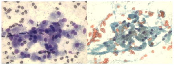
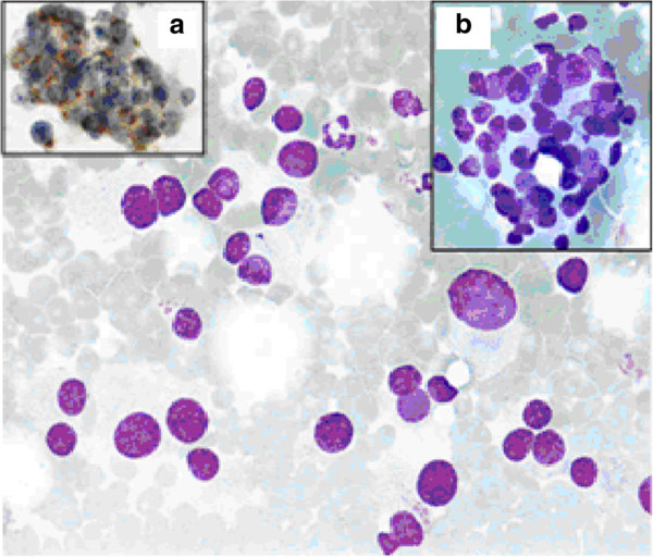
Granular cell tumor on FNA. A, a loose cluster of large cells with abundant, finely granular cytoplasm and small, round, bland nuclei (Diff-Quik, 400×). B, the same aspirate with granular, pale cytoplasm and indistinct cell borders (Papanicolaou stain, 400×).Image: Saleh Husain A et al., PMC open access (Cytojournal. 2008 Mar 28;5:4. doi: 10.1186/1742-6413-5-4) Figure 5, CC BY.Carotid body paraganglioma on FNA. Dissociated tumor cells with marked anisonucleosis, some plasmacytoid. Inset a, synaptophysin-positive tumor cells (immunocytochemistry); inset b, a small group of tumor cells without an organoid pattern. Giemsa stain, 60× objective.Image: Peric Barbara et al., PMC open access (World J Surg Oncol. 2014 Aug 20;12:267. doi: 10.1186/1477-7819-12-267) Figure 1, CC BY.
Sinonasal and Odontogenic Tumors on Cytology
Definition. Sinonasal tumors arise in the nasal cavity, paranasal sinuses and skull base. Odontogenic tumors and cysts arise from tooth-forming tissues of the jaws. Both groups are classified in the WHO Classification of Head and Neck Tumours, 5th edition (2022) (Thompson and Bishop 2022; Vered and Wright 2022). In cytology, sinonasal tumors are sampled most often at metastatic sites, especially cervical lymph nodes, and less often by FNA or scrape of the primary tumor (Okano et al. 2021; Roy et al. 2023; Satomi et al. 2026). Jaw lesions are sampled by FNA, and cell blocks from these aspirates are examined as small biopsies (Zaidi et al. 2021). The WHO 5th edition sinonasal chapter describes cytology and FNA findings where they aid diagnosis and triage (Thompson and Bishop 2022).
WHO 5th edition classification of sinonasal tumors: changes and entity features
Entity in the 5th edition
Change
Previous rule (4th edition, 2017)
Source
SWI/SNF complex-deficient sinonasal carcinoma
New entity; SMARCB1-deficient and SMARCA4-deficient subtypes
Provisionally included
Thompson and Bishop 2022
HPV-related multiphenotypic sinonasal carcinoma
New entity (no longer provisional)
Provisionally included as HPV-related carcinoma with adenoid cystic-like features
Thompson and Bishop 2022
Sinonasal tract angiofibroma
Moved to the sinonasal chapter
In the nasopharynx chapter
Thompson and Bishop 2022
Adamantinomatous craniopharyngioma
Moved to the sinonasal chapter
In the nasopharynx chapter
Thompson and Bishop 2022
Pituitary neuroendocrine tumor (PitNET)
Ectopic or invasive PitNET placed in the neuroendocrine chapter
Pituitary adenoma
Thompson and Bishop 2022
NUT carcinoma. NUT carcinoma accounts for up to 18% of poorly differentiated carcinomas of the upper aerodigestive tract. NUTM1 fuses mainly with BRD4, and the NUT antibody shows speckled nuclear staining (Thompson and Bishop 2022).
Sinonasal undifferentiated carcinoma (SNUC). A diagnosis of exclusion. A significant subset of the remaining SNUC group harbors IDH2 hotspot mutations, and this subset has a better prognosis (Thompson and Bishop 2022).
Olfactory neuroblastoma. Hyams grading is advocated because grades differ in outcome. Most tumors express somatostatin receptors (Thompson and Bishop 2022).
Teratocarcinosarcoma. Recurrent biallelic SMARCA4 inactivation and activating CTNNB1 mutations; germ cell markers are negative (Thompson and Bishop 2022).
Biphenotypic sinonasal sarcoma. Most cases harbor PAX3::MAML3, and SOX10 is always negative (Thompson and Bishop 2022).
Sinonasal squamous cell carcinoma and papillomas
Entity
Molecular and viral features
Source
Keratinizing SCC
Only rarely associated with high-risk HPV
Thompson and Bishop 2022
Non-keratinizing SCC
Diffusely p40 positive; transcriptionally active high-risk HPV in up to 60%; DEK::AFF2 in up to half
Thompson and Bishop 2022
Inverted sinonasal papilloma
EGFR mutations in about 90%; low-risk HPV in a subset
Thompson and Bishop 2022
Oncocytic sinonasal papilloma
KRAS hotspot mutations; HPV is not an etiologic factor
Thompson and Bishop 2022
Exophytic sinonasal papilloma
Usually low-risk HPV types 6 and 11
Thompson and Bishop 2022
Malignant progression in inverted or oncocytic papilloma
TP53 and/or CDKN2A alterations
Thompson and Bishop 2022
DEK::AFF2-associated carcinoma.DEK::AFF2-associated non-keratinizing SCC is often bland and can be mistaken for inverted papilloma (Thompson and Bishop 2022). In a scrape cytology report from a 26-year-old woman, features shared with sinonasal papilloma were a perivascular arrangement and intracytoplasmic nuclear debris–like structures. Features that differed were a mainly discohesive pattern, nuclear enlargement with anisonucleosis, prominent nucleoli, stippled chromatin and crackled cytoplasm. AFF2 immunohistochemistry, DEK break-apart FISH and RT-PCR confirmed the diagnosis (Satomi et al. 2026).
HPV testing of sinonasal SCC. The CAP 2025 guideline recommends routine high-risk HPV testing of sinonasal SCC, directly for transcriptionally active HPV such as RNA in situ hybridization. p16 may be used as a screen, and HPV DNA PCR is not used as a stand-alone test. About 26% of sinonasal SCCs are HPV associated. The 2018 guideline did not recommend routine HPV testing of nonoropharyngeal primaries, including sinonasal SCC (Faquin et al. 2025; Lewis et al. 2018).
HPV-related multiphenotypic sinonasal carcinoma. High-risk HPV is required, and HPV 33 is the most common type. p16 is always positive but is not specific. Myoepithelial markers such as smooth muscle actin are abluminal, and CD117 highlights ducts. Prognosis is favorable despite aggressive histology (Thompson and Bishop 2022).
Poorly differentiated and small round cell sinonasal tumors on cytology
Tumor
Cytologic findings
Confirmatory test
Source
Olfactory neuroblastoma
Small round cells with scant cytoplasm, round to oval nuclei, fine to granular chromatin, inconspicuous nucleoli; clusters and single cells
Okano et al. 2021; Roy et al. 2023; Faragalla and Weinreb 2009
NUT carcinoma
Hypercellular; small to medium round to oval cells; dusty chromatin; prominent nucleoli; scant, strongly basophilic cytoplasm; many mitoses; necrosis and apoptosis
Sampling sites. In 8 patients with olfactory neuroblastoma sampled by FNA, 7 specimens came from metastatic lesions, mostly cervical lymph nodes, and 1 from the nasal cavity tumor (Okano et al. 2021).
Grade and morphology. A two-cell pattern of large viable cells and small apoptotic cells was present in 3 of 4 high-grade tumors and in none of 4 low-grade tumors. Rosettes and paranuclear blue bodies were seen in 1 patient (Okano et al. 2021).
Differential diagnosis at nodal sites. Malignant lymphoma and small cell neuroendocrine carcinoma (Okano et al. 2021).
Immunoprofile. Olfactory neuroblastoma is keratin negative and positive for neuroendocrine markers, with S100-positive sustentacular cells surrounding the tumor nests. Patchy staining and immunophenotypic diversity occur (Faragalla and Weinreb 2009).
Metastatic aspirates. Seven aspirates from 5 patients showed small round cells with fine to coarse granular chromatin, moderate to high cellularity, loosely cohesive clusters and dispersed cells. Two-cell pattern, nuclear streaking and molding were frequent. Neuropil, fibrillary cytoplasm and rosettes were uncommon (Roy et al. 2023).
INSM1. INSM1 on destained alcohol-fixed smears was strongly positive in 30–100% of tumor cells (mean 62%) in all aspirates tested. All non-neuroendocrine malignant round cell tumors were negative (Roy et al. 2023).
NUT carcinoma and SWI/SNF-deficient carcinoma in cytology
NUT carcinoma. NUT carcinoma occurs mainly in children and young adults and is highly lethal. Cytology material can be used for morphologic and molecular diagnosis (Klijanienko et al. 2016).
SMARCB1-deficient carcinoma. An ethmoid tumor first diagnosed as non-keratinizing SCC because of strong p40 staining was reclassified as SMARCB1-deficient carcinoma after additional stains. Its lung metastasis on EBUS-FNA lost INI1 and was negative for CK5/6, CAM5.2, p40, p63, CK7, AE1/AE3 and TTF-1 (McHugh and Policarpio-Nicolas 2019).
Morphologic range. SMARCB1-deficient sinonasal carcinoma can mimic small round blue cell and spindle cell tumors on cytology (McHugh and Policarpio-Nicolas 2019).
Outcome. More than 50% of patients with SWI/SNF complex-deficient sinonasal carcinoma die within 2 years (Thompson and Bishop 2022).
Changes to odontogenic and maxillofacial bone tumors in the WHO 5th edition
Essential and desirable criteria. The 5th edition is the first to give essential and desirable diagnostic features for each entity (Vered and Wright 2022).
New entities. Surgical ciliated cyst was added to the odontogenic cysts. Adenoid ameloblastoma was newly recognized as a benign epithelial odontogenic tumor. Segmental odontomaxillary dysplasia was added to fibro-osseous tumors and dysplasias. Rhabdomyosarcoma with TFCP2 rearrangement was added to malignant jawbone tumors. None of these was an entity in the 4th edition (Vered and Wright 2022).
Deleted from this chapter. Melanotic neuroectodermal tumor of infancy and osteoid osteoma were removed from the benign bone and cartilage tumors, and solitary plasmacytoma of bone was removed from the chapter. All three were in the 4th edition chapter (Vered and Wright 2022). Melanotic neuroectodermal tumor of infancy is now covered in the oral cavity chapter under tumors of uncertain histogenesis (Muller and Tilakaratne 2022).
Molecular pathways. Genetic alterations in odontogenic cysts and tumors fall mainly into the MAPK/ERK pathway (mostly BRAF and KRAS), the Wnt/β-catenin pathway (mostly CTNNB1) and the Hedgehog pathway (PTCH1) (Guimarães, Odell and Gomes 2026).
Ameloblastoma on cytology
Frequency. Ameloblastomas account for about 1% of tumors and cysts of the jaw and are among the most common odontogenic neoplasms (Zaidi et al. 2021).
Cytomorphology. Cohesive epithelial clusters of small basaloid cells with peripherally placed tall columnar cells, and occasional large squamous cells adjoining the basaloid clusters or in isolated groups (Kaliamoorthy et al. 2013). Clusters of basaloid cells with high N/C ratio and dense chromatin; focal squamoid differentiation and cystic change (Zaidi et al. 2021).
Cell block. Cribriform and trabecular arrangements with peripheral nuclear palisading, squamoid foci and cystic change are better seen on cell block sections (Zaidi et al. 2021).
Accuracy. In 26 intraosseous jaw lesions, 15 were ameloblastomas. FNA had a sensitivity of 86.6% and a specificity of 100% for ameloblastoma (Kaliamoorthy et al. 2013).
Molecular genetics of ameloblastoma
Alteration
Distribution
Source
Hedgehog pathway or MAPK pathway mutations
Over 80% of ameloblastomas
Sweeney et al. 2014
SMO mutation (including p.L412F)
Common in maxillary ameloblastoma
Sweeney et al. 2014
BRAF p.V600E
Predominant in mandibular ameloblastoma
Sweeney et al. 2014
BRAF mutation, pooled
72% of 937 patients; more likely in younger patients, the mandible and unicystic ameloblastoma
Zhang et al. 2024
Therapeutic relevance. Ameloblastoma cells with BRAF p.V600E were sensitive to vemurafenib, and the SMO p.L412F alteration was inhibited by arsenic trioxide in experimental systems (Sweeney et al. 2014).
Testing method. Concordance between molecular tests and immunohistochemistry for BRAF mutation was strong (Zhang et al. 2024).
Odontogenic keratocyst on cytology
Aspirate. Liquid or viscous contents. Smears show many isolated or grouped keratinocytes with normal or ill-defined nuclei, numerous anucleate squamous cells and keratinous debris (Vargas et al. 2007).
Immunohistochemistry. Keratin lamellae were positive for pan-cytokeratin and CK19 and negative for CK14 (Vargas et al. 2007).
Value of FNA. FNA was a reliable and safe tool for the preoperative diagnosis of odontogenic keratocyst in 8 consecutive patients (Vargas et al. 2007).
Differential diagnosis of basaloid and squamous jaw aspirates
Lesion
Cytologic findings
Source
Ameloblastoma
Basaloid clusters with peripheral columnar palisading; squamous cells; cystic change
Kaliamoorthy et al. 2013; Zaidi et al. 2021
Odontogenic keratocyst
Keratinocytes, anucleate squames and keratinous debris
Vargas et al. 2007
Metastatic or primary SCC
Keratinized malignant cells with nuclear atypia; p16 status by site
Basaloid carcinoma with ducts and abluminal myoepithelial cells; HPV 33 most common
Thompson and Bishop 2022
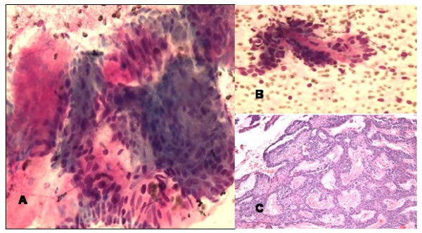
Ameloblastoma of the maxillary sinus on FNA. A and B, groups of basaloid epithelial cells with peripheral palisading, mixed with myxoid mesenchymal material (Diff-Quik, 400×). C, the resected tumor with anastomosing islands of basaloid cells in myxoid stroma (H&E, 100×).Image: Saleh Husain A et al., PMC open access (Cytojournal. 2008 Mar 28;5:4. doi: 10.1186/1742-6413-5-4) Figure 1, CC BY.
Oral Brush Cytology
Definition. Oral brush cytology is the collection of cells from the oral mucosa with a brush for cytologic examination. Baby toothbrushes, cytobrushes and the OralCDx and Orcellex brushes have been used to obtain transepithelial samples for conventional and liquid-based cytology (Alsarraf, Kujan and Farah 2018). Its main uses are the evaluation of oral potentially malignant disorders (OPMD) and oral squamous cell carcinoma (SCC), and the rapid diagnosis of vesiculobullous and infectious lesions (Verdugo-Paiva et al. 2026; Noormohammadpour et al. 2020). Biopsy remains the first choice for a definitive diagnosis of OPMD and oral SCC (Urquhart et al. 2026).
Oral potentially malignant disorders and dysplasia in the WHO 5th edition
Changes to the OPMD list. Oral lichenoid lesions were added, defined as lesions that resemble oral lichen planus but lack its typical clinical or histopathologic appearance. Chronic candidiasis, syphilitic glossitis and actinic keratosis were deleted. All three deleted disorders were listed as OPMD in the 4th edition (2017) (Muller and Tilakaratne 2022).
Smokeless tobacco keratosis. The WHO 5th edition lists smokeless tobacco keratosis with a note that its risk varies with the type of tobacco (Muller and Tilakaratne 2022).
New sections. Non-neoplastic lesions, OPMD and oral epithelial dysplasia (OED) as separate sections, submucous fibrosis and HPV-associated dysplasia. OPMD and OED were not separate sections in the 4th edition (Muller and Tilakaratne 2022).
Grading of OED. A three-tier OED grading system is kept. The binary system is described but lacks validation against malignant transformation (Muller and Tilakaratne 2022).
Carcinoma in situ. The term is not recommended in the oral cavity and is treated as synonymous with severe dysplasia (Muller and Tilakaratne 2022).
Proliferative verrucous leukoplakia. Verrucous morphology without cytologic atypia is considered mild dysplasia (Muller and Tilakaratne 2022).
HPV-associated oral epithelial dysplasia. High-risk HPV, usually type 16, is the cause. HPV is not a significant etiologic factor in oral SCC (Muller and Tilakaratne 2022).
Sampling and preparation of oral brush specimens
Transepithelial sampling. Brushes are used to obtain cells from the full thickness of the epithelium (Alsarraf, Kujan and Farah 2018).
Conventional smears. Cells are smeared onto a slide, fixed in 95% ethanol and stained by the Papanicolaou method (Sukegawa et al. 2020).
Liquid-based cytology (LBC). The brush is rinsed in an alcohol-based preservative, and the cells are processed by density centrifugation and sedimentation onto coated slides before Papanicolaou staining (Sukegawa et al. 2020).
Cell blocks. Cell blocks prepared from oral liquid-based brush samples allow immunostaining. In clinically lichenoid lesions, a Ki-67-based model detected the cases with epithelial dysplasia with 100% accuracy (Idrees et al. 2022).
Surface sampling limit. False-negative results occurred in well-differentiated SCC with keratinized surfaces and little atypia, because cytology collects cells mainly from the surface layer (Sukegawa et al. 2020).
Reporting systems for oral cytology
System
Categories
Source
Japanese Society of Clinical Cytology (Bethesda-based)
Negative for intraepithelial lesion or malignancy (NILM); oral low-grade squamous intraepithelial lesion (OLSIL); oral high-grade squamous intraepithelial lesion (OHSIL); SCC; indefinite for neoplasia
Sukegawa et al. 2020
Bethesda-type categories in a multirater study
NILM; LSIL; HSIL; SCC and other malignancy
Sekine et al. 2017
Modified 2014 Bethesda cervical system
Cervical Bethesda categories adapted to oral samples; the system used in most studies of oral classification systems
Sood et al. 2025
OralCDx computer-assisted brush biopsy
Negative; atypical; positive
Sciubba 1999
No uniform system. The absence of a uniform oral cytologic grading system is one reason oral cytology has not been accepted for mass screening (Sood et al. 2025).
Pooled accuracy of classification systems. In 9 studies with 3,163 samples, pooled sensitivity was 0.74 and specificity 0.84, with high heterogeneity (Sood et al. 2025).
Bethesda terminology and HPV. In 63 oral leukoplakias, oral SCCs and oropharyngeal SCCs, 36.5% were PCR positive for HPV 16. The Bethesda system gave higher sensitivity and specificity for HPV-related changes than Papanicolaou grading (Sivakumar et al. 2021).
Diagnostic accuracy of oral brush cytology
Study
Design
Result
Source
ADA living systematic review, 2026
36 studies; pooled estimate from 35 studies and 7,038 lesions in adults
Sensitivity 0.89, specificity 0.89; low certainty; large proportion of false-positive results
LBC sensitivity 79.4%, specificity 85.1%; conventional 76.7% and 69.2%
Sukegawa et al. 2020
Multirater study
327 cases, 10 raters
NILM as negative: sensitivity 93.5%, specificity 50.6%, NPV 89.8%; NILM and LSIL as negative: sensitivity 77.8%, specificity 83.9%
Sekine et al. 2017
OralCDx multicenter study
945 patients
Sensitivity 100% for dysplasia and carcinoma; specificity 100% for "positive" and 92.9% for "atypical"
Sciubba 1999
OralCDx follow-up study
152 scalpel biopsies after abnormal brush results
Positive predictive value 7.9% for an abnormal result and 7.4% for "atypical"; false-positive proportion up to 92.1%
Bhoopathi, Kabani and Mascarenhas 2009
Liquid-based brush cytology, oral and oropharyngeal SCC
75 patients
Before treatment: sensitivity 88%, specificity 100%; after treatment: sensitivity 71%, specificity 77%
Castillo et al. 2022
Clinically benign-appearing lesions. OralCDx found dysplasia or carcinoma in 4.5% of clinically benign-appearing lesions. The study recommended scalpel biopsy for all "atypical" and "positive" results (Sciubba 1999).
Borderline lesions. False-positive and false-negative results clustered in the LSIL and HSIL categories. Histopathologic examination is recommended for cytologic LSIL, HSIL and SCC (Sekine et al. 2017).
LBC. LBC was superior to conventional smears, especially for OLSIL and OHSIL (Sukegawa et al. 2020).
Evidence base. A 2018 systematic review of 36 studies with 4,302 samples found that the published evidence did not support firm recommendations for brush cytology in screening, and it called for studies with validated cytologic criteria (Alsarraf, Kujan and Farah 2018).
Guidelines on cytology adjuncts for oral lesions
Question
ADA 2026 (current)
ADA 2017 (previous)
Source
Cytology in adults without mucosal abnormalities
Conditional recommendation against cytology adjuncts
No adjunct had sufficient accuracy to support routine use as a triage tool for lesions
Urquhart et al. 2026; Lingen et al. 2017
Cytology in adults with mucosal abnormalities
Conditional recommendation against; reserved for specific circumstances when biopsy is not possible or not indicated and clinician and patient agree
Immediate biopsy or referral; cytology suggested in exceptional cases, such as a patient who declines biopsy or lives in a rural area with limited access, to start the diagnostic process until biopsy
Urquhart et al. 2026; Lingen et al. 2017
Clinical oral examination
Good practice statement: examine all adult patients
Good practice statement on remaining alert to OPMD
Urquhart et al. 2026; Lingen et al. 2017
Value of a negative result. In adults with oral lesions, negative cytology may accurately identify healthy patients and reduce unnecessary referrals and biopsies (Verdugo-Paiva et al. 2026).
False positives. Cytology produces a considerable proportion of false-positive results, which lead to unnecessary biopsies and patient anxiety (Verdugo-Paiva et al. 2026).
No randomized evidence. The 2026 review found no randomized trials of cytology on outcomes that matter to patients and no studies in adults without lesions (Verdugo-Paiva et al. 2026).
Oral cytology of vesiculobullous, infectious and lichenoid lesions
Lichen planus and lichenoid lesions. Cytomorphologic assessment of oral liquid-based brush samples separated lichen planus and lichenoid lesions from dysplastic lesions with an accuracy of 77.27%. Adding BAX immunostaining raised accuracy to 95% (Idrees et al. 2022).
Pemphigus vulgaris and the Tzanck smear
Cytologic features. Acantholytic cells, complete or incomplete. Rounding is the most consistent feature, followed by nuclear enlargement. Incomplete acantholytic cells predominated in 60.32% of pemphigus cases (Chaudhari et al. 2025).
Accuracy in oral erosions. In 68 patients with erosive or ulcerated oral lesions, acantholytic cells had a sensitivity of 80.5% for pemphigus vulgaris. Specificity was 84.6% with Giemsa staining and 96.3% with hematoxylin-eosin (Noormohammadpour et al. 2020).
Comparison with serology. In a dental clinic series, the Tzanck smear had a sensitivity of 96.7% and a specificity of 60%. Combining the Tzanck smear with ELISA gave 82% sensitivity and 98.7% specificity (Zhou et al. 2016).
Confirmation. Pemphigus is routinely diagnosed by histopathology and direct immunofluorescence (Noormohammadpour et al. 2020).
Pemphigoid. Tzanck smears from bullous pemphigoid blisters contained no epithelial cells. Acantholytic cells from pemphigus vulgaris showed IgG on their membranes by immunofluorescence (Verma, Khaitan and Singh 1993).
Viral and fungal infection in oral cytology
Herpes simplex virus. In oral scraping cytology, HSV-infected cells showed ground-glass nuclei and multinucleation. HSV-1 DNA was confirmed by PCR on the Papanicolaou slides, and HSV coexisted with pemphigus vulgaris in the same smear (Takahashi et al. 1998). Herpes virus cytopathic effect is also recognized on Tzanck smears, as is molluscum contagiosum (Chaudhari et al. 2025).
Candida. In 62 clinically diagnosed cases of oral candidiasis, PAS-stained smears were the most sensitive method for detecting Candida (sensitivity 100%, specificity 66.7%). Papanicolaou smears under light microscopy had a sensitivity of 67.9% (Kumaraswamy Naik et al. 2016). Chronic candidiasis is no longer listed as an OPMD in the WHO 5th edition (Muller and Tilakaratne 2022).
HPV testing on oral and oropharyngeal brush samples
Oropharyngeal and oral SCC. HPV DNA detection on liquid-based brush cytology before treatment had a sensitivity of 85%, a specificity of 100% and an accuracy of 91%. After treatment, accuracy was 100% (Castillo et al. 2022).
Dysplasia or carcinoma possible; low positive predictive value in follow-up studies
Scalpel biopsy
Sciubba 1999; Bhoopathi, Kabani and Mascarenhas 2009
LSIL, HSIL or SCC category
Borderline and malignant categories
Histopathologic examination
Sekine et al. 2017
Negative result on a keratinized exophytic lesion
False negative from surface sampling is possible
Biopsy when clinical suspicion persists
Sukegawa et al. 2020; Urquhart et al. 2026
Rounded acantholytic keratinocytes in an erosion
Pemphigus vulgaris
Histopathology and direct immunofluorescence
Noormohammadpour et al. 2020; Chaudhari et al. 2025
Ground-glass nuclei with multinucleation
Herpes simplex virus infection
Clinical correlation; HSV PCR on the smear
Takahashi et al. 1998
Lichenoid lesion
Lichen planus versus dysplasia
Cell block immunostaining or biopsy
Idrees et al. 2022
References
Alsarraf AH, Kujan O, Farah CS. The utility of oral brush cytology in the early detection of oral cancer and oral potentially malignant disorders: a systematic review. J Oral Pathol Med. 2018;47(2):104–116. doi:10.1111/jop.12660. PMID 29130527
Badoual C. Update from the 5th edition of the World Health Organization classification of head and neck tumors: oropharynx and nasopharynx. Head Neck Pathol. 2022;16(1):19–30. doi:10.1007/s12105-022-01449-2. PMID 35312986
Barry E, Gilley DR, Tassone P. A case of vagal paraganglioma initially diagnosed as metastatic thyroid cancer from fine-needle aspiration. Am J Otolaryngol. 2023;44(2):103744. doi:10.1016/j.amjoto.2022.103744. PMID 36628912
Bhoopathi V, Kabani S, Mascarenhas AK. Low positive predictive value of the oral brush biopsy in detecting dysplastic oral lesions. Cancer. 2009;115(5):1036–1040. doi:10.1002/cncr.24089. PMID 19165806
Castillo P, de la Oliva J, Alos S, et al. Accuracy of liquid-based brush cytology and HPV detection for the diagnosis and management of patients with oropharyngeal and oral cancer. Clin Oral Investig. 2022;26(3):2587–2595. doi:10.1007/s00784-021-04228-5. PMID 34839418
Chamberlain BK, McClain CM, Gonzalez RS, Coffin CM, Cates JM. Alveolar soft part sarcoma and granular cell tumor: an immunohistochemical comparison study. Hum Pathol. 2014;45(5):1039–1044. doi:10.1016/j.humpath.2013.12.021. PMID 24746209
Chandan VS, Faquin WC, Wilbur DC, Khurana KK. The utility of GLUT-1 immunolocalization in cell blocks: an adjunct to the fine needle aspiration diagnosis of cystic squamous lesions of the head and neck. Cancer. 2006;108(2):124–128. doi:10.1002/cncr.21714. PMID 16435376
Chaudhari S, Meshram N, Bhatkule M, et al. Tzanck smear: old handy tool in modern dermatology. Diagn Cytopathol. 2025;53(7):333–343. doi:10.1002/dc.25470. PMID 40129299
Chen CC, Hsiao KY, Lin HL. Clinical utility of oral cytology for detecting oral epithelial dysplasia or worse: a diagnostic meta-analysis. Cancer Cytopathol. 2026;134(4):e70088. doi:10.1002/cncy.70088. PMID 41846325
Childers EL, Fanburg-Smith JC. Congenital epulis of the newborn: 10 new cases of a rare oral tumor. Ann Diagn Pathol. 2011;15(3):157–161. doi:10.1016/j.anndiagpath.2010.10.003. PMID 21393037
Daoud A, Ronen O. Parathyroid cystic adenoma: a systematic review and meta-analysis. Endocr Pract. 2023;29(1):2–10. doi:10.1016/j.eprac.2022.11.004. PMID 36370985
Fanburg-Smith JC, Meis-Kindblom JM, Fante R, Kindblom LG. Malignant granular cell tumor of soft tissue: diagnostic criteria and clinicopathologic correlation. Am J Surg Pathol. 1998;22(7):779–794. doi:10.1097/00000478-199807000-00001. PMID 9669341
Faquin WC, Lewis JS Jr, Beadle B, et al. High-risk HPV testing in head and neck carcinoma: key updates from the 2025 College of American Pathologists guideline. Head Neck Pathol. 2025;19(1):115. doi:10.1007/s12105-025-01847-2. PMID 41021082
Faragalla H, Weinreb I. Olfactory neuroblastoma: a review and update. Adv Anat Pathol. 2009;16(5):322–331. doi:10.1097/PAP.0b013e3181b544cf. PMID 19700942
Field AS, Pitman MB, Rekhi B, et al. WHO Reporting Systems in cytopathology: a review of current status and the future. Acta Cytol. 2026 (online ahead of print). doi:10.1159/acy/abjag010. PMID 42814652
Fine SW, Li M. Expression of calretinin and the alpha-subunit of inhibin in granular cell tumors. Am J Clin Pathol. 2003;119(2):259–264. doi:10.1309/GRH4-JWX6-J9J7-QQTA. PMID 12579997
Fleming MV, Oertel YC, Rodríguez ER, Fidler WJ. Fine-needle aspiration of six carotid body paragangliomas. Diagn Cytopathol. 1993;9(5):510–515. doi:10.1002/dc.2840090508. PMID 8287758
Goldenberg D, Begum S, Westra WH, et al. Cystic lymph node metastasis in patients with head and neck cancer: an HPV-associated phenomenon. Head Neck. 2008;30(7):898–903. doi:10.1002/hed.20796. PMID 18383529
Guimarães LM, Odell EW, Gomes CC. Update on the molecular pathology and classification of odontogenic cysts and tumours. Semin Diagn Pathol. 2026;43(4):151030. doi:10.1016/j.semdp.2026.151030. PMID 42361775
Hammarstedt Nordenvall L, Jörtsö E, von Beckerath M, et al. Prevalence of cystic metastases in a consecutive cohort of surgically removed branchial cleft cysts. Acta Otolaryngol. 2022;142(1):100–105. doi:10.1080/00016489.2021.2016951. PMID 34962438
Han LM, Saunders TA, Calkins SM. The utility of high-risk human papillomavirus in situ hybridization in cytology cell block material from cystic head and neck lesions. Cancer Cytopathol. 2022;130(2):154–160. doi:10.1002/cncy.22529. PMID 34758205
Handa U, Kundu R, Mohan H. Cytomorphologic spectrum in aspirates of extra-adrenal paraganglioma. J Cytol. 2014;31(2):79–82. doi:10.4103/0970-9371.138667. PMID 25210234
Hernandez-Prera JC. Update from the 5th edition of the World Health Organization classification of head and neck tumors: the neck and lymph nodes, metastasis, and melanocytic tumors. Head Neck Pathol. 2022;16(1):110–122. doi:10.1007/s12105-022-01433-w. PMID 35312983
Holmes BJ, Sokoll LJ, Li QK. Measurement of fine-needle aspiration thyroglobulin levels increases the detection of metastatic papillary thyroid carcinoma in cystic neck lesions. Cancer Cytopathol. 2014;122(7):521–526. doi:10.1002/cncy.21413. PMID 24591483
Idrees M, Shearston K, Farah CS, Kujan O. Immunoexpression of oral brush biopsy enhances the accuracy of diagnosis for oral lichen planus and lichenoid lesions. J Oral Pathol Med. 2022;51(6):563–572. doi:10.1111/jop.13301. PMID 35460123
Ippolito G, Palazzo FF, Sebag F, Sierra M, De Micco C, Henry JF. A single-institution 25-year review of true parathyroid cysts. Langenbecks Arch Surg. 2006;391(1):13–18. doi:10.1007/s00423-005-0579-y. PMID 16180046
Jin B, Saleh H. Pitfalls in the diagnosis of adult rhabdomyoma by fine needle aspiration: report of a case and a brief literature review. Diagn Cytopathol. 2009;37(7):483–486. doi:10.1002/dc.21033. PMID 19217050
Jörtsö E, Marklund L, Harper Hysek M, et al. Fine needle aspiration cytology including the analysis of human papilloma virus (HPV) DNA enhances the diagnostic workup of solitary cystic neck lesions in a population with a high incidence of HPV positive oropharyngeal cancer. Acta Oncol. 2025;64:276–283. doi:10.2340/1651-226X.2025.42078. PMID 39962709
Kaliamoorthy S, Venkatapathy R, Babu P, Veeran V. Practical significance of utilizing fine needle aspiration cytology as an adjunct diagnostic aid in the preoperative presumptive diagnosis of ameloblastoma. J Cytol. 2013;30(4):247–251. doi:10.4103/0970-9371.126652. PMID 24648668
Klijanienko J, Le Tourneau C, Rodriguez J, et al. Cytological features of NUT midline carcinoma arising in sino-nasal tract and parotid gland: report of two new cases and review of the literature. Diagn Cytopathol. 2016;44(9):753–756. doi:10.1002/dc.23506. PMID 27338676
Koh ES, Dabsha A, Rahouma M, et al. Succinate dehydrogenase mutations in head and neck paragangliomas: a systematic review and meta-analysis of individual patients' data. Head Neck. 2024;46(7):1795–1808. doi:10.1002/hed.27652. PMID 38273766
Koshy J, Schnadig V, Nawgiri R. Is fine needle aspiration cytology a useful diagnostic tool for granular cell tumors? A cytohistological review with emphasis on pitfalls. Cytojournal. 2014;11:28. doi:10.4103/1742-6413.143304. PMID 25379050
Kumaraswamy Naik LR, Shetty P, Krishna Prasad MS, Karnaker VK, Shroff SE, Madathil LP. Fluorescence of Candida in diagnosis of oral candidiasis. Indian J Dent Res. 2016;27(6):618–622. doi:10.4103/0970-9290.199592. PMID 28169259
Layfield LJ, Esebua M, Schmidt RL. Cytologic separation of branchial cleft cyst from metastatic cystic squamous cell carcinoma: a multivariate analysis of nineteen cytomorphologic features. Diagn Cytopathol. 2016;44(7):561–567. doi:10.1002/dc.23461. PMID 26956661
Lewis JS Jr, Beadle B, Bishop JA, et al. Human papillomavirus testing in head and neck carcinomas: guideline from the College of American Pathologists. Arch Pathol Lab Med. 2018;142(5):559–597. doi:10.5858/arpa.2017-0286-CP. PMID 29251996
Lewis JS Jr, Beadle B, Bishop JA, et al. Human papillomavirus testing in head and neck carcinomas: guideline update. Arch Pathol Lab Med. 2025;149(6):e115–e150. doi:10.5858/arpa.2024-0388-CP. PMID 40126379
Li H, Huang C, Chen Q, et al. Lymph-node Epstein-Barr virus concentration in diagnosing cervical lymph-node metastasis in nasopharyngeal carcinoma. Eur Arch Otorhinolaryngol. 2020;277(9):2513–2520. doi:10.1007/s00405-020-05937-5. PMID 32240363
Lingen MW, Abt E, Agrawal N, et al. Evidence-based clinical practice guideline for the evaluation of potentially malignant disorders in the oral cavity: a report of the American Dental Association. J Am Dent Assoc. 2017;148(10):712–727.e10. doi:10.1016/j.adaj.2017.07.032. PMID 28958308
Lydiatt WM, Patel SG, O'Sullivan B, et al. Head and neck cancers—major changes in the American Joint Committee on Cancer eighth edition cancer staging manual. CA Cancer J Clin. 2017;67(2):122–137. doi:10.3322/caac.21389. PMID 28128848
Macdonald MR, Freeman JL, Hui MF, et al. Role of Epstein-Barr virus in fine-needle aspirates of metastatic neck nodes in the diagnosis of nasopharyngeal carcinoma. Head Neck. 1995;17(6):487–493. doi:10.1002/hed.2880170606. PMID 8847207
McHugh KE, Policarpio-Nicolas MLC. Metastatic SMARCB1 (INI-1)-deficient sinonasal carcinoma diagnosed by endobronchial ultrasound-guided fine-needle aspiration (EBUS-FNA): a potential diagnostic pitfall and review of the literature. Acta Cytol. 2019;63(5):431–437. doi:10.1159/000500351. PMID 31132763
Mete O, Asa SL, Gill AJ, et al. Overview of the 2022 WHO classification of paragangliomas and pheochromocytomas. Endocr Pathol. 2022;33(1):90–114. doi:10.1007/s12022-022-09704-6. PMID 35285002
Mete O, Wenig BM. Update from the 5th edition of the World Health Organization classification of head and neck tumors: overview of the 2022 WHO classification of head and neck neuroendocrine neoplasms. Head Neck Pathol. 2022;16(1):123–142. doi:10.1007/s12105-022-01435-8. PMID 35312985
Moore M, Goldfaden JS, Okhuaihesuyi O, et al. The utility of PU.1 immunohistochemical marker in differentiating granular cell tumor from oral histiocytic lesions. Oral Surg Oral Med Oral Pathol Oral Radiol. 2026;142(2):260–265. doi:10.1016/j.oooo.2026.02.008. PMID 41833496
Muller S, Tilakaratne WM. Update from the 5th edition of the World Health Organization classification of head and neck tumors: tumours of the oral cavity and mobile tongue. Head Neck Pathol. 2022;16(1):54–62. doi:10.1007/s12105-021-01402-9. PMID 35312982
Noormohammadpour P, Kamyab-Hesari K, Mousavian MS, et al. Diagnostic value of cytology in oral ulcer and comparison with histopathology and direct immunofluorescence. Dermatol Ther. 2020;33(6):e13929. doi:10.1111/dth.13929. PMID 32602587
Okano K, Ishida M, Sandoh K, et al. Review of the cytological features of olfactory neuroblastoma: a retrospective single-center study. Diagn Cytopathol. 2021;49(8):E301–E306. doi:10.1002/dc.24728. PMID 33629822
Pai RK, Erickson J, Pourmand N, Kong CS. p16(INK4A) immunohistochemical staining may be helpful in distinguishing branchial cleft cysts from cystic squamous cell carcinomas originating in the oropharynx. Cancer Cytopathol. 2009;117(2):108–119. doi:10.1002/cncy.20001. PMID 19365840
Pan JJ, Mai HQ, Ng WT, et al. Ninth version of the AJCC and UICC nasopharyngeal cancer TNM staging classification. JAMA Oncol. 2024;10(12):1627–1635. doi:10.1001/jamaoncol.2024.4354. PMID 39388190
Papavramidis TS, Chorti A, Pliakos I, Panidis S, Michalopoulos A. Parathyroid cysts: a review of 359 patients reported in the international literature. Medicine (Baltimore). 2018;97(28):e11399. doi:10.1097/MD.0000000000011399. PMID 29995785
Pareja F, Brandes AH, Basili T, et al. Loss-of-function mutations in ATP6AP1 and ATP6AP2 in granular cell tumors. Nat Commun. 2018;9(1):3533. doi:10.1038/s41467-018-05886-y. PMID 30166553
Pynnonen MA, Gillespie MB, Roman B, et al. Clinical practice guideline: evaluation of the neck mass in adults. Otolaryngol Head Neck Surg. 2017;157(2 suppl):S1–S30. doi:10.1177/0194599817722550. PMID 28891406
Rana RS, Dey P, Das A. Fine needle aspiration (FNA) cytology of extra-adrenal paragangliomas. Cytopathology. 1997;8(2):108–113. doi:10.1111/j.1365-2303.1997.tb00592.x. PMID 9134336
Rayess HM, Monk I, Svider PF, et al. Thyroglossal duct cyst carcinoma: a systematic review of clinical features and outcomes. Otolaryngol Head Neck Surg. 2017;156(5):794–802. doi:10.1177/0194599817696504. PMID 28322121
Roy D, Sriram S, Kakkar A, et al. Cytological diagnosis of olfactory neuroblastoma at metastatic sites, with emphasis on role of insulinoma-associated protein 1 immunocytochemistry. Diagn Cytopathol. 2023;51(2):146–152. doi:10.1002/dc.25074. PMID 36369994
Satomi H, Honma K, Kukita Y, et al. Scrape cytology of DEK::AFF2 fusion-associated papillary squamous cell carcinoma of the sinonasal tract masquerading as Schneiderian papilloma: a case report. Acta Cytol. 2026 (online ahead of print). doi:10.1159/acy/abjag004. PMID 42607006
Satturwar SP, Rossi ED, Maleki Z, et al. Thyroid paraganglioma: a diagnostic pitfall in thyroid FNA. Cancer Cytopathol. 2021;129(6):439–449. doi:10.1002/cncy.22390. PMID 33232572
Sciubba JJ. Improving detection of precancerous and cancerous oral lesions: computer-assisted analysis of the oral brush biopsy. U.S. Collaborative OralCDx Study Group. J Am Dent Assoc. 1999;130(10):1445–1457. doi:10.14219/jada.archive.1999.0055. PMID 10570588
Sekine J, Nakatani E, Hideshima K, Iwahashi T, Sasaki H. Diagnostic accuracy of oral cancer cytology in a pilot study. Diagn Pathol. 2017;12(1):27. doi:10.1186/s13000-017-0618-3. PMID 28298213
Sheahan P, O'Leary G, Lee G, Fitzgibbon J. Cystic cervical metastases: incidence and diagnosis using fine needle aspiration biopsy. Otolaryngol Head Neck Surg. 2002;127(4):294–298. doi:10.1067/mhn.2002.128600. PMID 12402008
Shen J, Wei X, Bian Y, et al. A systematic review of 33 cases of branchiogenic carcinoma: clinicopathological features and prognostic outcomes. Head Neck. 2026;48(8):2087–2098. doi:10.1002/hed.70209. PMID 41793169
Sivakumar N, Narwal A, Kumar S, et al. Application of the Bethesda system of reporting for cervical cytology to evaluate human papilloma virus induced changes in oral leukoplakia, oral squamous cell carcinoma, and oropharyngeal squamous cell carcinoma: a cytomorphological and genetic study. Diagn Cytopathol. 2021;49(9):1036–1044. doi:10.1002/dc.24813. PMID 34165918
Sood A, Narayan B, Raghavan S, et al. Diagnostic accuracy of oral cytology classification systems for oral cancer and potentially malignant disorders: systematic review and meta-analysis. J Am Soc Cytopathol. 2025;14(6):444–453. doi:10.1016/j.jasc.2025.07.011. PMID 40975707
Stelow EB, Bishop JA. Update from the 4th edition of the World Health Organization classification of head and neck tumours: tumors of the nasal cavity, paranasal sinuses and skull base. Head Neck Pathol. 2017;11(1):3–15. doi:10.1007/s12105-017-0791-4. PMID 28247233
Sukegawa S, Ono S, Nakano K, et al. Clinical study on primary screening of oral cancer and precancerous lesions by oral cytology. Diagn Pathol. 2020;15(1):107. doi:10.1186/s13000-020-01027-6. PMID 32912249
Sweeney RT, McClary AC, Myers BR, et al. Identification of recurrent SMO and BRAF mutations in ameloblastomas. Nat Genet. 2014;46(7):722–725. doi:10.1038/ng.2986. PMID 24859340
Tabet P, Saydy N, Letourneau-Guillon L, et al. Cystic masses of the lateral neck: diagnostic value comparison between fine-needle aspiration, core-needle biopsy, and frozen section. Head Neck. 2019;41(8):2696–2703. doi:10.1002/hed.25755. PMID 30945785
Takahashi I, Kobayashi TK, Suzuki H, Nakamura S, Tezuka F. Coexistence of pemphigus vulgaris and herpes simplex virus infection in oral mucosa diagnosed by cytology, immunohistochemistry, and polymerase chain reaction. Diagn Cytopathol. 1998;19(6):446–450. doi:10.1002/(sici)1097-0339(199812)19:6<446::aid-dc8>3.0.co;2-2. PMID 9839135
Thompson LDR, Bishop JA. Update from the 5th edition of the World Health Organization classification of head and neck tumors: nasal cavity, paranasal sinuses and skull base. Head Neck Pathol. 2022;16(1):1–18. doi:10.1007/s12105-021-01406-5. PMID 35312976
Thompson LDR, Herrera HB, Lau SK. Thyroglossal duct cyst carcinomas: a clinicopathologic series of 22 cases with staging recommendations. Head Neck Pathol. 2017;11(2):175–185. doi:10.1007/s12105-016-0757-y. PMID 27704385
Urquhart O, Bhosale AS, Martins-Pfeifer C, et al. Living evidence-informed guideline on the early detection of oral squamous cell carcinoma and potentially malignant disorders: cytology adjuncts to determine the need for biopsy, version 2026 1.0. J Am Dent Assoc. 2026;157(3):224–234. doi:10.1016/j.adaj.2025.12.017. PMID 41781073
Valero C, Shah JP. Staging of human papilloma virus related cancers of the oropharynx. J Surg Oncol. 2021;124(6):931–934. doi:10.1002/jso.26584. PMID 34622962
van Nederveen FH, Gaal J, Favier J, et al. An immunohistochemical procedure to detect patients with paraganglioma and phaeochromocytoma with germline SDHB, SDHC, or SDHD gene mutations: a retrospective and prospective analysis. Lancet Oncol. 2009;10(8):764–771. doi:10.1016/S1470-2045(09)70164-0. PMID 19576851
Vargas PA, da Cruz Perez DE, Mata GM, et al. Fine needle aspiration cytology as an additional tool in the diagnosis of odontogenic keratocyst. Cytopathology. 2007;18(6):361–366. doi:10.1111/j.1365-2303.2007.00444.x. PMID 17388930
Vazquez Salas S, Pedro K, Balram A, et al. Head and neck cystic lesions: a cytology review of common and uncommon entities. Acta Cytol. 2022;66(5):359–370. doi:10.1159/000525144. PMID 35717936
Verdugo-Paiva F, Martins-Pfeifer C, Bhosale AS, et al. Cytology adjuncts to determine the need for biopsy for early detection of oral squamous cell carcinoma and potentially malignant disorders: an evidence summary of a living systematic review, version 2026 1.0. J Am Dent Assoc. 2026;157(3):235–246. doi:10.1016/j.adaj.2025.12.008. PMID 41781074
Vered M, Wright JM. Update from the 5th edition of the World Health Organization classification of head and neck tumors: odontogenic and maxillofacial bone tumours. Head Neck Pathol. 2022;16(1):63–75. doi:10.1007/s12105-021-01404-7. PMID 35312978
Wakely PE Jr. Benign peripheral nerve tumors: analysis of 89 FNA cases emphasizing diagnostic accuracy and their morphologic spectrum. Cancer Cytopathol. 2023;131(5):300–312. doi:10.1002/cncy.22680. PMID 36650522
Weiner MF, Miranda RN, Bardales RH, et al. Diagnostic value of GLUT-1 immunoreactivity to distinguish benign from malignant cystic squamous lesions of the head and neck in fine-needle aspiration biopsy material. Diagn Cytopathol. 2004;31(5):294–299. doi:10.1002/dc.20127. PMID 15468132
Westra WH, Lewis JS Jr. Update from the 4th edition of the World Health Organization classification of head and neck tumours: oropharynx. Head Neck Pathol. 2017;11(1):41–47. doi:10.1007/s12105-017-0793-2. PMID 28247229
Zaidi A, Srinivasan R, Rajwanshi A, Dey P, Gupta K. Ameloblastoma diagnosis by fine-needle aspiration cytology supplemented by cell block samples. Diagn Cytopathol. 2021;49(3):E93–E98. doi:10.1002/dc.24600. PMID 32841532
Zanoni DK, Patel SG, Shah JP. Changes in the 8th edition of the American Joint Committee on Cancer (AJCC) staging of head and neck cancer: rationale and implications. Curr Oncol Rep. 2019;21(6):52. doi:10.1007/s11912-019-0799-x. PMID 30997577
Zhang AB, Zhang JY, Liu YP, et al. Clinicopathological characteristics and diagnostic accuracy of BRAF mutations in ameloblastoma: a Bayesian network analysis. J Oral Pathol Med. 2024;53(6):393–403. doi:10.1111/jop.13542. PMID 38777565
Zhang Y, Yu X, Yang Y, et al. Diagnostic utility of immunohistochemical analysis and Epstein-Barr virus-encoded small RNAs in situ hybridisation of cell block sections obtained using fine-needle aspiration in nasopharyngeal carcinoma with lymph node metastasis. Diagn Cytopathol. 2019;47(3):200–205. doi:10.1002/dc.24074. PMID 30593738
Zhou T, Fang S, Li C, Hua H. Comparative study of indirect immunofluorescence, enzyme-linked immunosorbent assay, and the Tzanck smear test for the diagnosis of pemphigus. J Oral Pathol Med. 2016;45(10):786–790. doi:10.1111/jop.12439. PMID 27005471
Zidar N, Gale N. Update from the 5th edition of the World Health Organization classification of head and neck tumors: hypopharynx, larynx, trachea and parapharyngeal space. Head Neck Pathol. 2022;16(1):31–39. doi:10.1007/s12105-021-01405-6. PMID 35312977
Image credits
Granular cell tumor on FNA. A, a loose cluster of large cells with abundant, finely granular cytoplasm and small, round, bland nuclei (Diff-Quik, 400×). B, the same aspirate with granular, pale cytoplasm and indistinct cell borders (Papanicolaou stain, 400×). Image: Saleh Husain A et al., PMC open access (Cytojournal. 2008 Mar 28;5:4. doi: 10.1186/1742-6413-5-4) Figure 5, CC BY.
Carotid body paraganglioma on FNA. Dissociated tumor cells with marked anisonucleosis, some plasmacytoid. Inset a, synaptophysin-positive tumor cells (immunocytochemistry); inset b, a small group of tumor cells without an organoid pattern. Giemsa stain, 60× objective. Image: Peric Barbara et al., PMC open access (World J Surg Oncol. 2014 Aug 20;12:267. doi: 10.1186/1477-7819-12-267) Figure 1, CC BY.
Ameloblastoma of the maxillary sinus on FNA. A and B, groups of basaloid epithelial cells with peripheral palisading, mixed with myxoid mesenchymal material (Diff-Quik, 400×). C, the resected tumor with anastomosing islands of basaloid cells in myxoid stroma (H&E, 100×). Image: Saleh Husain A et al., PMC open access (Cytojournal. 2008 Mar 28;5:4. doi: 10.1186/1742-6413-5-4) Figure 1, CC BY.